ARAŞTIRMA
KRONIK BLEFARITLI GERIATRIK HASTALARDA
MEIBOMIAN BEZ MORFOLOJISI VE GÖZ YAŞI
FILM TABAKASININ DEĞERLENDIRILMESI
A
BSTRACTÖ
Z Turkish Journal of GeriatricsDOI: 10.31086/tjgeri.2019.108 2019; 22(3): 324-330 Cafer TANRIVERDI1
Burcu NUROZLER TABAKÇI1
CORRESPONDANCE Cafer TANRIVERDI
Istanbul Medipol University, Ophthalmology Department, Istanbul, Turkey
Phone: +905056578732 e-mail: [email protected] Received: 19/03/2019 Accepted: 17/08/2019
1 Istanbul Medipol University, Ophthalmology Department, Istanbul, Turkey.
GERIATRIC PATIENTS WITH CHRONIC
BLEPHARITIS
Introduction: To investigate the changes in meibomian gland morphology and their
effects on tear parameters in geriatric patients with chronic blepharitis.
Materials and Method: The study included 70 eyes of 35 healthy subjects in the control
group and 60 eyes of 30 patients with chronic blepharitis. The patients were requested to complete a symptom questionnaire [Ocular surface disease index] before clinical examination. Non-contact meibography and non-invasive tear break-up time tests were performed with the Sirius Scheimpflug camera. Meibography scoring (Meiboscore) of the upper and lower lids was performed in all patients. Invasive tear break up time was performed 1 min after putting a single drop of fluorescein in all patients.
Results: The mean age of the patients was 74.6±7.1 and 73.9±6.3 years in the blepharitis
and control groups, respectively (p = 0.669). The total meiboscore, non-invasive first and mean tear break up time, invasive tear break up time, and ocular surface disease index score were 3.6±1.7, 8.0±4.8, 9.7±4.0, 7.1±3.0, and 27.4±11.7, respectively, for the blepharitis group and 3.0±1.4, 10.4±5.7, 11.4±5.0, 8.6±3.1, and 18.0±6.4, respectively for the control group (respectively p = 0.03, 0.01, 0.03, 0.00, 0.00).
Conclusion: Chronic blepharitis in patients aged ≥65 years was associated with
morphological changes in meibomian gland, decreased tear break up time, and increased ocular surface disease index scores. In these patients, meibomian gland atrophy score is considered as a valuable method consistent with clinical findings.
Keywords: Blepharitis; Tears; Dry eye syndromes; Meibomian gland; Aged
Giriş: Bu çalışmada kronik blefariti olan geriatrik hastalarda meibomian bez morfolojisini
ve bunun gözyaşı parametrelerine olan etkisini değerlendirmek amaçlanmıştır.
Gereç ve Yöntem: Bu çalışmaya 30 kronik blefaritli hastanın 60 gözü ve kontrol grubuna
35 sağlıklı bireyin 70 gözü dahil edildi. Klinik muayene öncesinde tüm hastalardan semptom anketini tamamlaması istendi [Oküler yüzey hastalık indeksi]. Non-kontakt meibografi ve non-invaziv göz yaşı kırılma zamanı ölçümleri Sirius Scheimpflug Camera ile yapıldı. Tüm olgularda her iki gözde alt ve üst kapaklarda meibografi skorlaması (Meiboskor) yapıldı. Tüm olgularda invaziv gözyaşı kırılma zamanı bir damla floresein damlatıldıktan 1 dakika sonra ölçüldü.
Bulgular: Çalışmaya alınan olgularda yaş ortalaması blefarit grubunda 74.6±7.1, kontrol
grubunda 73.9±6.3 yıldı (p = 0.669). Sırasıyla toplam meiboskor, non-invaziv ilk ve ortalama gözyaşı kırıma zamanı, invaziv gözyaşı kırılma zamanı, oküler yüzey hastalık indeksi skoru blefarit grubunda 3.6±1.7, 8.0±4.8, 9.7±4.0, 7.1±3.0, 27.4±11.7 iken kontrol grubunda 3.0±1.4, 10.4±5.7, 11.4±5.0, 8.6±3.1, 18.0±6.4 bulundu (sırasıyla p = 0.03, 0.01, 0.03, 0.00, 0.00).
Sonuç: Altmışbeş yaş ve üzerindeki kronik blefaritli hastalarda meibomian bezlerde
morfolojik değişiklikler, azalmış gözyaşı kırılma zamanı ve artmış oküler yüzey hastalık indeksi skorları olduğu görülmektedir. Bu hastalarda, meibomian bez atrofi skorunun klinik bulgularla uyumlu değerli bir yöntem olduğu düşünüldü.
INTRODUCTION
Blepharitis is one of the common diseases encountered by ophthalmologists and can be defined as a large group of diseases that lead to the inflammation of the eyelid margin and ocular surface and can affect children and adults (1,2). The symptoms associated with this inflammatory condition are burning, irritation of the eyelid, crusting, and injection of the eyelid margin and conjunctiva. The exact cause of blepharitis is unknown, thereby making this condition difficult to treat (3).
Blepharitis is categorized in two different ways by Lindsley et al. (4). The first categorization is based on the disease duration: acute or chronic blepharitis. The second categorization is based on the anatomical position of the disease: anterior and posterior blepharitis. Notably, posterior blepharitis is more common and often involves the meibomian gland (MG) on the internal surface of the eyelid. Prolonged obstruction of the MG results in inflammation and hyperkeratinization of the ductal epithelium. Therefore, meibomian gland dysfunction (MGD) is thought to be a complication of posterior blepharitis (5).
MG provides the oily components of the tear film, which helps to reduce tear fluid evaporation. MGD is one of the most common causes of evaporative dry eye disease (DED), which is more common than the aqueous-deficient dry eye. Changes in MG morphology can be seen in patients
invasive meibography helps to evaluate the status of MG in vivo, and owing to this method, the structure of MG, including channels and acini, can be completely observed. Additionally, non-invasive meibography provides photographic documentation of MG (7,8).
Conversely, the prevalence of DED has increased (9). The Asia Dry Eye Society recently reviewed the criteria for dry eye diagnosis and defined DED as follows: “dry eye is a multifactorial disease characterized by unstable tear film causing a variety of symptoms and/or visual impairment, potentially accompanied by ocular surface damage.” This new definition emphasized the significance of the tear film in DED as well as the diagnostic importance of non-invasive meibography and tear film break-up time (TBUT) tests (10).
Several studies have shown that MGD increases with age (11-13). In addition, a study by McCann et al. (3) revealed a significant difference in tear physiology and MG function between patients with blepharitis and healthy individuals.
These reasons may point to a significant correlation between aging, blepharitis, MGD, and DED. Therefore, the present study investigated the MG morphology and its effect on the tear film layer in geriatric patients with chronic blepharitis.
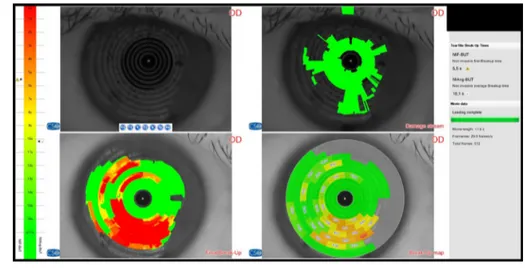
detected with Sirius Scheimpflug camera (Figure 1). The first and mean non-invasive TBUT were recorded. The non-invasive TBUT measurements were performed first based on the assumption that the use of fluorescein drops in patients may affect TBUT duration. Thereafter, invasive TBUT was performed 1 min after a single drop of fluorescein was used in all patients. The invasive TBUT was evaluated using a slit lamp with a cobalt blue exciter filter. The first invasive TBUT was recorded. Finally, non-invasive meibography was performed in all patients. The lipids could be discharged from the MG orifices to the ocular
surface by manipulating the lid during non-invasive meibography. This manipulation could have led to errors in TBUT measurement. Therefore, non-invasive meibography was performed after TBUT measurements. Meibography scoring (meibomian gland atrophy score or meiboscore) in the upper and lower lids of both the eyes was performed for all patients. First, MG of the upper eyelid was examined and graded. The grading was based on the criteria proposed by Arita et al. (7,11), where in the atrophy is graded as follows: 0 point for no atrophy, 1 point for less than one-third atrophy, 2 points for more than one-third atrophy,
Figure 1. Sirius Scheimpflug camera imaging in a representative patient; non-invasive first and mean tear film break-up time.
and 3 points for more than two-third atrophy. All eyelids were examined and graded in the same way (Figure 2). The values obtained for the upper and lower eyelids were then added to the total meiboscore ranging from 0–6. Patients who were <65 years; using contact lens; undergoing eyelid surgery; or having rosacea, glaucoma, chronic ocular disease, eyelid abnormalities, and systemic disease with dry eyes were excluded from the study. The control group included subjects over 65 years of age who had no ocular and systemic disease and had no ocular surgery.
Statistical analysis was performed using the Statistical Package for the Social Sciences version 20.0 (SPSS Inc., Chicago, IL, USA). Normal distribution was assessed using the Kolmogorov– Smirnov test. Independent samples t-test was used to test the statistical significance, and a p value of <0.05 was considered statistically significant.
RESULTS
The blepharitis group comprised 22 women and 8 men, and the control group comprised 28 women and 7 men. The mean age of the patients included in the study was 74.6 ± 7.1 and 73.9 ± 6.3 years in the blepharitis and control group, respectively. No statistically significant difference was found regarding age and sex between the groups (p = 0.669).
Total meiboscore value was 3.6 ± 1.7 and 3.0 ± 1.4 for the blepharitis and control group,
was 2.8 ± 1.3 in women and 3.7 ± 1.7 in men. The difference was statistically significant (p = 0.03). There was no statistically significant difference in all other parameters between men and women in the healthy group (p > 0.05). In the blepharitis group, there was no statistically significant difference in the parameters according to gender (p > 0.05).
DISCUSSION
The primary goal of our study was to investigate the effects of chronic blepharitis on MG and eventually on the tear film layer in the geriatric patients. Additionally, we determined whether the upper or lower lid MG is more affected in patients with chronic blepharitis and healthy subjects in the geriatric patients. Furthermore, we observed the effects of tear film changes on the activities of daily living in the blepharitis and control groups through OSDI scoring.
Blepharitis is a chronic disease that significantly affects the ocular surface. Posterior blepharitis often results in significant eyelid margin abnormalities, such as telangiectasia, swelling, and hyperemia (2,14). Jester et al. (14) reported that these findings are associated with the keratinization abnormalities of the ductal epithelium that led to increased obstruction of the MG orifices and evaporation of tear film.
Meibography was first described in 1977 by Tapie using infrared light (15). Over the years, meibography has gained importance and has been
Previous studies have shown that MGD increases with age (5,11). Moreover, the adverse effects of MGD in blepharitis are well known (2,3,5). However, to the best of our knowledge, no studies have been performed regarding the changes in MG in patients with chronic blepharitis and geriatric patients. Considering that DED is more common in the geriatric patients, we believe that this study is useful to better understand the underlying etiological factors. This effect can be understood well in the geriatric patients and can prove critical regarding preventive medicine use. Meticulous treatment of chronic blepharitis observed in these patients can significantly help with MGD and DED prophylaxis in the future.
The present study had few limitations. First, it was a retrospective study. Second, only a limited number of cases were found as per the study criteria. Third, only meibography and TBUT were used to evaluate MGD and the tear film layer, and the OSDI score was used to assess the effects of these on the activities of daily living.
The findings of the present study suggest that meiboscore values and TBUT durations are significantly reduced in geriatric patients with chronic blepharitis. In addition, these changes led to significant limitations in the activities of daily living of the patients. Aging, blepharitis, MGD, and DED were found to be correlated. Meibography
and TBUT tests are thought to be useful tools in clinical practice. Moreover, meibography can be considered a beneficial diagnostic method to observe the changes in MG morphology.
Our study is concordant with the literature. A study by McCann et al. (3) found significantly lower tear and MG function in patients with blepharitis. Additionally, they found that the tear evaporation rate was markedly higher in the blepharitis group than the group of healthy individuals. Alsuhaibani et al. (17) explained that MG could range from mild to total dropout in patients with blepharitis. Finis et al. (18) and Arita et al. (11) showed that the total meiboscore increased significantly with age.
Lately, in vivo confocal microscopy (IVCM) has been used to demonstrate changes in MG. Randon et al. (19) showed that significant changes in MG could be found in patients with blepharitis using IVCM. One of the most significant findings of this study is the strong correlation between IVCM scores and meibography scores. Their study considered IVCM to be another valuable method that could demonstrate MGD.
Recently Arita et al. (20) showed that male sex was associated with MGD and female sex was associated with dry eye. On the other hand, Amano et al. (21) didn’t find a significant relationship between sex with MGD in their study. Viso et al. (22) explained that the prevalence of asymptomatic
Table 1. The results of the study are summarized.
Blepharitis group Control group *P
Total meiboscore 3.6 ± 1.7 3.0 ± 1.4 0.03
Non-invasive first TBUT 8.0 ± 4.8 10.4 ± 5.7 0.01
Non-invasive mean TBUT 9.7 ± 4.0 11.4 ± 5.0 0.03
Invasive TBUT 7.1 ± 3.0 8.6 ± 3.1 0.00
OSDI scores 27.4 ± 11.7 18.0 ± 6.4 0.00
REFERENCES
1. McCulley JP, Dougherty JM, Deneau DG. Classification of chronic blepharitis. Ophthalmology. 1982; 89(10): 1173-80. (PMID: 6218459).
2. AlDarrab A, Alrajeh M, Alsuhaibani AH. Meibography for eyes with posterior blepharitis. Saudi J Ophthalmol. 2017; 31(3): 131-4. (PMID:28860908).
3. McCann LC, Tomlinson A, Pearce EI, Diaper C. Tear and meibomian gland function in blepharitis and normals. Eye Contact Lens. 2009; 35(4): 203-8. (PMID:19516145).
4. Lindsley K, Matsumura S, Hatef E, Akpek EK. Interventions for chronic blepharitis. Cochrane Database Syst Rev. 2012 May 16; (5): CD005556. (PMID:22592706).
5. Soh R, Tong L. Healthcare delivery in meibomian gland dysfunction and blepharitis. Ocul Surf. 2018 Nov 17. pii: S1542-0124(18)30160-5. (PMID:30458245). 6. Geerling G, Tauber J, Baudouin C, et al. The international
workshop on meibomian gland dysfunction: report of the subcommittee on management and treatment of meibomian gland dysfunction. Invest Ophthalmol Vis
Diagnosis: A Consensus Report by the Asia Dry Eye Society. Ocul Surf. 2017;15(1):65-76. (PMID:27725302). 11. Arita R, Itoh K, Inoue K, Amano S. Noncontact infrared
meibography to document age-related changes of the meibomian glands in a normal population. Ophthalmology. 2008; 115(5):911-5. (PMID:18452765). 12. Ban Y, Shimazaki-Den S, Tsubota K, Shimazaki J.
Morphological evaluation of meibomian glands using noncontact infrared meibography. Ocul Surf. 2013;11(1):47-53. (PMID:23321359).
13. Yeotikar NS, Zhu H, Markoulli M, Nichols KK, Naduvilath T, Papas EB. Functional and Morphologic Changes of Meibomian Glands in an Asymptomatic Adult Population. Invest Ophthalmol Vis Sci. 2016;57(10):3996-4007. (PMID:27490319).
14. Jester JV, Rife L, Nii D, Luttrull JK, Wilson L, Smith RE. In vivo biomicroscopy and photography of meibomian glands in a rabbit model of meibomian gland dysfunction. Invest Ophthalmol Vis Sci. 1982;22(5):660-7. (PMID:7076409).
15. Tapie R. Etude biomicroscopique des glandes de
MGD is more common than symptomatic MGD. In this study, MGD was found to be higher in men in asymptomatic cases but there was no statistically significant a difference between two genders in symptomatic cases.
In our study, subgroup analyses showed that only meiboscore was higher in males among all parameters in healthy group, whereas there was no difference between women and men in blepharitis group.
We firmly believe that non-invasive
meibography and TBUT tests are useful tools in the etiological evaluation of MGD and DED, which are common nowadays. Furthermore, non-invasive meibography can prove beneficial as a standard diagnostic method alongside other diagnostic tests to evaluate patients with MGD and DED. Nonetheless, further prospective randomized studies with more participants are required to validate the data of the present study.
of Meibomian Gland Atrophy by Non-contact Infrared Meibography. Curr Eye Res. 2015;40(10):982-9. (PMID:25330304).
19. Randon M, Aragno V, Abbas R, Hong L, Labbé A, Baudouin C. In vivo confocal microscopy classification in the diagnosis of meibomian gland dysfunction. Eye (Lond). 2018 Dec 12. (PMID:30542066).
20. Arita R, Mizoguchi T, Kawashima M, et al. Meibomian Gland Dysfunction and Dry Eye are Similar, but Different based on a Population-Based Study
(Hirado-Takushima Study) in Japan. Am J Ophthalmol. 2019 Mar 6. (PMID: 30851269).
21. Amano S, Inoue K. Clinic-Based Study on Meibomian Gland Dysfunction in Japan. Invest Ophthalmol Vis Sci. 2017;58(2):1283-7. (PMID: 28241316).
22. Viso E, Rodríguez-Ares MT, Gude F. Prevalence and associations of external punctal stenosis in a general population in Spain. Cornea. 2012;31(11):1240-5. (PMID:22367046).