Non-epithelial tumors of the larynx: a single
institution review
☆
S. Karatayli-Ozgursoy, MD
a,b, J.A. Bishop, MD
a,c, A.T. Hillel, MD
a,
L.M. Akst, MD
a, S.R. Best, MD
a,⁎
aDepartment of Otolaryngology–Head and Neck Surgery, Johns Hopkins School of Medicine, Baltimore, MD, USA b
Department of Otolaryngolgy, Ufuk University, Ankara, Turkey
cDepartment of Pathology, Johns Hopkins School of Medicine, Baltimore, MD, USA
A R T I C L E I N F O A B S T R A C T
Article history:
Received 1 December 2015
Aim: Non-epithelial tumors of the larynx are rare and encompass a wide range of pathology. We present the decade-long experience of a single institution to define clinical presentations and outcomes.
Material and methods: This is a ten year retrospective chart review of a tertiary head and neck cancer center. Index patients were identified from a review of a pathology database, and patient demographics, presenting signs and symptoms, treatment modalities, and clinical outcomes were extracted from electronic medical records. Epithelial tumors (squamous cell carcinoma, spindle cell carcinoma, and salivary tumors), granulomas, sarcoidosis, papilloma, and amyloidosis were all excluded.
Results: Twenty-four patients with ages ranging from 2 months-old to 84 years were identified. Malignant lesions (11) included chondrosarcoma (6), Kaposi's sarcoma (2), metastatic melanoma, synovial cell sarcoma, and T cell neoplasm. Six were operated upon endolaryngeally, but four required either upfront or salvage total laryngectomy. Two received adjuvant therapy. Benign lesions (13) included hemangioma (4), granular cell tumor (3), myofibroblastic tumor (2), schwannoma (2), chondroma, and ossifying fibromyxoid tumor. Nine underwent endolaryngeal operations, and four were managed medically or with observation. None have required aggressive open resection or total laryngectomy.
Conclusion: Treatment approach of non-epithelial tumors of the larynx depends on the site and extent of the tumor, histology, and sensitivity of adjuvant therapy. Benign tumors can be managed without need for aggressive resection thereby sparing laryngeal function.
© 2016 Elsevier Inc. All rights reserved.
1.
Introduction
Benign and malignant laryngeal neoplasms are almost exclu-sively epithelial in origin. The most frequently occurring
malignant (squamous cell carcinoma), premalignant (squamous dysplasia), and benign (papilloma) lesions of the larynx all arise from the squamous epithelium which covers the true vocal cords. Even rare forms of laryngeal malignancies such as spindle
☆ This work was presented at the 10th Congress of the European Laryngology Society, April 9–12, 2014, Antalya, Turkey.
⁎ Corresponding author at: Department of Otolaryngology–Head and Neck Surgery, Johns Hopkins School of Medicine, 601 N. Caroline Street, Room 6210, Baltimore, MD 21287. Tel.: + 1 410 955 1654; fax: +1 410 955 0035.
http://dx.doi.org/10.1016/j.amjoto.2016.01.005 0196-0709/© 2016 Elsevier Inc. All rights reserved.
A v a i l a b l e o n l i n e a tw w w . s c i e n c e d i r e c t . c o m
ScienceDirect
cell carcinoma and salivary gland tumors are epithelial in origin. However, there is a small subset of laryngeal tumors that are non-epithelial in origin, arising from a diversity of cell types, including cartilage, muscle, or blood vessels. These tumors are usually reported as small series or isolated cases, making it difficult to assess incidence and management strategies.
In this study, we aim to present our experience with non-epithelial neoplasms over a decade at a large cancer to provide data on the incidence, management, and outcomes for these rare tumors. We further aim to provide a guide for otolaryngologists in their approach to these rare tumors by reviewing the literature to discuss approaches and treatment strategies for rarely encountered pathologies.
2.
Materials and methods
In an IRB-approval protocol, the pathology archives of the Johns Hopkins Hospital and clinical records of all patients with laryngeal lesions were reviewed to identify non-epithe-lial laryngeal tumors between January 2004 and December 2013. Age, gender, presenting symptoms, location of the tumor, pathology, treatment modality (surgery, radiation and/or chemotherapy), and disease status were then extract-ed from the patient charts. Epithelial tumors (squamous cell, spindle cell, and salivary tumors), granulomas, sarcoidosis, cystic lesions, papilloma, and amyloidosis were all excluded.
3.
Results
Seven hundred seventy eight (778) patient records were identified with laryngeal tumors over the decade-long study period. When all epithelial tumors (squamous cell carcinoma, spindle cell, and salivary tumors) were excluded, twenty-four patients were identified with non-epithelial tumors (Table 1). Eleven malignant tumors were classified as non-epithelial in origin, making up 1% of all malignant laryngeal lesions. These tumors presented in adulthood, with ages at presentation ranging from 41 to 84 years old. The majority (8; 72%) were male, and the most common presenting symptom was dysphonia. Only four (36%) were smokers. The most common malignant pathology was chondrosarcoma, which presented exclusively in the cricoid cartilage. There were two cases of Kaposi's sarcoma, both in patients with long-standing HIV infections. Three lesions presented as a site of metastasis from malignancies primary to other locations– melanoma, synovial cell sarcoma, and a T cell neoplasm diagnosed in the larynx as an extra-cutaneous site from a known cutaneous malignancy.
Chondrosarcoma was the most common non-epithelial malignancy (6 patients). Initial management was divided between open and endoscopic approaches. Two patients underwent up-front total laryngectomy, two patients had endoscopic laser excisions, and one underwent an open partial resection of the cricoid cartilage. One elderly patient underwent biopsy and palliative tracheotomy and elected no further treatment. None were treated with adjuvant therapy. The recurrence rate for these lesions was high, including one patient with a total laryngectomy who then required a partial
pharyngectomy and has persistent lesions in the posterior pharyngeal wall and soft palate. Both endoscopic resections had recurrence/persistence of disease, one treated with total laryngectomy and another with serial endoscopic excisions. The patient with partial resection of the cricoid ultimately required total laryngectomy. The overall rate of larynx preservation is therefore only 33%, with no patients treated with conservative surgery experiencing long-term disease-free intervals.
Both Kaposi's sarcoma patients were HIV positive, male, and smokers. A subglottic lesion caused dyspnea and was treated with endoscopic excision and adjuvant chemoradiation due to positive margins and potential for airway obstruction. Ten years of disease-free survival was achieved, with the patient ultimately expiring from issues related to the underlying immunosuppression. The lesion in the supraglottis (Figs. 1–4) caused globus sensation without airway obstruction or dys-phonia, and was successfully treated endoscopically with negative margins. No adjuvant therapy was recommended.
Three patients presented with laryngeal metastases from other known primaries– melanoma, synovial cell carcinoma, and cutaneous T cell malignancy. All lesions caused symp-tomatic dysphonia, and were biopsied or excised endoscop-ically to achieve diagnosis and relieve symptoms. Malignant melanoma was diagnosed as a metastasis to the supraglottis causing airway obstruction and dysphonia in a 49 year-old woman and was completely excised endoscopically. Unfortu-nately the patient died within six months from the underlying disease. A lesion of the thyroid cartilage causing destruction of the laryngeal framework was diagnosed as a metastasis from mediastinal synovial cell sarcoma in a 48-year old man. Total laryngectomy was recommended but he elected treat-ment with combined chemoradiation and was lost to follow-up. A 60 year-old man presented with an interarytenoid mass in the setting of a cutaneous T cell malignancy. Pathology on excision was consistent with his known primary tumor and no further laryngeal treatment was required.
A total of thirteen benign non-epithelial tumors were identified in the cohort (Table 1). Five of the patients (38%) presented in childhood, with ages ranging from 2 months to 15 years-old, and age range of entire cohort was 2 months to 73 years old. The most common lesion was laryngeal heman-gioma (4), followed by granular cell tumors (3), myofibroplastic tumors (2), schwannomas (2), chrondroma (1) and ossifying fibromyxoid tumor (1). Only one patient (8%) was a smoker. Eight were operated endolaryngeally with intent to treat, two were followed-up with conservative approach, and two underwent only biopsy for diagnostic purposes.
The most common benign non-epithelial tumor of the larynx in this series was hemangioma. All four presented with airway symptoms– stridor in two infants (subglottic), and exertional dyspnea in two adults (supraglottic and transglottic). The two infants were approached conservatively, one with observation where the hemangioma has been stable for five years, and the second treated with propranolol, which caused significant disease regression. Neither has required excision to maintain an adequate airway without symptoms. One of the adults has presented with upper lip and soft palate hemangiomas in addition to dyspnea, hemoptysis, and a transglottic hemangio-ma. He was treated endoscopically with excision for hemostasis
only and has been stable for 7 years with a tracheostomy for airway protection. The second adult patient was treated with partial endoscopic resection twice before achieving long-term stability without progression or recurrent airway symptoms.
All three patients presenting with granular cell tumors were African American females. One presented in childhood, with an intra-arytenoid and glottic tumor that was managed endoscopically and has been disease-free for over three years. One patient was incidentally found to have supraglottic lesion identified during a work-up for a mediastinal mass, after biopsy and diagnosis of granular cell tumor she was lost to follow-up. The other patient was similarly lost to follow-up after initial diagnosis and excision.
Both of the patients with myofibroblastic tumors present-ed in childhood, ages 8 and 15 respectively. These were true vocal fold lesions causing dysphonia and were managed endoscopically with laser excisions (Figs. 5–8). Both have achieved complete disease remission with a single procedure. Our series includes two young women with laryngeal schwannoma, one filling the subglottic/paraglottic space, and the second involving the aryepiglottic fold and arytenoid. The subglottic lesion presented in a young adult and has been managed with endoscopic debulking of the lesion to maintain airway patency, as attempted exenteration of the paraglottic space could potentially devastate her normal voice. After the initial debulking surgery she had no symptoms for 11 years until
Table 1– Non-epithelial tumors of the larynx: patient demographics, presentation, and treatment modalities.
Age Gender Histology Tumor
location Tobacco Clinical presentation Initial treatment Operative technique Status Follow-up period Malignant tumors
1 79 M Chondrosarcoma Cricoid Y Dysphonia Surgery TL NED 2 y 2 84 M Chondrosarcoma Cricoid N Dyspnea Biopsy Trach,
declined Tx
LTF LTF 3 65 M Chondrosarcoma Cricoid N Dysphonia Surgery Endo Recurrence,
salvage TL 5 y 4 54 F Chondrosarcoma Cricoid Y Dysphonia Surgery Endo
debulking
AWD 4 mo 5 78 M Chondrosarcoma Cricoid NI Dyspnea Surgery TL Recurrence,
AWD
4 y 6 49 M Chondrosarcoma Cricoid N Dysphonia Surgery PL Recurrence,
salvage TL 7 y 7 41 M Kaposi's sarcoma Subglottic Y Dyspnea Surgery–
RT/CT
Endo DOC 10 y 8 43 M Kaposi's sarcoma Supraglottic Y Globus Surgery Endo NED 1 y 9 48 M Synovial cell sarcoma Thyroid cartilage NI Dysphonia RT/CT – LTF LTF 10 49 F Metastatic Melanoma Supraglottic N Dysphonia, dyspnea
Surgery Endo DOD 3 mo 11 60 F T cell neoplasm Transglottic N Dysphonia,
dyspnea
Biopsy – AWD 1.5 y
Benign tumors
12 2 mo F Hemangioma Subglottic N Stridor Observation – Stable 5 y 13 2 mo F Hemangioma Subglottic N Stridor Propranolol – Stable 1 y 14 51 F Hemangioma Supraglottic N Dyspnea Surgery Endo Stable 7 y 15 32 M Hemangioma Transglottic N Dyspnea Surgery Endo, Trach Stable 7 y 16 11 F Granular cell
tumor
Glottic N Dysphonia Surgery Endo NED 3 y 17 40 F Granular cell tumor Glottic Y Incidental finding Biopsy – LTF LTF 18 54 F Granular cell tumor
Glottic NI Dysphonia Surgery Endo LTF LTF 19 15 F Myofibroblastic
tumor
Glottic N Dysphonia Surgery Endo NED 3 y 20 8 M Myofibroblastic
tumor
Glottic N Dysphonia Surgery Endo NED 1.5 y 21 29 F Schwannoma Subglottic NI Dyspnea Surgery Endo Recurrence,
Serial Endo 14 y 22 47 F Schwannoma Supraglottic N Dysphonia Surgery Endo NED 6 mo 23 62 F Chondroma Cricoid N Incidental
finding
Biopsy – Stable 1 y 24 73 M Ossifying
fibromyxoid tumor
Supraglottic N Dysphonia Surgery Endo Recurrence 6 mo
Abbreviations: TL– total laryngectomy; Trach – tracheotomy; Tx – treatment; AWD – alive with disease; PL – partial laryngectomy; Endo – endoscopic; RT– radiation therapy; CT – chemotherapy; DOC – dead other cause; DOD – dead of disease; LTF – lost to follow-up; NED – no evidence of disease; NI– no information.
the lesion recurred and was removed in a similar fashion. She has been symptom free for another 3 years, highlighting the slow-growing nature of this disease. The second schwannoma was completely excised endoscopically with negative margins and has not recurred.
A cricoid mass was detected incidentally in a 62 year old woman undergoing an imaging work-up for hearing loss. The lesion in the cricoid cartilage did not display imaging character-istics of invasive chondrosarcoma or symptoms of airway obstruction. Biopsy revealed bland cartilaginous tissue consistent with chondroma and she is managed with serial observation (Figs. 9–12). One case of ossifying fibromyxoid tumor was diagnosed in the supraglottis at age of 73, which was likely a recurrence of a solitary fibrous tumor excised 10 years prior at an outside hospital. He underwent endoscopic resection with recurrence at the same site six months later requiring reopera-tion. After the second surgery he was lost to follow-up.
4.
Discussion
In this report we document a large series of non-epithelial tumors of the larynx. Malignant non-epithelial tumors made up 1% of the
malignant laryngeal tumors over a decade at our institution, and presented uniformly in adulthood. Despite aggressive surgery and adjuvant treatment, there was a high rate of recurrence and salvage surgery in this group. Benign non-epithelial tumors on the other hand, presented in young children and adults, and were uniformly managed by endoscopic excision, medical manage-ment, or observation with 100% laryngeal preservation.
Non-epithelial tumors of the larynx are rare, and there are only three other reports that review large series of tumors, all reporting cumulative 10-year experiences[1–3]. A Chinese study [1] reported 23 cases of laryngeal malignant non-epithelial tumors; 11 of which were sarcomas, 9 lymphomas, 2 malignant melanomas, and 1 extra-medullary plasmocytoma. As in our series, males made up the majority of the patients in the malignant non-epithelial tumor cohort. Surgical resection was the primary treatment in most cases, and according to the histopathologic features, adjuvant chemotherapy and/or radia-tion were applied. Six cases of sarcomas, two supraglottic lymphomas, and two malignant melanomas of the larynx survived for 4–10 years. The second report of the experience on non-epithelial tumors in the literature was from Spain in 2009[2]. They reported 8 non-epithelial tumors, 6 of which was malignant (2 chondrosarcomas, 1 liposarcoma, 1 synovial sarcoma, 1 plasmocytoma, and 1 diffuse B-cell lymphoma), and 2 were benign (1 neurinoma, and 1 inflammatory pseudotumor). Initial Fig. 1– Office indirect laryngoscopy, demonstrating an
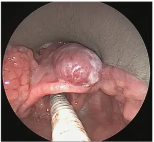
exophytic, violaceous mass of the right epiglottis.
Fig. 2– Operative direct laryngoscopy showing the mass pedicled off of the vallecular surface of the epiglottis.
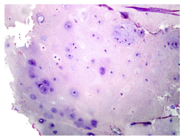
Fig. 3– Kaposi sarcoma consisting of a proliferation of slit-like vascular spaces with no significant cellular atypia.
Fig. 4– One year post-operative view showing healed epiglottis with no recurrence of lesion.
treatment depended on the histopathologic type; conservative surgical approaches were applied in available patients.
The largest series in the literature is a series of 38 patients [3], 22 of which were malignant (11 chondrosarcoma, 4 synovial cell sarcoma, 2 lymphoma, 2 neuroendocrine carcinoma, 1
adenoid cystic carcinoma, 1 rhabdomyosarcoma, 1
myofibroblastic sarcoma), and 16 of which were benign (5 granular cell tumor, 2 lipoma, 2 neurofibroma, 1 rhabdomyoma, 1 benign spindle cell neoplasm, 1 vascular malformation, 1 hemangioma, 1 lymphangioma, 1 oncocytoma, and 1 osteoma). Again, males were the majority of patient (59%). The majority of patients were treated with endoscopic surgical resection (24/ 36), while 11/36 underwent open partial laryngectomy. Eight of the 10 patients with malignancies other than chondrosarcoma received radiation with or without chemotherapy in an adjuvant or neo-adjuvant manner.
In all of these large series, as in the current report, the most common non-epithelial malignant tumor of the larynx was chondrosarcoma of the cricoid cartilage [4]. The remaining pathologies are all rare individually, but as a group malignant
epithelial tumors are more common than benign non-epithelial neoplasms. Males made up the majority of patients in all series. Although dependent on specific pathologic charac-teristics, there is a high rate of adjuvant therapy reported in the literature for these malignant non-epithelial tumors. In select-ed cases, endoscopic or open partial laryngectomy followselect-ed by adjuvant therapy can lead to long-term disease control and spare total laryngectomy.
Benign lesions were less common than malignant lesions in our series and in other published reports. Unlike malignant tumors, there were a significant number of patients present-ing in childhood. In particular, subglottic hemangioma is a well-described benign tumor of infanthood, and was the most common benign non-epithelial tumor in our series. Granular cell tumor was the second most common tumor in our report, and the most common in the report from Friedman et al.[3]. These and other benign tumors can be managed almost Fig. 5– Office indirect laryngoscopy of right true vocal cord
mass.
Fig. 6– Direct laryngoscopy demonstrating a bulky, firm, exophytic, subepithelial mass of the true vocal cord.
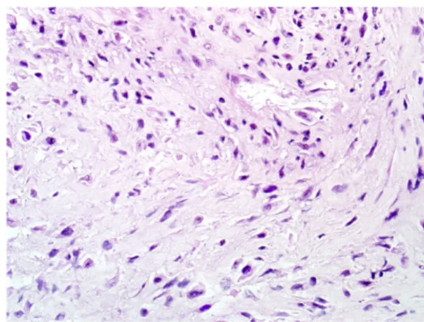
Fig. 7– This lesion consisted of a proliferation of
hypochromatic myofibroblastic cells with a "tissue-culture" like appearance and mild inflammatory cell infiltrate.
Fig. 8– One year post-operative view after laser excision. Anterior vocal cord web at site of lesion but no recurrence of subepithelial mass.
exclusively with endolaryngeal approaches, medical manage-ment, and observation in order to preserve laryngeal function. In order to highlight the different pathologies, the approach to selected non-epithelial pathologies is discussed below:
4.1. Malignant neoplasms 4.1.1. Chondrosarcoma
Laryngeal chondrosarcomas constitute the majority of malig-nant submucosal lesions both in our series, and represent the most common type of submucosal neoplasm (benign and malign) in the larynx overall. They constitute 0.5–1% of all laryngeal tumors[4]. The mean age at diagnosis is between the 6th and 7th decades of life and they have a male predominance of 3–4:1 [5]. Laryngeal chondrosarcomas arise in the cricoid cartilage in 70–75% of the cases, thyroid cartilage in 20%, and in the arytenoids, the epiglottis, and the corniculate cartilage in the decreasing order of frequency. Because most are low-grade and unlikely to spread locally or distantly, surgery is the treatment of choice and can vary from endoscopic to open partial or total laryngectomy, depending on the extent, mobility
of the cricoarytenoid joints and the histological grade of the cancer. The vast majority can be treated without total laryn-gectomy oncologically, but reconstruction of the larynx after excision of a significant portion of the cricoid can be challenging [6]. In a total of 111 laryngeal chondrosarcoma cases, wide excision or voice-sparing surgery was performed in 73 patients, whereas 37 patients had laryngectomy [4]. Recurrences oc-curred in 20 (18%) of the patients, 10 of whom underwent salvage laryngectomy. In our small series here, there was a high recurrence rate for partial laryngeal surgery, but this can almost always be salvaged with total laryngectomy, as laryngeal chondrosarcomas have a low propensity for metastasis (1.9%).
4.1.2. Kaposi's sarcoma
Kaposi's sarcoma is usually an HIV-associated neoplasm, and the upper aerodigestive tract is a rare but reported location for this lesion. Mochloulis et al.[7]reported 17 cases of laryngeal Kaposi's sarcoma, most (11) of them had supraglottic location, and treatment was mostly conservative; five patients received low dose radiotherapy to the larynx and 10 were treated with systemic chemotherapy for disseminated Kaposi's sarcoma. They reported that laryngeal Kaposi's sarcoma did not contribute to patient mortality. Treatment with intralesional bevacizumab has also been reported as a possible treatment Fig. 9– Office indirect laryngoscopy demonstrating a
submucosal mass of the right cricoid.
Fig. 10– Axial CT scan demonstrating expansile lesion in the right postero-lateral cricoid.
Fig. 11– This lesion consisted of a proliferation of
hypochromatic myofibroblastic cells with a "tissue-culture" like appearance and mild inflammatory cell infiltrate.
Fig. 12– Post-operative view shows reduced bulk of the lesion, and ability to monitor lesion for signs of encroach-ment upon airway.
for mucosal lesions [8]. Our two cases were treated with endoscopic excision with no evidence of disease in one year and 12 years of follow-up respectively.
4.1.3. Melanoma
Review of the literature shows that 0.6% of patients with cutaneous melanoma metastasize to the mucosa of the upper aerodigestive tract, and of those metastatic sites, 12% will be laryngeal[9]. Given the rarity of the problem, optimal manage-ment is not clear, but there may be benefit to excising sites of isolated recurrence and metastasis, particularly if they cause airway symptoms[10]. Endoscopic excision was performed in our case but the patient died of overall disease burden within three months.
4.2. Benign neoplasms 4.2.1. Hemangiomas
Infantile hemangioma is the most common benign tumor of infancy, affecting 1–2% of infants[11]. Hemangiomas demon-strate a proliferative phase during the first year of life, followed by involution. Infantile hemangiomas are usually found to occur in the subglottis, adult hemangiomas occur commonly in the supraglottic regions of the larynx, as in our cases. Various medical and surgical treatment strategies for airway hemangioma have been utilized depending on the age of the patient, the site and the size of the lesion, and the hemodynamic flow of the hemangioma, which include systemic and injectable steroids, chemotherapeutic agents (vincristine, interferon), serial endoscopic laser excision, open resection and temporary tracheotomy. Propranolol is now a common treatment for infantile hemangioma that appears to be effective in the vast majority of patients[11]. Conservative approach with close follow-up is another option if the lesion is not obstructive, especially in infantile hemangiomas, which will eventually undergo involution.
4.2.2. Granular cell tumors
Half of all granular cell tumors occur in head and neck region; 7–10% occur in the larynx[12]. More than 50% of these tumors involve the true vocal cords. The treatment modality is generally local excision; in large tumors, laryngofissure can be performed. Our cases were all excised or biopsied with an endoscopic approach, and long-term disease remission was obtained in cases not lost to follow-up.
5.
Conclusion
Mesenchymal tumors of the larynx are rare. Treatment approach of non-epithelial tumors depends on the site and extent of the tumor, histology, and sensitivity of adjuvant therapy. Benign tumors can be managed without need for aggressive resection thereby sparing laryngeal function.
R E F E R E N C E S
[1]Cai X, Wang T, Shi L. Nonepithelial malignant neoplasms of the larynx and hypopharynx. Zhonghua Er Bi Yan Hou Ke Za Zhi 1998;33:113–6.
[2]Fernandez-Acenero MJ, Larach F, Ortega-Fernandez C. Non-epithelial lesions of the larynx: review of the 10-year experience in a tertiary Spanish hospital. Acta Otolaryngol 2009;129:108–12.
[3]Friedman AD, Burns JA, Lutch MJ, et al. Submucosal neoplasms of the laryngeal introitus. J Laryngol Otol 2012; 126:706–13.
[4]Thompson LD, Gannon FH. Chondrosarcoma of the larynx: a clinicopathologic study of 111 cases with a review of the literature. Am J Surg Pathol 2002;26:836–51.
[5]Tiwari R, Mahieu H, Snow G. Long-term results of organ preservation in chondrosarcoma of the cricoid. Eur Arch Otorhinolaryngol 1999;256:271–6.
[6]Zeitels SM, Burns JA, Wain JC, et al. Function preservation surgery in patients with chondrosarcoma of the cricoid cartilage. Ann Otol Rhinol Laryngol 2011;120:603–7. [7]Mochloulis G, Irving RM, Grant HR, et al. Laryngeal Kaposi's
sarcoma in patients with AIDS. J Laryngol Otol 1996;110: 1034–7.
[8]Ablanedo-Terrazas Y, Alvarado-de la Barrera C, Ormsby CE, et al. Intralesional bevacizumab in patients with human immunodeficiency virus-associated Kaposi's sarcoma in the upper airway. Laryngoscope 2015;125:E132–7.
[9]Henderson LT, Robbins KT, Weitzner S. Upper aerodigestive tract metastases in disseminated malignant melanoma. Arch Otolaryngol Head Neck Surg 1986;112:659–63.
[10] Mifsud M, Padhya TA. Metastatic melanoma to the upper aerodigestive tract: a systematic review of the literature. Laryngoscope 2014;124:1143–9.
[11] Elluru RG, Friess MR, Richter GT, et al. Multicenter evaluation of the effectiveness of systemic propranolol in the treatment of airway hemangiomas. Otolaryngol Head Neck Surg 2015; 153:452–60.
[12] Sataloff RT, Ressue JC, Portell M, et al. Granular cell tumors of the larynx. J Voice 2000;14:119–34.