Ş B OYUN CERRAHİSİDE
A rare cause of nasal obstruction:
giant invasive nonfunctioning pituitary adenoma
Nadir bir burun tıkanıklığı nedeni: Dev invaziv nonfonksiyonel hipofiz adenomu
Güçlü Kaan Beriat, M.D.,1 Cem Doğan, M.D.,1 Şefik Halit Akmansu, M.D.,1Demet Karadağ, M.D.,2 Handan Doğan, M.D.3
Nasal obstruction is a very rare symptom caused by a pituitary adenoma. A 57-year-old man admitted to our clinic with bilateral nasal obstruction for the last six months. Endoscopic examination revealed soft pinkish pulsatile tissues in both nasal cavities. Radiologic investigation revealed a suprasellar mass extending to the frontal lobes, spheno-ethmoidal sinuses and nasal cavities. He had bitemporal supe-rior quadrant hemianopsia. Pituitary hormone levels were normal. Biopsies were taken from the patient endonasally. Pathological evaluations and labora-tory findings were compatible with nonfunctioning pituitary adenoma.
Key Words: Biopsy; nasal obstruction; pituitary adeno-ma.
Burun tıkanıklğı hipofiz adenomun neden olduğu çok nadir bir semptomdur. Elli yedi yaşında erkek hasta altı aydır süren iki taraflı burun tıkanıklığı ile kliniğimize başvurdu. Endoskopik muayenesinde her iki burun boşluğunda yumuşak pembemsi, pul-satil doku izlendi. Radyolojik incelemelerinde frontal loba, sfeno-ethmoidal sinüslere ve burun boşluk-larına uzanım gösteren suprasellar kitle izlendi. Hastada bitemporal üst kadran hemianopsi vardı. Hipofiz hormon düzeyleri normal idi. Biyopsiler has-tadan endonazal yol ile alındı. Patolojik değerlendir-mesi ve laboratuvar bulguları nonfonksiyonel hipofiz adenomu ile uyumluydu.
Anahtar Sözcükler: Biyopsi; burun tıkanıklığı; hipofiz ade-nomu.
Pituitary adenomas account for 10-20% of all intra-cranial tumors.[1] Small pituitary tumors have a prevalence of 14.4% in autopsy studies and 22.5% in radiologic studies.[2,3] They arise from suprasel-lar hypophyseal adenoid tissue in the supraselsuprasel-lar space. They appear as an expansion of the sella turcica in lateral cephalic radiograms or are direct-ly visualised as a soft tissue mass in computed tomography (CT) or magnetic resonance images (MRI). They can cause anterior pituitary hormonal imbalance, structural problems related to
inva-sion of surrounding structures, or syndromes of hormone excess.[4] Sometimes pituitary adenomas extend to the suprasellar region, cavernous sinus-es, paranasal sinussinus-es, nasopharynx and nasal cav-ity. Locally infiltrating adenomas are called inva-sive pituitary adenomas.[1,5,6] These tumors must be distinguished from other malignant aggressive tumors with an invasive behavior.
The frequency of nasopharyngeal extension of a pituitary tumor is estimated to be approximately 2% of all the pituitary adenomas.[6] By contrast, Departments of 1Otolaryngology, 2Radiology, 3Pathology, Medicine Faculty of Ufuk University, Ankara, Turkey
Received / Geliş tarihi: April 12, 2010 Accepted / Kabul tarihi: June 10, 2010
Correspondence / İletişim adresi: Güçlü Kaan Beriat, M.D. Ufuk Üniversitesi Tıp Fakültesi Kulak Burun Boğaz Hastalıkları Anabilim Dalı, 06520 Balgat, Ankara, Turkey. Tel: +90 312 - 204 41 75 Fax (Faks): +90 312 - 204 40 88 e-mail (e-posta): [email protected]
ectopic pituitary adenomas located in the sphe-noid sinus and nasopharynx region are extremely uncommon. They may arise from the embryonal remnant of the extrasellar pituitary tissue in the sphenoid sinus or nasopharyngeal regions during the migration process of Rathke’s pouch.[7]
Pituitary tumors were classified as basophil-ic, acidophilbasophil-ic, or chromophobic on the basis of whether or not they took up the stains hema-toxylin and eosin. This classification has fallen into disuse. At present, classification of pituitary tumors is based on radiologic findings, immu-nohistochemical staining and plasma hormone levels.[4] Corticotropic adenomas, thyrotropic ade-nomas and gonadotropic adeade-nomas are gener-ally basophilic; somatotropic adenomas, lactotropic adenomas are acidophilic; null cell adenomas may be stained with synaptophysin.[4]
We herein describe a rare case of giant invasive nonfunctioning pituitary adenoma (NFPA) origi-nating from the pituitary gland and exclusively involving the sphenoid sinus, nasopharynx, pos-terior ethmoidal cells, pospos-terior nasal cavity along with an invasion of bilateral frontal lobes and cav-ernous sinus.
We primarily focused on the diagnosis with rhi-nologic examination, immunohistochemical stud-ies and radiologic examinations.
CASE REPORT
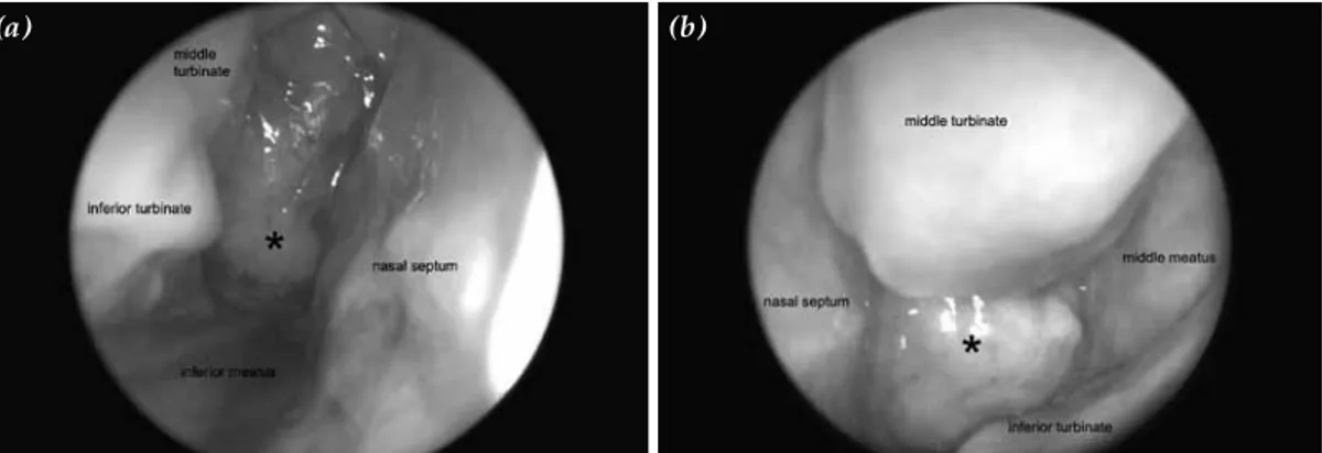
A fifty-seven-year-old male patient was admitted to our clinic with complaints of nasal obstruc-tion for the last six months. No other symptoms were noted. Nasal endoscopic examination was performed on the patient in both nasal cavi-ties. Smooth surfaced, pinkish, pulsatile masses hanging from the spheno-ethmoid recess to
both nasal cavities were seen (Figures 1a, b). Eye examination detected bitemporal superior quad-rant hemianopsia. No other pathological find-ings were found in the patient in his systemic examination. Laboratory examinations showed no abnormalities.
Radiologic examinations, including CT and MRI were performed. Axial high-resolution CT images revealed the mass completely filling the sphenoid sinus and posterior ethmoidal cells. Gadolinium-enhanced coronal MRI T1-weighted image demonstrated the mass arising from the sella region and invading bilateral frontal lobes and cavernous sinus, whereas sagittal T1-weighted image showed the mass eroding the floor of the sella turcica and extending to the right posterior nasal cavity. Axial T2-weighted image revealed the mass projecting into the posterior nasal cavity (Figures 2, 3).
In consultation with the neurosurgery clinic, we decided on endonasal biopsy of the tumor because of the possibility of confronting with chordoma, chondrosarcoma, meningioma or pituitary adeno-ma. Biopsies were taken from both nasal cavities under local anesthesia.
Pathologic findings suggested a diagnosis of null cell pituitary adenoma and revealed histologi-cally that the tumor was composed of monomor-phic round cells, arranged diffusely with a char-acteristic sinusoidal pattern around the capillaries. The tumor cells had round and oval nuclei, con-spicuous nucleoli and chromophobic cytoplasm. No mitotic activity was found (Figure 4). The Ki-67 proliferative index using MIB-1 antibody was 1.7%. The Ki-67 proliferative index was less than 3%. And tumor cells had no mitotic activity or pleomor-phism (Figure 5).
Figure 1. Right and left nasal cavities with a viewes of the zero degree endoscope. (a) Right middle and lower nasal meatus filled by mass. (b) Left middle meatus filled by mass.
Hormonal examination showed normal lev-els of serum thyroid stimulating hormone (TSH), and gonadotropins such as follicle-stimulating hormone (FSH) and luteinizing hormone (LH), prolactin (PRL), growth hormone (GH) and adreno-corticotropic hormone (ACTH). So this tumor was a null cell giant pituitary adenoma without atypical features.
As a complementary surgical treatment, the patient was referred to the Neurosurgery Department for the excision of the remaining intra-cranial portion of the tumor. However, he declined
an intracranial operation and is being followed up with no other complaints.
DISCUSSION
Pituitary tumors can also be classified by size and invasive characteristics. Microadenomas are <10 mm whereas macroadenomas are >10 mm. Intrapituitary adenomas are within the substance of the pituitary gland; intrasellar adenomas are con-fined to the sella; diffuse adenomas fill the sella and cause focal sellar bone erosions. Invasive adenomas erode sellar and sphenoid bone walls, and spread into surrounding soft tissues like the cavernous sinuses, optic chiasm, third ventricle and brain.[4]
Nonfunctioning pituitary adenomas account for approximately 30% of pituitary tumors.[1] These Figure 2. Gadolinium-enhanced sagital T1-weighted image
shows the mass which eroding the floor of the sella turcica, extending to the right posterior nasal cavity.
Figure 3. Axial T2-weighted image reveals the mass projecting into the posterior nose.
Figure 4. Monomorphic round adenoma cells with round and oval nuclei, conspicuous nucleolus and chromophobic
tumors do not cause clinical hormone hyper-secretion. Despite the lack of clinical hormone hypersecretion immunocytochemical staining of hormones reveals evidence for hormone expression in up to 79% of these tumors. This situation can be explained by non-effective hormone synthesis.[1,6]
Nonfunctioning pituitary adenomas can be classified into two main groups: null cell adeno-mas and silent adenoadeno-mas. Null cell adenoadeno-mas are divided into non-oncocytic and oncocytic adeno-mas. Oncocytomas contain large numbers of mito-chondria, show focal immunostaining for anterior pituitary gland hormones, and produce hormones in vitro. Silent adenomas consist of three morpho-logical subtypes: silent corticotrope, silent somato-trope and gonadosomato-trope NFPAs.[6]
Enlargement of a tumor into the suprasellar area results in optic chiasmal compression which may cause visual field deficits. The initial visual field deficits are frequently bitemporal superi-or quadrant defects. Occasionally these tumsuperi-ors extend into the cavernous sinus.
Enlargement of a nonfunctioning adenoma can cause progressive loss of pituitary function over months or years. Gonadotropic hormone func-tion is usually lost first, while adrenocorticotropic hormone is lost last. Loss of antidiuretic hormone function is almost never a presenting symptom.[7]
Although progressive bitemporal visual field loss and progressive hypopituitarism are the typi-cal presenting clinitypi-cal manifestations, they may rarely be obvious symptoms. Occasionally, there may be cranial hemorrhage or infarction, in which case patients may complain of headache, visual loss and present acute hormonal insufficiency or loss of consciousness.[8]
If the enlarged adenoma is asymmetrical, the lateral radiograph may give the impression of a double sellar floor. The sella becomes more round-ed. Also the dorsum sella may be thin, pushed back and anterior clinoids may be undercut.
The CT and MRI shows the exact anatomical configuration of the adenoma.[9] In our case, the CT and MRI clearly depicted the presence of a large, soft mass in the sphenoid sinus and its extensive invasion to the peripheric tissues.
In particular, MRI was found useful in delin-eating precise anatomic relationships between the sphenoid sinus, tumor and the pituitary fossa. It was useful both in defining anatomic relationships
among the sphenoid sinus, tumor and the sella turcica, and in differentiating from nontumorous mucosal lesions.
Radiologic differential diagnosis of a pituitary adenoma is a very extensive spectrum. Tuberculum sella meningiomas do not show enlargement of the sella, internal carotid artery aneuysm can be diag-nosed as a flow void seen on an MRI, malignant tumor metastasis to the sella is frequently associ-ated with extraocular muscle palsies or diabetes inspitus. Rathke’s pouch cleft cyst presents as sellar or suprasellar cystic masses. Tuberculoma, giant cell hypophysitis and sarcoidosis may mimic NFPA.
A review of the literature indicates very few cases of pituitary adenomas of ectopic origin. In this case the mass was completely within the pituitary gland; it reached the widest diameter in this region and demolished all the sellar walls. Therefore, the tumor was thought to originate from the sellar region.
Pituitary tumors, which primarily infiltrate the sphenoid sinus and nasopharynx, are there-fore uncommon disorders. They frequently pro-duce such unique symptoms as nasal obstruction, recurrent epistaxis, or intermittent mucoid nasal discharge. In addition to these nasal symptoms, the majority of tumors also develop visual field defects, decreased visual acuity, and oculomotor or trochlear nerve palsy, which are attributable to simultaneous suprasellar and parasellar exten-sions of the tumor. In this case, in spite of massive tumor tissue, the patient’s only complaint was nasal obstruction. Patient had no other com-plaints and no other pathological examination results apart from the sellar mass extending to the sphenoid sinus and beyond nasopharynx and oropharynx.
It was recently demonstrated by Scheithauser et al.[10] on the basis of immunocytochemical and ultrastructural analysis of 365 pituitary adeno-mas that there was a correlation between the frequency and the nature of invasiveness and the functional type of the adenoma. Prolactin produc-ing adenomas and mixed GH- and PRL-producproduc-ing adenomas are prone to grow in any direction, but the preferential pathway is infrasellar. They often invade the sphenoid bone and extend further into the nasopharynx.[10] In the present report, however, despite its nonfunctioning nature, the current ade-noma appears to show a radiographic evidence of paranasal and posterior nasal cavity invasiveness.
Recently cDNA (Complementary deoxyribo-nucleic acid) microarray analysis has revealed that the folate receptor (FR-alfa) is significantly over-expressed in clinically non-functional adenomas. Folate receptor-alfa is a high affinity folate trans-porter. Folate receptor-alfa is significantly over-expressed by NFPAs. Overexpression of FR-alfa provides growth advantage to tumor cells and this may be detected by Western Blot technique.[11]
The usual treatment of a NFPA is microscopic or endoscopic transsphenoidal removal of the tumor.[2] Sellar and suprasellar tumors can be removed through a transsphenoidal approach by either a sublabial, transseptal or direct endonasal route using the endo-scope or microendo-scope. Relative contraindications to transsphenoidal surgery include a dumbbell tumor, especially if the neck is narrow and the upper part of dumbbell tumor is large. Occasionally, craniotomy will be needed in patients for whom transsphenoidal surgery is contraindicated.
Radiation therapy can be used in patients with NFPAs. It controls tumor growth in 80-98% of patients with NFPAs. If the patient is medically unstable, radiation therapy may be the sole option. The patient who postoperatively has a significant amount of residual tumor or who shows regrowth may be a candidate for fractionated radiation therapy.[12,13]
REFERENCES
1. Burger PC, Scheithauer BW, Vogel FS. Region of the sella turcica. In: Burger PC, Scheithauer BV, Vogel FS, editors. Surgical pathology of the nervous system and its coverings. 4th ed. New York: Churchill Livingstone; 2002. p. 437-97.
2. Ezzat S, Asa SL, Couldwell WT, Barr CE, Dodge WE, Vance ML, et al. The prevalence of pituitary adenomas: a systematic review. Cancer 2004;101:613-9.
3. Asa SL. Practical pituitary pathology: what does the pathologist need to know? Arch Pathol Lab Med 2008;132:1231-40.
4. Isselbacher KJ, Eugene B, Wilson JD, Martin JB, Anthony SF, Kasper DL. Endocrinology and metabo-lism. In: Fauci AS, Braunwald E, Wilson JD, Harrison TR, editors. Harrison’s Principles of Internal Medicine. 14th ed. New York: Mc-Graw Hil; 1998. p. 917-78. 5. Godey B, Morandi X, Le Gall F, Feat S, Brassier G, Le
Clech G. Pituitary adenomas with infra-sellar exten-sion into the nasopharynx. J Laryngol Otol 1999; 113:1109-11.
6. van der Mey AG, van Seters AP, van Krieken JH, Vielvoye J, van Dulken H, Hulshof JH. Large pitu-itary adenomas with extension into the nasopharynx. Report of three cases with a review of the literature. Ann Otol Rhinol Laryngol 1989;98:618-24.
7. Nelson PB, Goodman ML, Flickenger JC, Richardson DW, Robinson AG. Endocrine function in patients with large pituitary tumors treated with operative decompression and radiation therapy. Neurosurgery 1989;24:398-400.
8. Biousse V, Newman NJ, Oyesiku NM. Precipitating factors in pituitary apoplexy. J Neurol Neurosurg Psychiatry 2001;71:542-5.
9. Mikhael MA, Ciric IS. MR imaging of pituitary tumors before and after surgical and/or medical treatment. J Comput Assist Tomogr 1988;12:441-5.
10. Scheithauer BW, Kovacs KT, Laws ER Jr, Randall RV. Pathology of invasive pituitary tumors with special reference to functional classification. J Neurosurg 1986;65:733-44.
11. Evans CO, Young AN, Brown MR, Brat DJ, Parks JS, Neish AS, et al. Novel patterns of gene expression in pituitary adenomas identified by complementary deoxyribonucleic acid microarrays and quantitative reverse transcription-polymerase chain reaction. J Clin Endocrinol Metab 2001;86:3097-107.
12. Marcou Y, Plowman PN. Stereotactic radiosurgery for pituitary adenomas. Trends Endocrinol Metab 2000;11:132-7.
13. Becker G, Kocher M, Kortmann RD, Paulsen F, Jeremic B, Müller RP, et al. Radiation therapy in the mul-timodal treatment approach of pituitary adenoma. Strahlenther Onkol 2002;178:173-86.