ORIGINAL ARTICLE
Evaluation of hematological parameters in children with FMF
Alaaddin Yorulmaz1 &Hikmet Akbulut1&Suna Adeviye Taş1&Merve Tıraş1&İbaa Yahya1&Harun Peru2
Received: 1 February 2018 / Revised: 23 September 2018 / Accepted: 12 October 2018 / Published online: 16 October 2018 # International League of Associations for Rheumatology (ILAR) 2018
Abstract
In this study, we aimed to investigate whether neutrophil/lymphocyte ratio (NLR), platelet/lymphocyte ratio (PLR), and mean platelet volume (MPV) might be helpful in the diagnosis of subclinical inflammation of familial Mediterranean fever (FMF). Clinical, laboratory, and genetic results of the patients who were followed up with the diagnosis of FMF were obtained retrospectively from the hospital files and recorded to standardized form. Age- and sex-matched healthy subjects were included as the control group. Eighty-three of the 143 patients (58.0%) were male and 60 (42.0%) were female. The mean age of our patients was 164.62 ± 51.20 months and the mean age of the control group was 164.92 ± 51.10 months. The mean diagnosis age of our patients was 98.10 ± 49.11 months. The mean follow-up time of the patients was 66.03 ± 36.37 months. 91.60% of our patients had abdominal pain, 78.32% fever, and 28.67% joint pain. The mean NLR of the patients was significantly higher than the mean levels at attack-free period and the control group. However, no statistically significant difference was found between the mean levels at attack-free period and the control group. MPV levels were statistically significantly high during acute attack when compared with the control group. However, they showed no statistically significant difference between acute attack and attack-free period. NLR is a useful marker to predict inflammation in FMF patients. However, our results did not support the idea that MPV might reflect acute attack and attack-free period.
Keywords Attack . Child . Familial Mediterranean fever . Inflammation
Introduction
Familial Mediterranean fever (FMF) is an autosomal recessive inherited disorder characterized by recurrent inflammatory fe-brile episodes and polyserositis [1]. The characteristics of these episodes include high fever lasting from several hours to 3–4 days and serositis (90%), fever (90%), arthritis (33%), pleuritis (31%), scrotum (5%), and pericardium (1%). Erysipelas-like rash is usually associated with arthritis and tends to involve the distal end of the lower extremities, usually between the knee and the ankle, and on the dorsum of the proximal foot near the ankle [2,3]. Patients are completely healthy between the attacks of FMF [1]. Although FMF is
mainly prevalent among people of Mediterranean origin, it is observed worldwide due to extensive population movements of the twentieth century [4]. The estimated prevalence and carrier rate of FMF in Turkey are 1/1000 and 1:5, respectively. With a population of more than 70 million, a considerable amount of FMF cases throughout the world live in Turkey [5]. FMF is caused by mutations in the MEFV gene which encodes the pyrin protein which is thought to be associated with interleukin-1-related inflammation process. During the attacks of FMF, serositis usually involves the abdomen, chest, or joints. Fever and elevated acute phase reactants are com-mon characteristics. Erythrocyte sedimentation rate (ESR), C-reactive protein (CRP), serum amyloid A (SAA), and fibrin-ogen levels increase during these attacks which usually turn to normal in attack-free periods [6]. The duration of attacks is usually 1 to 3 days which end spontaneously. However, it has been shown that subclinical inflammation may continue, dur-ing attack-free periods. Because there is increased risk of de-veloping amyloidosis with this type of inflammation, defining this subclinical inflammation in FMF patients is essential [7–9]. The parameters absolute neutrophil-to-absolute lym-phocyte ratio (NLR), platelet count-to-absolute lymlym-phocyte
* Alaaddin Yorulmaz [email protected]
1 Department of Pediatrics, Selçuk University Medical School,
Konya, Turkey
2
Department of Pediatric Rheumatology, Selçuk University Medical School, Konya, Turkey
ratio (PLR), mean platelet volume (MPV), and red cell distri-bution width (RDW) may be used as markers of systemic inflammation [10,11]. These have been associated with con-ditions including cardiovascular diseases, malignancies, ulcer-ative colitis, hepatic cirrhosis, and systemic lupus erythema-tosus. NLR and MPV have also been showed to be signifi-cantly higher in patients with FMF [10–12].
In this study, we aimed to determine if this association exists between RDW levels and FMF, investigate whether MPV and platelet distribution width (PDW) could be used as an inflammatory marker in FMF patients, and investigate whether NLR, PLR, MPV, PDW or RDW might be helpful in the diagnosis of subclinical inflammation of FMF.
Material and methods
This study included 143 patients aged between 1 month and 18 years who were followed up with the diagnosis of FMF in Selçuk University Medical Faculty Pediatric Nephrology and Rheumatology Clinic between January 2014 and June 2017. Age- and sex-matched healthy subjects were included as the control group. Clinical, laboratory, and genetic results of the patients were obtained retrospectively from the hospital files and recorded to standardized form. The age, gender, height, weight, age of diagnosis, family history of FMF and consan-guineous marriage, and presence of fever, abdominal pain, joint pain were also recorded and the relationship of these between genetic mutation was investigated.
The Tel-Hashomer criteria was used for the diagnosis of FMF: the presence of at least 1 of 4 major criteria, 2 of 5 minor criteria, 1 minor criterion plus 5 of 10 supportive criteria, or 4 of 5 specific supportive criteria [13]. FMF attack was defined when the following criteria were met: (1) applying with clin-ical symptoms (fever, abdominal pain, chest pain, arthralgia, serositis, pleuritis, pericarditis, arthritis, peritonitis, myalgia, erysipelas-like erythema) in the first 72 h, (2) exclusion of other febrile etiologies, (3) fever should be above 37 °C and last at least 12 h, (4) presence of the following laboratory findings: erythrocyte sedimentation rate (ESR)≥ 30 mm/h, C-reactive protein (CRP)≥ 5 mg/dL, fibrinogen ≥ 350 mg/h, white blood cell (WBC) count≥ 10.000/mm3. Clinical and laboratory findings of patients at the time of FMF attack were recorded. The attack-free period was defined as at least 2 weeks after the end of the last FMF attack.
Patients with splenomegaly, diabetes mellitus, asthma, he-matologic disorder, renal or liver failure, uncontrolled hyper-tension, or proteinuria were excluded from the study. In addi-tion, patients who received anticoagulant therapy or nonste-roidal anti-inflammatory drugs were not included to the study. Laboratory analyses including hemogram, CRP, ESR, fi-brinogen, serum electrolytes, blood glucose, urea, and liver function tests at the time of attack and at least 1 month after
the onset of the attack were reviewed. Hemogram analyses were made by an automatic blood count device. WBC, neu-trophil count (K/μL), lymphocyte count (K/μL), platelet count (K/μL), NLR, PLR, MPV (fL), PDW, RDW, CRP, and ESR values were recorded from the hemogram results. The fre-quency of exon 2 and exon 10 mutations of the MEFV gene was determined by DNA sequencing method in all patients. All blood samples were studied in the same regularly checked analyzer (Abbott CELL-DYN 3700, USA).
Statistical analysis
SPSS 21.0 (IBM SPSS statistics 21) packet computer program was used for statistical analysis of the data. Kolmogorov-Smirnov and Shapiro-Wilk normality tests were carried out to determine whether the data showed normal distribution. Descriptive data were presented with mean ± standard devia-tion. Two dependent numerical variables showing normal dis-tribution were tested by paired-samplet test. Student’s t test was used if the comparison of two independent numerical variables showed a normal distribution, and Mann-Whitney U test was used if not. Chi-square test was used for categorical variables. Significance was assessed at p < 0.05 level. Correlations between parameters with normal and abnormal distribution were computed through Pearson’s and Spearman’s correlation analyses, respectively.
Results
Eighty-three of the 143 patients (58.0%) were male and 60 (42.0%) were female. The M/F ratio was 1.38. Demographic characteristics of patients are shown in Table1. The mean age of our patients was 164.62 ± 51.20 (35–252) months and the mean age of the control group was 164.92 ± 51.10 (38–253) months. Age and gender distributions were not statistically different between the groups. The mean diagnosis age of FMF of our patients was 98.10 ± 49.11 (12–204) months. The mean follow-up time of the patients was 66.03 ± 36.37 (8–190) months. None of our patients had amyloidosis. All FMF patients were receiving colchicine treatment one to three times a day with a dose ranged between 0.5 and 2 mg. 91.60% (n, 131) of our patients had abdominal pain, 78.32% fever, and 28.67% joint pain. The distribution of clinical findings of our patients is shown in Table2. When the MEFV gene mutations of the patients were examined, 50 (34.96%) patients were homozygous; 47 (32.86%) compound heterozygotes and 34 (23.77%) heterozygous mutations were detected (Table3). No mutation was detected in 12 (8.39%) patients.
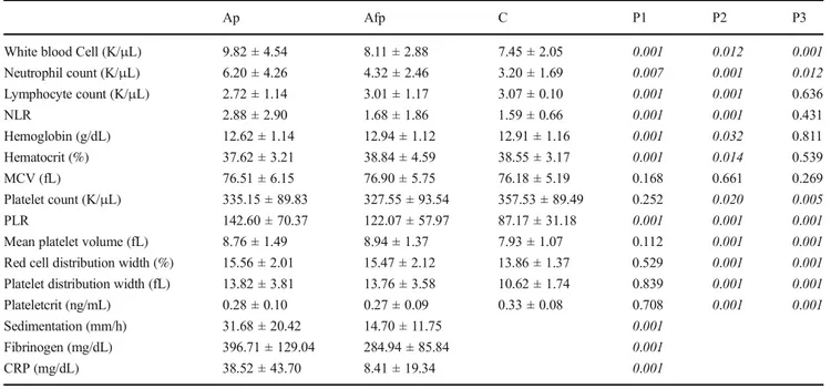
The laboratory characteristics of the patients are given in Table4. ESR, fibrinogen, and CRP levels were found statisti-cally significantly high in acute attack when compared with attack-free period. There was a statistically significant
increase in total leukocyte counts, absolute neutrophil counts, and platelet count levels during acute FMF attack when com-pared with attack-free period and the control group. These levels also showed a statistically significant difference in pa-tients with FMF at attack-free period and the control group. The mean absolute lymphocyte counts, NLR, hemoglobin, and hematocrit of the patients were significantly higher than the mean levels at attack-free period and the control group. However, no statistically significant difference was found be-tween the mean levels at attack-free period and the control group. MPV, RDW, and PDW levels were statistically signif-icantly high during acute attack when compared with the con-trol group. However, they showed no statistically significant difference between acute attack and attack-free period.
Patients carrying the M694V gene mutation (heterozygous or homozygous) and other mutation-detected patients were compared (Table5). There was no statistically significant ference for NLR and PLR levels in both groups, but the dif-ference was found in terms of RDW levels.
CRP values of patients with FMF acute attack were positively correlated with NLR, PLR, MPV, and RDW. The correlation between NLR and CRP was strong (r = 504, p < 0.001).
Discussion
The main aim of our study was to determine the most useful marker for subclinical inflammation in children with FMF by comparing NLR, PLR, MPV, and RDW. Although PLR, MPV, and RDW demonstrated changes with subclinical in-flammation in FMF, NLR had the strongest correlation with subclinical inflammation within these markers. Based on these
Table 1 Demographic characteristics of the study population
Patients Control group
N % N %
Gender
Male 83 58.0 83 58.0
Female 60 42.0 60 42.0
Mean ± SD (min–max) Mean ± SD (min–max) Age (month) 164.62 ± 51.20 (35–252) 164.92 ± 51.10 (38–253) Weight 30.09 ± 15.26 (9–84)
Height 127.07 ± 22.46 (72–177) Age at diagnosis (month) 98.10 ± 4911 (12–204) Duration of follow-up (month) 66.03 ± 36.37 (8–190)
Table 3 The distribution of MEFV gene mutations
Mutation Number Percent
M694V homozygous 36 25.17 M680I homozygous 5 3.49 E148Q homozygous 4 2.79 R202Q homozygous 3 2.09 V726A homozygous 2 1.39 M694V heterozygous 21 14.68 E148Q heterozygous 8 5.59 M680I heterozygous 3 2.09 P369S heterozygous 1 0.69 R202Q heterozygous 1 0.69 M694V/M680I 18 12.58 M694V/V726A 9 6.29 M680I/V726A 4 2.79 M680I/E148Q 3 2.09 M694V/R202Q 2 1.39 M680I/R202Q 2 1.39 E148Q/M694V 2 1.39 E148Q/V726A 2 1.39 M694V/R761H 1 0.69 E148Q/P369S 1 0.69 R202Q/J339F 1 0.69 R202Q/R761H/M680I 1 0.69 R202Q/V726A/M694V 1 0.69 No mutation 12 8.39
Table 2 The distribution of the clinical findings of patients with FMF Clinical findings Number Percent Abdominal pain 131 91.60
Fever 112 78.32
Family history for FMF 69 48.25
Joint pain 41 28.67 History of appendectomy 22 15.38 Chest pain 15 10.48 Rash 4 2.79 Hepatomegaly 3 2.09 Splenomegaly 1 0.69
data, the cutoff value for NLR can be used to detect subclinical inflammation in FMF.
Amyloidosis is the most destructive complication of FMF. The main cause of amyloidosis in FMF is subclinical inflam-mation despite the treatment of colchicine. Many inflammato-ry markers have been studied in FMF. ESR, CRP, fibrinogen, serum amyloid A protein, and WBC levels are the markers which are used to determine the acute phase response (APR)
in FMF [13]. Levels of these markers increase during acute attack periods and return to normal ranges in attack-free pe-riods [6]. Subclinical inflammation has been reported to be continued in up to 30% of the patients with FMF during the attack-free period. Persistent elevation of these markers is im-portant in terms of reflecting the subclinical inflammation, which play the main role for the development of amyloidosis and other complications including anemia, splenomegaly, and osteopenia. Several studies have been conducted to discover a new marker which reflects subclinical inflammation in FMF [6,8].
FMF is an autoinflammatory disease resulting from im-mune system abnormalities and decreased or complete loss of pyrin function [2]. Pyrin mutations and FMF have been linked before and it has been suggested that pyrin has both pro- and anti-inflammatory effects. Inflammation is caused by the release of inflammatory cytokines from macrophages and monocytes. Pyrine dysfunction causes autoinflammatory dis-ease which results with the abnormal production of interleukin (IL)-1β and IL-18 [14]. These cytokines further lead to in-creased amounts of tumor necrosis factor alpha (TNF-α) and IL-6 [15]. It is known that elevated serum amyloid A and C-reactive proteins and inflammatory cytokines including IL-6 and IL-18 are implicated in the disease activity of FMF [16]. However, a specific biomarker for FMF is not available up to date. IL-6 is an inflammatory cytokine which is known to play role in autoimmune and chronic inflammatory diseases. Increased levels of IL-6 during FMF attacks have been report-ed [17]. IL-1β, IL-6, IL-8, and TNF-a levels have been shown to be increased in FMF patients during the attack-free period
Table 4 The comparison of laboratory characteristics of patients with FMF
Ap Afp C P1 P2 P3
White blood Cell (K/μL) 9.82 ± 4.54 8.11 ± 2.88 7.45 ± 2.05 0.001 0.012 0.001 Neutrophil count (K/μL) 6.20 ± 4.26 4.32 ± 2.46 3.20 ± 1.69 0.007 0.001 0.012 Lymphocyte count (K/μL) 2.72 ± 1.14 3.01 ± 1.17 3.07 ± 0.10 0.001 0.001 0.636 NLR 2.88 ± 2.90 1.68 ± 1.86 1.59 ± 0.66 0.001 0.001 0.431 Hemoglobin (g/dL) 12.62 ± 1.14 12.94 ± 1.12 12.91 ± 1.16 0.001 0.032 0.811 Hematocrit (%) 37.62 ± 3.21 38.84 ± 4.59 38.55 ± 3.17 0.001 0.014 0.539 MCV (fL) 76.51 ± 6.15 76.90 ± 5.75 76.18 ± 5.19 0.168 0.661 0.269 Platelet count (K/μL) 335.15 ± 89.83 327.55 ± 93.54 357.53 ± 89.49 0.252 0.020 0.005 PLR 142.60 ± 70.37 122.07 ± 57.97 87.17 ± 31.18 0.001 0.001 0.001 Mean platelet volume (fL) 8.76 ± 1.49 8.94 ± 1.37 7.93 ± 1.07 0.112 0.001 0.001 Red cell distribution width (%) 15.56 ± 2.01 15.47 ± 2.12 13.86 ± 1.37 0.529 0.001 0.001 Platelet distribution width (fL) 13.82 ± 3.81 13.76 ± 3.58 10.62 ± 1.74 0.839 0.001 0.001 Plateletcrit (ng/mL) 0.28 ± 0.10 0.27 ± 0.09 0.33 ± 0.08 0.708 0.001 0.001 Sedimentation (mm/h) 31.68 ± 20.42 14.70 ± 11.75 0.001
Fibrinogen (mg/dL) 396.71 ± 129.04 284.94 ± 85.84 0.001 CRP (mg/dL) 38.52 ± 43.70 8.41 ± 19.34 0.001 Ap, attack period; Afp, attack-free period; C, control group
Pairwise comparisons: P1, Ap-Afp; P2, Ap-C; P3, Afp-C
Table 5 Comparison of clinical and laboratory values of FMF with homozygous or heterozygous M694V mutations and other mutations
M694V (+) M694V (−) P WBC (K/μL) 9.92 ± 4.90 9.66 ± 3.90 0.745 Neutrophil count (K/μL) 6.26 ± 4.71 6.11 ± 3.41 0.840 Lymphocyte count (K/μL) 2.80 ± 1.20 2.59 ± 1.02 0.305 NLR 2.87 ± 3.12 2.91 ± 2.51 0.933 Hemoglobin (g/dL) 12.49 ± 1.05 12.83 ± 1.25 0.079 Hematocrit (%) 37.47 ± 3.12 37.88 ± 3.37 0.463 MCV (fL) 75.42 ± 6.01 78.36 ± 5.98 0.005 PLT (K/μL) 345.54 ± 95.85 317.50 ± 76.22 0.071 PLR 143.00 ± 72.57 141.92 ± 67.13 0.928 MPV (fL) 8.29 ± 1.59 8.14 ± 1.34 0.549 RDW (%) 15.84 ± 2.12 15.09 ± 1.72 0.024 PDW (fL) 13.76 ± 3.91 13.93 ± 3.65 0.820 PCT (ng/dL) 0.29 ± 0.11 0.25 ± 0.07 0.023 Sedimentation (mm/h) 31.61 ± 20.79 23.85 ± 17.20 0.036 Fibrinogen (mg/dL) 415.21 ± 138.5 364.43 ± 122.31 0.060 CRP (mg/dL) 41.51 ± 45.12 33.50 ± 39.97 0.320
[18]. Contrastingly, serum levels of IL-1β, TNF-α, soluble interleukin-2 receptor (sIL-2R), IL-6, and IL-8 have been shown to be higher in patients during acute attack than attack-free periods and control groups [19,20]. In the study of Gang et al., levels of IL-1β were not elevated in FMF patients during attacks or attack-free periods [21]. The differ-ence between results may be due to the determination time of FMF phase, age group of patients, and processes and methods of laboratory analysis.
ESR is a nonspecific regularly ordered test for the diagno-sis of inflammatory diseases. CRP increase is a part of the acute phase response at acute and chronic inflammation [22]. Fibrinogen is a vital protein for the blood clotting pro-cess. In the present study, ESR, CRP, and fibrinogen levels were significantly higher in patients with FMF during acute attack when compared with attack-free period and the control group, consistent with the literature.
NLR which has been reported as an indicator of systemic inflammation is calculated by dividing neutrophil count to lymphocyte count [6,23]. NLR status in FMF has been stud-ied in various studies. Uslu et al. compared NLR in FMF patients and healthy controls. They found out that NLR was significantly higher in patients with FMF. They also reported that NLR was significantly higher in patients with amyloidosis when compared with amyloidosis-free patients [6]. Ahsen et al. suggested that NLR can be used as an acute phase re-sponse in FMF patients [7]. Both of these studies did not include FMF patients during attack period. Uluca et al. found that NLR levels were higher in patients in attack-free period and they concluded that NLR may be an indicator for attack period but not attack-free period [9]. Özer et al. studied NLR values of pediatric symptom-free FMF patients and healthy controls. They found that NLR had strong correlation with CRP and concluded that NLR could be a reliable marker for subclinical inflammation in patients with FMF [24]. Celikbilek et al. studied NLR in adult FMF patients during acute attack and attack-free period and they reported that NLR of FMF patients during acute attack was significantly higher than those of attack-free patients and control group [25]. Our results provide evidence which supports the thought that NLR may be a parameter to show the inflammation in attack period. However, it may not be useful to define the subclinical inflam-mation according to our study groups since there was no sig-nificant difference between attack-free patients and control group. As it is cost-effective, is available, and can be calculat-ed easily, NLR could be uscalculat-ed to prcalculat-edict systemic inflamma-tion in pediatric FMF patients during attack period.
MPV is a parameter detected during routine blood count which reflects platelet function and activation [26]. Larger platelets are hemostatically more active [27]. Thrombocyte activation is associated with increased risk of atherosclerosis. There is limiting data about the relationship between MPVand atherosclerosis in FMF patients. Although the relationship
between FMF and atherosclerosis is not clear, it has been suggested that attack periods of FMF may be associated with the increase of atherosclerosis due to platelet activation and by using colchicine prothrombotic pathways and atherosclerosis can be suppressed [28]. The first study conducted on this topic was by Makay et al. and they reported that MPV values were lower during attack in patients with FMF than in healthy con-trols [29]. This finding supports our study. While MPV was not found to be significant between the FMF group and healthy control group in the same study, it was found to be significant in our study. In the study by Arıca et al., MPV was found significantly higher both in acute attack and attack-free period patients than in healthy controls [30]. Coban and Adaniralso reported that MPV values were higher during attack-free period of FMF patients when compared with those of healthy controls [31]. Uluca et al. published two studies with opposite results, about the relationship of MPV and ath-erosclerosis in patients with FMF [9,24]. In the first study, they reported that MPV values were similar in patients with FMF and healthy controls and concluded that MPV did not predict atherosclerosis risk in pediatric patients with FMF [32]. In the later study, they compared the MPV values and epicardial adipose tissue thickness in children with FMF. They found significantly greater epicardial adipose tissue thickness and higher MPV values in children with FMF and they sug-gested that MPV values might indicate an increased risk of atherosclerosis in FMF [9]. In our study, both attack and attack-free patients had significantly higher MPV values com-pared to control group similar to most of the literature results. However, MPV values did not significantly differ between acute attack and attack-free periods. Therefore, while MPV is not useful to show the attack periods in FMF, it may be valuable to show the risk of atherosclerosis and subclinical inflammation in children with FMF.
PDW is another potential marker to indicate inflammation and atherothrombotic events which reflects thrombocyte ac-tivity like MPV. Our study groups had similar PDW values. While all of patients with FMF were under colchicine treat-ment, it may reduce inflammation and result in lack of signif-icant difference in PDW values between FMF patients and healthy controls.
RDW refers to the changeability in the size of the erythro-cytes in the circulation. It also reflects the degree of inflam-mation. Increased RDW has been demonstrated to be associ-ated with worse clinical outcomes in autoimmune diseases [33]. A strong correlation between RDW and widely used inflammatory markers including CRP and ESR have also been reported [34]. Cetin et al. have investigated the relationship between RDW levels in patients with FMF for treatment de-cision and to determine inflammatory condition. They con-cluded that high RDW levels may provide information about persistent subclinical inflammation in FMF patients during attack-free periods [35]. In our study, we found that RDW
levels were associated with subclinical inflammation and pos-itive correlation was found between RDW and ESR. We found higher RDW levels in FMF patients compared with those of controls in our study. Also, FMF patients with homozygous M694V mutation had higher RDW levels compared with those with other mutations. We suggest that RDW may be used as an inflammatory marker in FMF patients with espe-cially homozygous M694V mutation.
M694V mutation has been reported to be related with high disease activity and amyloidosis in FMF [7,9]. Although we could not find any significant difference of NLR and MPV values between FMF patients carrying M694V mutation, pa-tients with M694V homozygous and heterozygous mutations had higher ESR values.
A limitation of our study is that all of our patients with FMF were under colchicine treatment, which reduces inflam-mation status. Additionally, our study was one centered, so results do not reflect all population. Another limitation was that parameters were evaluated by cross section and follow-up values were not available.
Conclusions
NLR is a useful marker to predict inflammation in patients with FMF. On the other hand, our results did not support the idea that MPV might reflect acute attack and attack-free peri-od. While there are conflicting results in the literature about the role of MPV and NLR to show inflammation and subclin-ical inflammation, further investigations are needed to assess the validity of these parameters.
Compliance with ethical standards
All patients provided written informed consent. The study was approved by the local ethics committee.
Disclosures None.
References
1. Livneh A, Langevitz P (2000) Diagnostic and treatment concerns in familial Mediterranean fever. Baillieres Best Pract Res Clin Rheumatol 14:477–498
2. Savic S, Dickie LJ, Wittmann M, McDermott MF (2012) Autoinflammatory syndromes and cellular responses to stress: pathophysiology, diagnosis and new treatment perspectives. Best Pract Res Clin Rheumatol 26(4):505–533
3. Yilmaz R, Ozer S, Ozyurt H et al (2009) Familial Mediterranean fever gene mutations in the inner northern region of Turkey and genotype-phenotype correlation in children. J Paediatr Child Health 45(11):641–645
4. Samuels J, Aksentijevich I, Torosyan Y et al (1998) Familial Mediterranean fever at the millenium. Clinical spectrum, ancient
mutations and a survey of 100 American referrals to the National Institutes of Health. Medicine (Baltimore) 77:268–297
5. Turkish FMF Study Group (2005) Familial Mediterranean fever (FMF) in Turkey results of a nationwide multicenter study. Medicine 84:1–11
6. Uslu AU, Deveci K, Korkmaz SE et al (2013) Is neutrophil/ lymphocyte ratio associated with subclinical inflammation and am-yloidosis in patients with familial Mediterranean fever? Biomed Res Int 2013:185317
7. Ahsen A, Ulu MS, Yuksel S et al (2013) As a new inflammatory marker for familial Mediterranean fever: neutrophil-to-lymphocyte ratio. Inflammation 36(6):1357–1362
8. Bilginer Y, Akpolat T, Ozen S (2011) Renal amyloidosis in chil-dren. Pediatr Nephrol 26:1215–1227
9. Uluca Ü, Demir F, Ece A, Sen V, Günes A, Aktar F et al (2015) Assessment of epicardial adipose tissue thickness and the mean platelet volume in children with familial Mediterranean fever. Ital J Pediatr 41:15
10. Sakalli H, Kal O (2013) Mean platelet volume as a potential pre-dictor of proteinuria and amyloidosis in familial Mediterranean fe-ver. Clin Rheumatol 32(8):1185–1190
11. Akbas EM, Demirtas L, Ozcicek A, Timuroglu A, Bakirci EM, Hamur H, Ozcicek F, Turkmen K (2014) Association of epicardial adipose tissue, neutrophil-to-lymphocyte ratio and platelet-to-lymphocyte ratio with diabetic nephropathy. Int J Clin Exp Med 7(7):1794–1801
12. Taşoğlu İ, Sert D, Colak N et al (2013) Neutrophil-lymphocyte ratio and the platelet–lymphocyte ratio predict the limb survival in crit-ical limb ischemia. Clin Appl Thromb Hemost 20(6):645–650 13. Livneh A, Langevitz P, Zemer D et al (1997) Criteria for the diagnosis
of familial Mediterranean fever. Arthritis Rheum 40(10):1879–1885 14. Kim ML, Chae JJ, Park YH, de Nardo D, Stirzaker RA, Ko HJ, Tye
H, Cengia L, DiRago L, Metcalf D, Roberts AW, Kastner DL, Lew AM, Lyras D, Kile BT, Croker BA, Masters SL (2015) Aberrant actin depolymerization triggers the pyrin inflammasome and autoinflammatory disease that is dependent on IL-18, not IL-1beta. J Exp Med 212:927–938
15. Hoffman HM (2009) Therapy of autoinflammatory syndromes. J Allergy Clin Immunol 124:1129–1138
16. Ben-Zvi I, Livneh A (2011) Chronic inflammation in FMF: markers, risk factors, outcomes and therapy. Nat Rev Rheumatol 7:105–112
17. Manukyan GP, Ghazaryan KA, Ktsoyan Zh A et al (2008) Cytokine profile of Armenian patients with familial Mediterranean fever. Clin Biochem 41:920–922
18. Yildrim K, Uzkeser H, Keles M et al (2012) Relationship between serum interleukin-1β levels and acute phase response proteins in pa-tients with familial Mediterranean fever. Biochem Med 22:109–113 19. Oktem S, Yavuzsen TU, Sengul B et al (2004) Levels of
interleukin-6 and its soluble receptor in familial Mediterranean fe-ver (FMF) patients and their first degree relatives. Clin Exp Rheumatol 22:34–36
20. Baykal Y, Saglam K, Yilmaz MI, Taslipinar A, Akinci SB, Inal A (2003) Serum sIL-2r, IL-6, IL-10 and TNF-alpha level in familial Mediterranean fever patients. Clin Rheumatol 22:99–101 21. Gang N, Drenth JP, Langevitz P, Zemer D, Brezniak N, Pras M, van
der Meer J, Livneh A (1999) Activation of the cytokine network in familial Mediterranean fever. J Rheumatol 26:890–897
22. Marnell L, Mold C, Du Clos TW (2005) C-reactive protein: ligands, receptors and role in inflammation. Clin Immunol 117:104–111 23. Zahorec R (2001) Ratio of neutrophil to lymphocyte counts– rapid
and simple parameter of systemic inflammation and stress in criti-cally ill. Bratisl Lek Listy 102:5–14
24. Özer S, Yilmaz R, Sönmezgöz E, Karaaslan E, Taskin S, Bütün I et al (2015) Simple markers for subclinical inflammation in patients with familial Mediterranean fever. Med Sci Monit 21:298–303
25. Celikbilek M, Dogan S, Akyol L et al (2015) Neutrophil-lymphocyte ratio in patients with familial Mediterranean fever. J Clin Lab Anal 29:80–83
26. Park Y, Schoene N, Harris W (2002) Mean platelet volume as an indicator of platelet activation: methodological issues. Platelets 13:301–306
27. Makay B, Gücenmez ÖA, Duman M, Ünsal E (2014) The relation-ship of neutrophil-to-lymphocyte ratio with gastrointestinal bleed-ing in Henoch-Schonlein purpura. Rheumatol Int 34:1323–1327 28. Yuksel S, Ayvazyan L, Gasparyan AY (2010) Familial
Mediterranean fever as an emerging clinical model of ath-erogenesis associated with low-grade inflammation. Open Cardiovasc Med J 4:51–56
29. Makay B, Turkyilmaz Z, Unsal E (2009) Mean platelet volume in children with familial Mediterranean fever. Clin Rheumatol 28: 975–978
30. Arıca S, Ozer C, Arıca V, Karakuş A, Celik T, Güneşaçar R (2012) Evaluation of the mean platelet volume in children with familial Mediterranean fever. Rheumatol Int 32:3559–3563
31. Coban E, Adanir H (2008) Platelet activation in patients with fa-milial Mediterranean fever. Platelets 19:405–408
32. Uluca Ü, Ece A, Sen V, Karabel D, Yel S, Günes A et al (2014) Usefulness of mean platelet volume and neutrophil-to-lymphocyte ratio for evaluation of children with familial Mediterranean fever. Med Sci Monit 20:1578–1582
33. Cavusoglu E, Chopra V, Gupta A, Battala VR, Poludasu S, Eng C, Marmur JD (2010) Relation between red blood cell distribution width and all-cause mortality at two years in an unselected popula-tion referred for coronary angiography. Int J Cardiol 141:141–146 34. Lippi G, Targher G, Montagnana M, Salvagno GL, Zoppini G, Guidi GC (2009) Relation between red blood cell distribution width and inflammatory biomarkers in a large cohort of unselected out-patients. Arch Pathol Lab Med 133:628–632
35. Yildirim Cetin G, Gul O, Kesici-Metin F, Gokalp I, Sayarlıoglu M (2014) Evaluation of the mean platelet volume and red cell distri-bution width in FMF: are they related to subclinical inflammation or not? Int J Chronic Dis:1–5