CHEMICAL MODULATION OF SINGLET OXYGEN GENERATION RATES
IN THERMAL ENDOPEROXIDE DECOMPOSITION
&
NOVEL FLUORESCENT SENSORS FOR HYPERPHOSPORYLATED TAU
PROTEINS
A THESIS SUBMITTED TO
THE GRADUATE SCHOOL OF ENGINEEERING AND SCIENCE
OF BILKENT UNIVERSITY
IN PARTIAL FULLFILMENT OF THE REQUIREMENTS FOR
THE DEGREE OF
MASTER OF SCIENCE
IN
CHEMISTRY
By
Cansu Kaya
May 2016
i
CHEMICAL MODULATION OF SINGLET OXYGEN GENERATION RATES IN
THERMAL ENDOPEROXIDE DECOMPOSITION & NOVEL FLUORESCENT
SENSORS FOR HYPERPHOSPORYLATED TAU PROTEINS
By Cansu Kaya
May, 2016
We certify that we have read this thesis and that in our opinion it is fully adequate, in scope and in quality, as a
thesis for the degree of Master of Science.
Engin Umut Akkaya (Advisor)
B i l g e B a y t e k i n
Salih Özçubukçu
Approved for the Graduate School of Engineering and Science:
Levent Onural
ii
ABSTRACT
CHEMICAL MODULATION OF SINGLET OXYGEN GENERATION RATES IN THERMAL ENDOPEROXIDE DECOMPOSITION
&
NOVEL FLUORESCENT SENSORS FOR HYPERPHOSPORYLATED TAU PROTEINS
CANSU KAYA
M.S. in Chemistry
Supervisor: Prof. Dr. Engin Umut Akkaya May 2016
Chemical control over singlet oxygen generation is an open to improvement because of the importance of this reactive species in biological systems. In the first project, we aimed to synthesize a silylated 1,4-dimethylnaphthalene endoperoxide derivative which is expected to release singlet oxygen on thermolysis at a relatively slow rate at room temperature. Upon the deprotection of the silyl units with fluorine ions, it is expected it to release singlet oxygen at a much higher rate, giving rise to a control over the release of the product. The absorption and the fluorescence measurement with a trap molecule which consumes the generated singlet oxygen reveals promising results for the future work for the control of singlet oxygen generation rates. In the second part of this thesis, we focused on the synthesis of a novel fluorescent sensor of a BODIPY derivative which is capable of sensing Zinc cations. The zinc complex is also expected to have a further usage for the sensing of hyperphosphorylated tau proteins, which are commonly produced in the brains of people with Alzheimer’s disease. With this, it has a potential usage in the field of early detection of Alzheimer’s disease.
Keywords: Singlet Oxygen, Endoperoxide, Napthalene, Absorbance, Fluorescence, Photosensitizer,
iii
ÖZET
TERMAL ENDOPEROKSİT DEKOMPOZİSYONUNDAKI ÜRETİM HIZININ KİMYASAL MODÜLASYONU
&
HİPERFOSFORİLE OLMUŞ TAU PROTEİNLERİNİ ALGILAYABİLEN ÖZGÜN FLORESAN DUYAÇLARI
CANSU KAYA
Kimya Bölümü Yüksek Lisans Tezi Tez Yöneticisi: Prof. Dr. Engin Umut Akkaya
Mayıs 2016
Uyarılmış haldeki moleküler oksijenin (singlet oksijen) biyolojik sistemler üzerindeki önemi, bu molekülün kimyasal modülasyonla kontrollü bir şekilde üretilmesi konusundaki çalışmalara büyük bir önem kazandırmaktadır. Bu projenin ilk kısmında, sıcaklık etkisiyle kendiliğinden dekompoze olabilen ve silil gruplarıyla korunmuş 1, 4-dimetilnaftalin endoperoksitlerinin sentezlenmesi amaçlanmıştır. Florür anyonunun varlığında, deprotone olan molekülün singlet oksijen üretim hızının daha da artması ve böylelikle üretim üzerinde kimyasal modülasyonun sağlanması hedeflenmektedir. Bu amaçla, singlet oksijenle dönüşümsüz reaksiyona giren bir tuzak molekülü eşliğinde absorpsiyon ve floresans ölçümleri alınmıştır. Tezin ikinci bölümünde, floresans özelliği gösteren ve çinko katyonlarını algılayabilen Bodipy türevinin sentezlenmesi amaçlanmıştır. Çinko ile oluşturulan kompleksin, beyinde Alzheimer hastalığının bir sonucu olarak oluştuğu düşünülen hiperfosforile tau proteinlerinin tehşisinde de kullanılabilmesi hedeflenmektedir.
Anahtar Kelimeler: Singlet Oksijen, Endoperoksit, Naftalin, absorpsiyon, floresans, fotoduyarlaştırıcılar, Kimyasal Modülasyon, Termoliz
iv
v
ACKNOWLEDGEMENT
First of all, I would like to express my sincere gratitude to my advisor Prof. Engin Umut Akkaya for his continuous support and immense knowledge of science. His patience and his guidance helped me through three years of research and writing of this thesis. He is one of a kind not only scientifically, but also as a human. I feel privileged to have had the opportunity to study in his research group and know him personally.
I would then like to thank to my thesis committee, Ass. Prof. Bilge Baytekin and Ass. Prof. Salih Özçubukçu for their presence and the fruitful scientific discussions.
Next, I would like to thank Dr. Dilek Işık Taşgın for her continuous support and help. I am really happy to know her personally. I also would like to thank Ass. Prof. Dr. Fazlı Sözmen, Dr. Tuğba Özdemir Kütük and Dr. Safacan Kölemen for their endless support throughout my senior year and my master years.
I specially thank to my fellow labmates Darika Okeev, Tuğçe Karataş, Ceren Çamur, Hale Atılgan Bila, Jose Bila, and Melek Baydar not only for the inspiring scientific discussions during our time but also for their valuable friendship through all the good times and bad times. Without them, these time interval would be meaningless. I love you all.
I would like to thank our present and past group members including Özlem Seven, Tuğçe Durgut, Nisa Yeşilgül, Esma Uçar, Yiğit Altay, Veli Polat, Deniz Yıldız, Abdurrahman Türksoy, Bilal Kılıç, Sündüs Erbaş Çakmak, Yusuf Çakmak, Onur Büyükçakır, Esra Eçik Tanrıverdi and Taha Bilal Uyar, for their support, collaborations, and friendly ambiance in the laboratory. It was a memorable journey that I had with all of them.
I thank all of my classmates of 08’ including Ahmet Uçar, Kerem Emre Ercan, Lütfiye Hallıoğlu, Meryem Hatip and Sinem Gürbüz for their friendship through my years in Bilkent. I specially thank my BFF’s, Aykut Aydın and Esra Soner for paving my way on chemistry with challenging discussions and for their endless valuable friendship.
I finally thank to my family; my beloved mother and father for their continuous support and unrequited love they always have for me. I thank to my two brothers and sisters in law and my nephews for always being supportive and giving me a reason to carry on. I am lucky to be the smallest member of my family for they were always leading me for the best.
vi
LIST OF ABBREVIATIONS
BODIPY : 4, 4-difluoro-4bora-3a, 4a-diaza-s-indacene PDT : Photodynamic Therapy PS : Photosensitizer DCM : Dichloromethane DMF : Dimethylformamide DMSO : Dimethylsulfoxide THF : Tetrahydrofuran DPBF : 1, 3 Diphenylisobenzofuran
TBAF : Tetra-n- butylammonium fluoride
EtOAc : Ethyl acetate
TFA : Trifluoroacetic Acid
TBDMSCl : Tertbuthyldimethyl Silyl Chloride HOMO : Highest occupied molecular orbital LUMO : Lowest occupied molecular orbital ISC : Inter System Crossing
ICT : Internal Charge Transfer
PET : Photoinduced Electron Transfer
TLC : Thin Layer Chromatography NMR : Nuclear Magnetic Resonance MS : Mass Spectrometry
vii
TABLE OF CONTENTS
CHAPTER 1: INTRODUCTION ... 1
A. CHEMICAL MODULATION OF SINGLET OXYGEN GENERATION RATES IN THERMAL ENDOPEROXIDE DECOMPOSITION ... 1
1.1 Properties of Singlet Oxygen ... 1
1.1.1. Electronic Structure and the properties of Singlet Oxygen ... 1
1.1.2 Generation and Quenching of Singlet Oxygen ... 3
1.2 Polycyclic Aromatic Endoperoxides ... 6
1.2.1 General Information ... 6
1.2.2 Preparation of Endoperoxides ... 8
1.2.3 Thermal Dissociation of Endoperoxides... 10
1.2.4 In Situ Generation and Detection of Singlet Oxygen in Cell Studies ... 13
1.3 Silyl Protection of Alcohols and Phenols ... 15
1.4 Fluorine Mediated Deprotection of Protective Groups ... 17
B. NOVEL FLUORESCENT SENSORS FOR HYPERPHOSPORYLATED TAU PROTEINS 18 1.5 Supramolecular Chemistry and Molecular Recognition ... 18
1.5.1 Anion and Cation Recognition ... 18
1.5.2 Sensors ... 19
1.5.3 BODIPY Dyes ... 19
1.6 The Principles of Fluorescence and Fluorescent Chemosensors ... 20
1.7 Energy and Electron Transfer Processes ... 22
1.7.1 Photo induced Electron Transfer (PeT) ... 22
viii
CHAPTER 2: EXPERIMENTAL PROCEDURES ... 26
2.1 Methods and Materials ... 26
2.2 Synthetic Pathway (1) ... 27 2.3 Synthesis of Compound 3665 ... 28 2.4 Synthesis of Compound 3766 ... 28 2.5 Synthesis of Compound 3867 ... 29 2.6 Synthesis of Compound 3968 ... 29 2.7 Synthesis of Compound 4069 ... 30 2.8 Synthetic Pathway (2) ... 31 2.9 Synthesis of Compound 41 ... 32 2.10 Synthesis of Compound 42 ... 32 2.11 Synthesis of Compound 43 ... 33 2.12 Syntesis of Compound 4470 ... 33
CHAPTER 3: RESULTS AND DISCUSSION ... 35
CHAPTER 4: CONCLUSION ... 42
BIBLIOGRAPHY ... 43
APPENDIX A: 1H NMR and 13C NMR SPECTRA ... 48
ix
LIST OF FIGURES
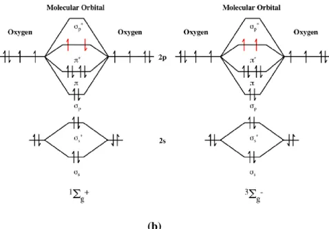
Figure 1: a) Schematic representation of 1Δ
g state of singlet state oxygen. b) Schematic representation
of 1Σ
g+ state of singlet oxygen and lowest triplet state of molecular oxygen………2
Figure 2. A schematic representation of different ways of generating oxygen………3
Figure 3. Structure of the commonly used singlet oxygen trap 1,3-diphenylisobenzofuran………….…4
Figure 4. Some examples to the commonly used photosensitizer molecules………..5
Figure 5. Examples of suitably substituted alkyl-napthalenes1………...6
Figure 6. Syntheses of water soluble napthalene carriers of 1O 2 ……….7
Figure 7. Steric effects on the 1, 8-dimethylnapthalene endoperoxide………8
Figure 8. Mechanism of [4+2] cycloaddition of Singlet Oxygen on aromatic hydrocarbons………9
Figure 9. Thermolysis pathways for aromatic endoperoxides………….………10
Figure 10. Activation parameters for the thermolysis of various aromatic endoperoxides and corresponding singlet oxygen yields………..….11
Figure 11. Temperature dependence of thermolysis of anthracene derivative. Reprinted with permission from reference 25. ………..12
Figure 12. 1-4 endoperoxide of 1-4 dimethoxy -9, 10-diphenylanthracene and 1, 4 dimethylnapthalene endoperoxide……….…………..12
Figure 13. Reaction scheme for the 9, 10 diphenyl anthracene endoperoxide………..…...13
Figure 14. Structure of the Si-DMEP endoperoxide for oxidation of DNA………..……..14
Figure 15. Structures of tert-butyldimethylsilyl chloride (21) and tert-butyldiphenlysilyl chloride (22)………...15
Figure 16. Silylation of various alcohols under different solvent and catalysts………16
Figure 17. Two different examples of fluorine mediated decomposition of silyl protecting groups..…17
Figure 18. Core structure of the BODIPY dye………20
Figure 19. Examples of BODIPY based fluorescent sensors. ………21
Figure 20. Some further examples to the BODIPY based sensors. ……….…..22
x
Figure 22. Examples of PeT based sensors..………..23
Figure 23. Schematic representation of ICT mechanism………...24
Figure 24. Some examples to ICT sensors..………...25
Figure 25. Synthetic Pathway for the synthesis of tert-butyl ((1, 4-dimethyl-1, 4-dihydro-1,4-epidioxynaphthalen-2-yl)oxy)dimethylsilane. ………...27
Figure 26. Synthesis of Compound 36..………..28
Figure 27. Synthesis of Compound 37..………..28
Figure 28. Synthesis of Compound 38..………..29
Figure 29. Synthesis of Compound 39………29
Figure 30. Synthesis of Compound 40………30
Figure 31. Schematic representation of the synthetic pathway for BODIPY based fluorescence sensor………...31
Figure 32. Synthesis of Compound 41……….…..32
Figure 33. Synthesis of Compound 42………32
Figure 34. Synthesis of Compound 43………33
Figure 35. Synthesis of Compound 44………33
Figure 36. Structure of the 1, 4-dimethylnapthalene ring………..35
Figure 37. The [4+2] addition reaction of singlet oxygen to Molecule 28………….….…...36
Figure 38. Absorption spectra of the Molecule 29..………...37
Figure 39. Absorption Change (abs) versus time (min) graph of the singlet oxygen release…………38
Figure 40. Structure of 2’, 7’- dichlorofluorescein………..……38
Figure 41. Structure of the Zn2+ binded target molecule…...………41
Figure 42. 1H-NMR spectrum of Compound 36. ………...48
Figure 43. 13C-NMR spectrum of Compound 36. ……….49
Figure 44. 1H-NMR spectrum of Compound 37……….…50
Figure 45. 13C-NMR spectrum of Compound 37.……….………..51
Figure 46. 1H-NMR spectrum of Compound 38……….…….. 52
xi
Figure 48. 1H-NMR spectrum of Compound 39.……….…..54
Figure 49. 13C-NMR spectrum of Compound 39. ……….55
Figure 50. 1H-NMR spectrum of Compound 40...………..56
Figure 51. 1H-NMR spectrum of Compound 40 in (CD3) 2SO……….………...57
Figure 52. 1H-NMR spectrum of Compound 40 with Fluoride anion. ………..58
Figure 53. 1H-NMR spectrum of Compound 41………59
Figure 54. 1HNMR spectrum of Compound 42.……….……… …60
Figure 55. 13C-NMR spectrum of Compound 42. ……….……….…....61
Figure 56. 1H-NMR spectrum of Compound 43..……….………..62
Figure 57. 1H-NMR spectrum of Compound 44..………..63
Figure 58.13C-NMR spectrum of Compound 44. ……….………..64
Figure 59. Mass spectrum of Compound 39.………..65
Figure 60. Mass spectrum of Compound 40.………..65
Figure 61. Mass spectrum of Compound 41.……….…...66
Figure 62. Mass spectrum of Compound 42………...66
xii
LIST OF TABLES
Table 1: Potential energy curves for three low lying electronic states of molecular oxygen……….2 Table 2: Schematic representation of photosensitizer excitation and singlet oxygen production…..……4 Table 3: Schematic Representation of concerted cycloreversion and homolytic cleaveage mechanism...9 Table 4: Reaction scheme for the silylation of alcohols………..16 Table 5: 1H-NMR of endoperoxide in (CD
3)2 SO without (top) and with (down) tetrabutylammonium
1
CHAPTER 1: INTRODUCTION
A. CHEMICAL MODULATION OF SINGLET OXYGEN GENERATION RATES IN
THERMAL ENDOPEROXIDE DECOMPOSITION
1.1 Properties of Singlet Oxygen
1.1.1. Electronic Structure and the properties of Singlet Oxygen
Oxygen, in its ground state, is essential to all organisms and biological processes in life. Since it is highly abundant on earth, investigation on its chemistry has been going for centuries. One of the most interesting discoveries about the molecular oxygen is its powerful oxidant excited version, singlet state oxygen, which gives a variety of reactions inside biological systems. Although its discovery goes back to 1924, singlet oxygen has become the focus of the laboratory studies after the work of Khan and Kasha on chemiluminescence of the hypochlorite-peroxide reaction as caused by liberated singlet oxygen2.
In order to understand the nature of this excited state, the electronic structure of the ground and the excited states of oxygen molecules should be investigated. Molecular oxygen (3Σ
g- ) has two
low-lying singlet excited states, above the triplet state3. The difference in these two states is the change in
the electronic configurations of л orbitals. These are represented as delta singlet oxygen 1Δ
g , which has
95 kJ /mol higher energy than ground state oxygen and sigma singlet oxygen 1Σ
g+, which has 158 kJ /mol
higher energy than the ground state oxygen4.The latter one has a very short lifetime and is quickly
converted to 1Δ
g state. 1Δg state has sufficient lifetime to react chemically in the biological environment5.
These states and their corresponding orbital assignment are represented in Figure 1.3
2
(b)
Figure 1: a) Schematic representation of 1Δg state of singlet state oxygen. b) Schematic representation of 1Σ
g+ state of singlet oxygen and lowest triplet state of molecular oxygen.
The potential energy diagram corresponding to different energetic states of molecular oxygen can be seen in Table 13.
Table 1: Potential energy curves for three low lying electronic states of molecular oxygen.
Singlet oxygen is a highly reactive and short-lived intermediate and because of its highly electrophilic nature, it exhibits a high reactivity towards electron rich molecules. Since its half-life is in the range of 1-50 μs in aqueous systems, it can travel appreciable distances in the cellular environment forming allylic hydro peroxides, dioxanes and endoperoxides.
3
Reaction of singlet oxygen with proteins is more selective yielding some toxic products for the cell, whereas the reaction of 1O
2 with DNA can lead to strand breaks and formation of altered bases.
Particularly, the photosensitized singlet oxygen has various applications, from photodynamic therapy to photooxidation. Like any other reactive species, it can be harmful at high concentrations, whereas at low concentration, it can act as a signaling molecule5. If 1O
2 can be selectively generated in a diseased tissue,
such as tumors, it can behave as a therapeutic agent. This characteristic is most commonly used in photodynamic therapy of the cancer cells.
Recent studies on singlet oxygen also revealed that it is also involved in the cell signaling, inducing a series of genes involved in signaling cascade and initiating three different pathways6.
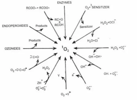
1.1.2 Generation and Quenching of Singlet Oxygen
Over the years, there have been numerous methods to produce singlet oxygen for investigating its photophysichal and chemical properties. Some of the earliest investigations at the beginning of 20th
century generated 1O
2 in a photosensitized project, such as the work by Fritzsche and Moureu, where
they independently investigated the photooxygeneration of naphthalene and rubrene respectively7.
Various ways which singlet oxygen can be generated is shown in Figure 24.
Figure 2: A schematic representation of different ways of generating oxygen.
In early 1940’s, researchers began to actively investigate the chemistry of singlet oxygen and its use in organic chemistry. The very first dye sensitized photooxygenation of α–terpinene, using chlorophyll as the sensitizer, was proposed by Schenck and Ziegler to synthesize the naturally occurring trans-annular peroxide, ascaridole8.
4
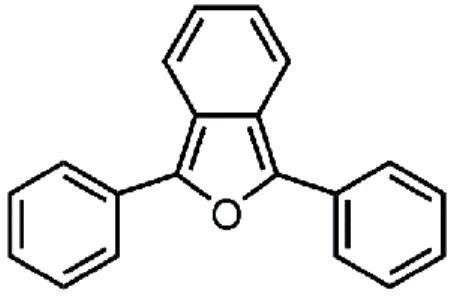
Dufraisse and Ecary worked with 3-diphenylisobenzofuran (DPBF), shown in Figure 3 demonstrating its efficiency as a singlet oxygen acceptor9. It is today among the commonly used
molecules in experiments to study the kinetics and photophysics of singlet oxygen sensitizers.
Figure 3: Structure of the commonly used singlet oxygen trap, 3-diphenylisobenzofuran.
The simplest and most controllable method for generating singlet oxygen is the photosensitized production3. It can be very efficient and is one of the most commonly used methods to generate 1O
2.
This method requires only oxygen, light of an appropriate wavelength and a photosensitizer which is capable of absorbing that energy and use it to excite oxygen to its singlet state. Excitation of sensitizer is generally achieved via one photon transition (hv) between the ground state and a singlet excited state. The basic representation of this method can be seen in Table 2.
Currently, there is a large number of photosensitizers that are used in 1O
2 generation and new
sensitizers are continuously being introduced. There are some specific requirements for these photosensitizers, in order to produce singlet oxygen effectively3.
Step (a) Absorption of light
Step (b) Intersystem crossing
Step (c) Energy transfer from triplet excited sensitizer to ground state (triplet) oxygen.
Table 2: Schematic representation of photosensitizer excitation and singlet oxygen production.
They should have a high absorption in the visible region and a triplet state of appropriate energy. The presence of a heavy atom increases the yield of intersystem crossing to the triplet state of the dye.
5
A high quantum yield, and a high photostability is also required. Methylene blue for example, has a strong absorption in the range of 550-770 nm and a significant quantum yield (Φ= 0.52). Some of the commonly used photosensitizers are methylene blue, fluorescein, Rose Bengal and hypericin and perylene diimide are shown together in Figure 310
Figure 4: Some examples to the commonly used photosensitizer molecules.
Fluorescein Hypericin 2 3 4 5 6 7 Methylene Blue Rose Bengal Perylenee Diimide
6
The choice of the sensitizer is dictated by the reaction conditions such as solubility, efficiency of 1O
2 formation, wavelength of excitation, as well as other conditions. Recently, borrondipyromethane
(BODIPY) dyes have also become attractive in this are with their high extinction coefficients and photostabilities. One example to these molecules was synthesized by Akkaya et al11, 7, shown in Figure 3. This molecule can be chemically modified and this allows attachment of chemical groups that would
enable water solubility, or increase photodynamic action.
1.2 Polycyclic Aromatic Endoperoxides
1.2.1 General Information
The unique chemistry of singlet oxygen as compared to triplet ground state oxygen makes it undergo addition reactions to form endoperoxides of different types. Many polycyclic compounds can trap singlet oxygen in which they give different types of endoperoxides that exhibit different features upon warming or light irradiation. The very first observation on endoperoxides was made in 1926 by Dufrassie and his friends in which dissociation occurs with the photo oxidation of the stil unknown rubrene12.
One of the first works on naphthalene endoperoxides was done by Wasserman et al1, which was
the synthesis 1, 4-dimethylnapthalene endoperoxide and its derivatives. Wasserman and his coworkers observed that, when two strongly electron releasing groups are present in the 1- and 4- positions of the naphthalene, endoperoxides that are generated from them were readily isolated and characterized. They also observed that, various derivatives have different half-life at different temperatures, although the decomposition of each endoperoxide yielded the original naphthalene and oxygen. The derivatives they have synthesized are shown in Figure 5.
7
After a while, this foundation is followed by the synthesis of 9,10-diphenylanthracene endoperoxide13. Further research revealed that at some instances, like in the case of 1, 4 dimethoxy-9,
and 10-diphenylanthracene), the dissociation to produce singlet oxygen can take place at the room temperature.
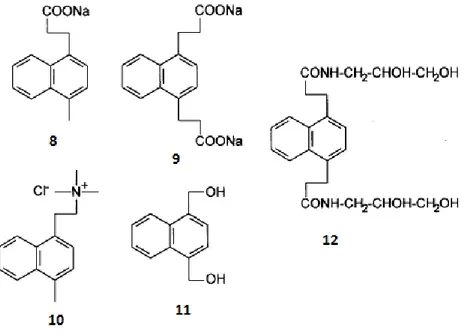
Figure 6: Syntheses of water soluble napthalene carriers of 1O 2.
After the observation of this new feature, water soluble naphthalene endoperoxide derivatives have been prepared and used as singled oxygen carriers as a source in biological environment by several research groups14,15. The very first example of water soluble carrier of singlet oxygen carries were
reported by Nieuwni et al and Saito et al. These endoperoxides, shown in Figure 6 as 8 and 9 have been used as chemical sources of singlet oxygen to assess the activity of towards biological or chemical targets.
Following these molecules, a second generation of carriers have been synthesized bearing specific groups as in the case of 10, 11 and 12. They bear some specific groups like quaternary ammonium groups or nonionic hydrophilic groups in order to assure particular affinity for specific sites16.
All of these examples demonstrated that, with different substation patterns, it is possible to control the rate and the temperature of thermal decomposition process of polycyclic aromatic endoperoxides which provide promising application areas for decomposition of singlet oxygen.
8
1.2.2 Preparation of Endoperoxides
Two main classes of reaction of singlet oxygen are particularly important. First of it is the addition to olefins, which produces allylic hydroperoxides. The second one is the additions to diene systems to produce endoperoxides which are analogous to Diels-Alder reactions17. The latter type is
most commonly referred as photosensitized oxygenation which involves [4+2] cycloaddition of singlet oxygen18,19.
Since 1O
2 has a strong electrophilic nature, the reactivity of aromatic hydrocarbons towards
singlet oxygen increases with increases electron donating groups involved. Thus, incorporation of groups which have high electron density on the reaction site of 1O
2 dramatically increases the rate
constant of the addition reaction. The rate order for various groups is H< C6H6< CH3<OCH313.
Unsubstituted naphthalene is known to be not reacting with singlet oxygen, whereas it is possible to observe endoperoxide with 1-methyl naphthalene.
Another structural effect on reactivity is the number of fused ring. Molecules that bare more than two rings like anthracene or tetrachene showed that as the fused ring number increases, reactivity also increases about two fold20.
Figure 7: Steric effects on the 1, 8-dimethylnapthalene endoperoxide.
Steric effects are also important parameters which can modify the reactivity of the molecule and change regioselectivity of the cycloaddition. In the case of the molecule in Figure 7, interactions between two neighboring methyl groups bound to a polycyclic aromatic hydrocarbon increases its reactivity towards singlet oxygen because steric strain is relieved in the transition state. Therefore compound 13 is 4 times more reactive than the 1, 5 isomer21.
Unlike common Diels Alder reactions, in the case of synthesis of endoperoxides from singlet oxygen, there is shown to be a strong solvent dependency of [4+2] cycloadditions. In order to understand this effect, singlet oxygen cycloaddition of 1,4 dimethylnapthalene was investigated with 28 solvents22.
9
This study have revealed that the reaction rate increases 100 fold from cyclohexane to dimethylformamide, DMF and becomes even faster with water soluble analogs.
The mechanism for the cycloaddition of singlet oxygen to the aromatic compounds is very similar to the classical Diels Alder type reactions, only with little alterations. Recent studies have shown that the mechanism goes through a concerted, single step reaction, exhibiting strong charge transfer from aromatic organic donor to singlet oxygen which is demonstrated on Figure 823.
Figure 8: Mechanism of [4+2] cycloaddition of singlet oxygen on aromatic hydrocarbons.
A reversible intermediate conjugate between aromatic ring and oxygen is formed in the singlet state during the reaction. Since singlet oxygen has a highly electrophilic character, this conjugate has a highly significant charge transfer character. After the intermediate is formed, the aromatic endoperoxide can be obtained through concerted mechanism. Charge transfer mediated inter system crossing (ISC) may also take place, switching the intermediate conjugate to the triplet state and triplet oxygen to be dissociated at the end.
Table 3: Schematic Representation of concerted cycloreversion and homolytic cleaveage mechanisms.
Inter system crossing of singlet state oxygen to triplet state oxygen can be seen on the Jablonski diagram on Table 324. It also shows the different electronic states of a molecule and the transitions that
10
1.2.3 Thermal Dissociation of Endoperoxides
Photolysis and thermolysis are two primary ways for endoperoxides to dissociate. Just as in photolysis, the dissociation of endoperoxides by heating involves two different mechanisms. First of them, is the cycloreversion, which leads to the substrate and molecular oxygen either in singlet or in triplet state and the cleavage of the peroxide bond. This cleaveage is often followed by rearrangements or decompositions to hydroxyketones or quinones as seen in Figure 925.
Figure 9: Thermolysis pathways for aromatic endoperoxides.
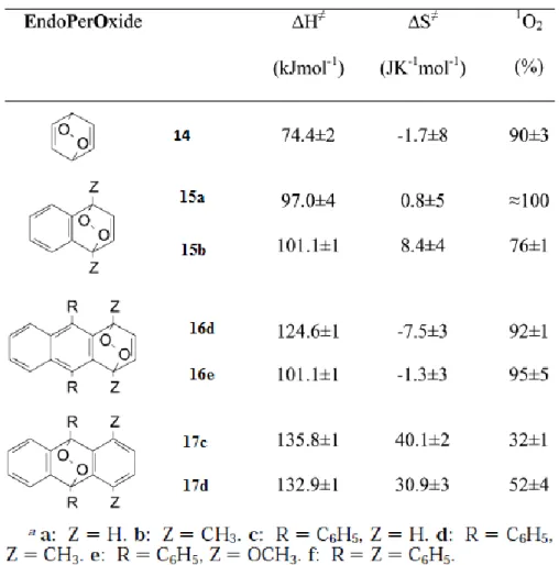
Activation enthalpy of each different endoperoxide molecule determines the ratio between cleavage and cycloreversion. In Figure 10, some activation parameters for the thermolysis of several aromatic endoperoxides are given13. It is seen that the activation enthalpy for cycloreversion, ΔH,
increases from benzenic to napthalenic and 1, 4 anthracene endoperoxides. In more condensed analogues, as a result, there is a competition between cleavage and cycloreversion.
Recent work on endoperoxides revealed that the incorporation of aromatic moieties to the bridgehead position of aromatic EPO’s, increases the possibility of cycloreversion process13. For thermal
cycloversion decomposition pathway, there are also two possible mechanisms. In a concreted cycloreversion, 1O
2 is produced quantitatively. On the other hand, homolytic cleavage involves ISC and
leads both molecular and singlet oxygen. In Figure 10, molecules 14, 15 and 16 are introduced by Turro and coworkers to decompose singlet oxygen in high yields, as compared to 9,10 substituted ones 17 which produce singlet and triplet state oxygen.
11
Figure 10: Activation parameters for the thermolysis of various aromatic endoperoxides and corresponding
singlet oxygen yields.
Further examples on naphthalene and anthracene endoperoxide derivatives are given in Figure
11 and Figure 12. In addition to small molecules, polymeric endoperoxides have also been prepared as
well, where naphthalene derivatives were successfully grafted on a various benzene moieties, and polymerization of methyl substituted vinylnapthalene26. In Figure 11, the temperature dependence of
12
Figure 11: Temperature dependence of thermolysis of anthracene derivative. Reprinted with permission from
reference 25.
In the case of Figure 12, although the molecule 19 is easily synthesized, its acetal structure can undergo hydrolysis in an aqueous medium27. The naphthalene derivative 20, however, is known to
release singlet oxygen at 37 with a % 76 yield25. This type of molecular structure can be compatible
with the biological conditions if water solubility can be attained with hydrophilic moieties.
Figure 12: 1-4 endoperoxide of 1-4 dimethoxy -9, 10-diphenylanthracene and 1, 4 dimethylnapthalene
endoperoxide.
13
1.2.4 In Situ Generation and Detection of Singlet Oxygen in Cell Studies
In aqueous biological systems, photogeneration and thermal generation of singlet oxygen play important roles, such as the formation of 1O
2 in natural surface waters or the photodynamic effect. Since 1O
2 has a critical role in cell killing mechanism of photodynamic therapy, studies have focused on
generating and monitoring singlet oxygen in vivo.
In order to understand whether singlet oxygen plays a key role in aforementioned reactions, it is necessary to develop a mechanism for specific detection and quantification of it. First attempts to do this were the studies made on aqueous solutions where detection can be maintained by fluorescent or near-IR phosphorescence molecules28,29. These molecules are most commonly known as chemical traps
and they have specific properties for providing the required features.
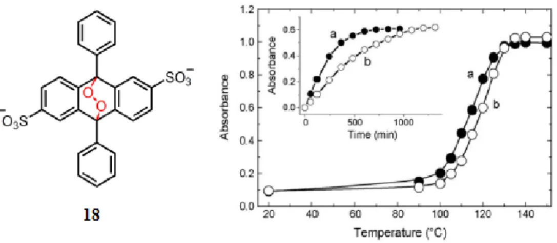
One of the first studies on intracellular production and detection of singlet oxygen was done by Karnovsky and his coworkers, where they tried to understand whether neutrophils are producing singlet oxygen during the process of phagocytosis. For this, they coated glass beads using both water soluble 9, 10 diphenyl anthracene and perylene30. Since the work of Khan2 has shown that the former molecule
which is shown in Figure 13, gives a faster reaction rate with singlet oxygen, they were able to obtain a quantitative information about the formation of the singlet oxygen in the cells.
Figure 13: Reaction scheme for the9, 10 diphenyl anthracene endoperoxide.
Later, measuring the lifetime of singlet oxygen in a single cell was done by Hatz et al31, in which
single neurons and HeLa cells are subjected to time resolved optical experiments upon a photosensitizer incorporated inside them. They concluded that the lifetime of singlet oxygen in metabolically functioning, H2O containing cells is about 3μs and this observation implies that it is best characterized
14
Investigation of singlet oxygen in vivo was further continued with the studies made on DNA. Modification of cellular DNA upon exposure to reactive oxygen was known to be the cause of induction of mutagenic oxidative stress reagents. A significant increase in the level of 8-oxo-7,8-dihydro-2’-deoxyguanosine was observed upon exposure the generation of singlet oxygen by an endoperoxide while the level of the other DNA bases remain unchanged32.
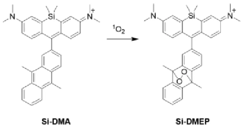
One of the most recent studies was done in 2014, in which a potential intracellular 1O 2
production is observed causing the oxidation of the cellular DNA33. For this purpose, water soluble and
thermally decomposable endoperoxide were synthesized. In order to understand the damage of singlet oxygen directly, the oxygen atom is radioactively labelled. The anthracene derivative endoperoxide that is used in the study can be seen in Figure 14. A cationic charge and lipophilicity of Si-DMA utilize the selective mitochondrial localization.
15
1.3 Silyl Protection of Alcohols and Phenols
Many protective groups were developed to block multifunctional organic molecules for specific synthetic reactions. Various requirements, however, change the choice of protecting group depending on the site they are protecting. A protective group should not have any more functional groups to react any further than the compound to be protected. It should be easily removed, by avoiding any side products that can affect the regenerated functional group34.
Silyl ether moieties are used most commonly for alcoholic or phenolic groups, because of their switchable reactivity on silicon atom. Among many trialkylsilyl reagents used to protect functional groups like hydroxyl groups, tert-butyldimethylsilyl chloride (TBDMSCl) and tert-butyldiphenlysilyl chloride (TBDPSCl) are the most commonly used reagents.
Figure 15: Structures of tert-butyldimethylsilyl chloride (21) and tert-butyldiphenlysilyl chloride (22).
The larger tert-butyl substituents disables the attack at the silicon atom, making the species a more stable substrate toward hydrolysis in weakly acidic or basic medium.
One of the properties that made silyl groups popular is the fact that they can be easily cleaved by fluoride anions. This is attributed to the fact that fluoride anion has a high affinity for silicon atom, and the Si-F bond strength is 30 kcal/mole more energetic than the Si-O bond34,35. There are many
example usages of these protective units.
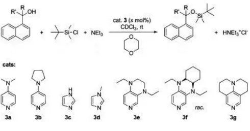
TBDMSCl was first introduced by Corey36. In his work, Corey showed that the
dimethyltert-butyl siloxy group is approximately 104 times more stable than trimethylsilylyl group. After this
observation, solvent and the catalyst effects were further investigated, and it is found that the use of imidazole as catalyst and dimethylformamide as solvent proved to be strongly effective and provide high yields under mild conditions. The basic reaction scheme is thus shown in Table 3.
16
Table 4: Reaction scheme for the silylation of alcohols.
The choice of solvent, catalyst and other additional reagents for various functional groups are still under investigation. Among the various factors that influence the silylation of primary and secondary alcohols for example, the choice of solvent and catalyst are interdependent. In a 2014 study on various alcohols demonstrated that, when DMF is used as solvent, there is no need for additional catalysts because of the high catalytic activity of this solvent37. The structure of the alcohol and different
catalysts can be seen in Figure 16.
17
1.4 Fluorine Mediated Deprotection of Protective Groups
Anion sensing has been a great interest for many reasons. The detection of simple anions in biological systems is highly important. Different halides are distinguished by different magnitudes of binding constants or unit responses38. Optical sensing of fluoride anion is a challenging topic because
of the high reactivity of fluoride anions in aqueous solution. They can easily be hydrated, reducing effective nuchleophilicty and basicity39. Since it is present enormously in organic environments, it is
important to monitor its concentration.
Fluorine mediated deprotection followed by a chemical change in molecule is one of the common deprotection methods for silyl groups. In biological systems it is important to use the presence of fluoride anions for such enchaining reactions. The efficiency of this type of deprotection was investigated on various nucleic acid derivatives and it is found that it is critically dependent on the amount of water present in the TBAF molecule40.
There are various examples in the literature, where upon deprotection with fluoride anion in the form of tetrabutylammonium fluoride (TBAF), there is a chemical process in the molecule. A recent study by Akkaya et al on a fluoride-mediated deprotection of the silyl-protecting group was investigated on a cyclic peroxide as shown in Figure 1741,42 as 23. Here, TBDMSCl protecting group was deprotected
easily on addition of tetrabutylammonium fluoride, yielding a very bright blue chemiluminescence.
Figure 17: Two different examples of fluorine mediated decomposition of silyl protecting groups.
Another example of a silyl protected group is a derivative of a Tetraphenylethylene (TPE) group which is protected by four sides shown also in Figure 1743 as 24. Fluoride sensing of this molecule was
accomplished by deprotection of the silyl groups from the TPE core. As a result of this, four phenoxide ions in full conjugation with the TPE moiety are available leading to a very strong intramolecular charge transfer.
18
B. NOVEL FLUORESCENT SENSORS FOR HYPERPHOSPORYLATED TAU
PROTEINS
1.5 Supramolecular Chemistry and Molecular Recognition
Supramolecular chemistry is one of the greatest interdisciplinary fields of science. It basically focuses on the chemical, physical and biological characteristics of molecular systems that are held together by non-covalent chemical interactions. Intermolecular interactions are weaker with respect to covalent bonds. That is the reason behind the fact that the supramolecular structures are thermodynamically less stable and kinetically more labile44.
Molecular recognition on the other hand, can be defined by the information and the energy carried by the selection of substrate by a certain receptor molecule45. Binding of the analyte (guest) and
the receptor (host) introduces a complex or a supramolecule. As a result of this interaction, molecular information can be stored at a molecular level. In 70’s, studies on selective complexation of metal ions initiated the use of notions of recognition and information which were used in relation with the biological systems46.
Biological molecular recognition is the most complex expression of molecular recognition leading to highly selective binding, reaction and transport of the different types of molecules. Both interactional and geometrical complementary is required between the associating species. Chelating or macrocyclic ligands are often employed due to the high thermodynamic stability of the complexes that contain chelate rings.
The balance between rigidity and stability is important because binding properties may change due to these different properties. Less flexible ligands have less disorder, and macrocyclic hosts requires less solvation energy. These different properties have been used in the design of many receptors which operate through various intermolecular forces.
1.5.1 Anion and Cation Recognition
Anions and cations both play very important roles in chemistry and the biology. Although cations have attained far more interest on research for many fields, the binding characteristics of anions have not fully understood. Anionic species like fluoride, bromide or chloride have various range of geometries are known to have formed different molecular complexes47,48.
19
Cation detection is very important for many biological activities. They play an important role such as enzyme activation (Zn2+, Mg2+, Mn2+) , transmission of nerve impulses ( Na+, Mg2+, Ca2+) or regulation
of the cell activity ( Zn2+, Mg2+, Ca2+) Also, detection of environmentally important cations are very
important.
1.5.2 Sensors
A sensor is a device that interacts with matter or energy in order to yield a measurable signal in response49. Today a wide range of molecular sensors can be designed for different sensing purposes.
They can signal a physical output, upon interaction with an analyte.
A molecular sensor basically is designed to have a selective interaction with different analytes. One can design a sensor that can contain binding sites that are selective and suitable for an analyte. This binding site should be able to induce a change in the signaling in terms of fluorescence emission change. There are also other designs like chemodosimer that forms a complex with a cation or anion.
1.5.3 BODIPY Dyes
4, 4-difluoro-4bora-3a, 4a-diaza-s-indacene molecule, which is most commonly known as BODIPY is first introduced by Treibs and Kreuzer in 196840. BODIPY dyes and their various derivatives
have wide applications from solar cells to biomolecular labelling and drug delivery systems. As previously mentioned, upon modification, these dyes are also strong candidates for photosensitizers in order to generate singlet oxygen.
The great interest on this molecules comes from their effective properties. As compared to other sensitizers, it has high fluorescence quantum yields and depending on the design, it may have high triplet quantum yields as well. Outside effects like solvent polarity or pH do not affect these molecules, therefore, they tend to maintain their structure and work in the therapeutic window. However, they can be easily decomposed under strongly acidic or basic conditions50.
20
Figure 18: Core structure of the BODIPY dye.
A core structure of the BODIPY molecule can be seen in Figure 18. It can be functionalized from any different positions, from 1 to 8. This property makes it a useful candidate for functionalizing it for any purpose.
1.6 The Principles of Fluorescence and Fluorescent Chemosensors
After the excitation of a molecule by a photon with an appropriate energy, electrons follow different pathways to relax. Firstly, they scatter their energy, which is called internal conversion until the S1 excited state. Then, from this state, they are ready to relax to the ground state via four different
competitive processes51. These mechanisms are shown in the Jablonski Diagram which was shown in Table 3. Fluorescence is probably the most important of them all, where there is an emission of a photon
from S1 state to S0. There is a difference between the absorbed and emitted energy. This is called Stokes
shift. In addition to this effect, some fluorescent molecules can display further Stokes shift due to the complex formation and energy transfer52.
A fluorophore is a molecule that emits light and fluorescent detection by fluorophore molecules is a highly sensitive, yet simple and cheap method for detecting the molecules or ions of interest. Binding of an analyte to the receptor may lead to significant changes in the photophysical properties of the fluorophore. A molecular level understanding of the biological processes in living organisms in vivo is important, because the detection of analytes in these organisms is a challenging area.
The design of the fluorescent molecular sensors have gained much attention after the work of Tsien’s calcium probe in 1980’s53,54,55. The most important property of a fluorescent sensor is to be able
to detect the presence of an anion or a cation in a complex environment like cell or organism.
Fluorescent sensors that are designed for detection of cations contains two essential features. Firstly, they have a metal chelating moiety and at least one fluorophore capable of absorbing and emitting light. They may be entirely chemical or they may contain nucleic acid or peptide components56.
21
Among all of the fluorescent sensors, BODIPY as a fluorescent dye, draws the greatest attention due to its unique properties. High fluorescence quantum yield, small stokes shift and high molar absorptivity makes it a great candidate for fluorescent probe. There are many examples of BODIPY based fluorescent probes in literature. Four different examples for detection of different cations are shown in Figure 19. Molecules 26 and 27 are specifically designed for the detection of zinc cation57.
Molecules 28 and 29 are designed for the detection of the Cu+ and Cd2+ cations respectively56,58. Water
soluble moieties can be attached as in the case of 27 and 29 in order to increase the hydrophilicity.
Figure 19: Examples of BODIPY based fluorescent sensors.
Further examples can be given to BODIPY based sensor with different properties. In Figure
20,for example, molecule 30 was introduced by Negano et al59, has been known to have high triplet
quantum yields. Molecule 31 was introduced by Hamachi and his coworkers in 200960. This molecule,
other than being a fluorescent Zinc sensor, has been used as molecular probe for selective detection of neurofibrially tangles in the brains of Alzheimer disease patients.
26 27
28
22
Figure 20: Some further examples to the BODIPY based sensors.
As a result of these examples, other derivatives of BODIPY molecules which can both act as a sensor and imaging probe have gained great attention.
1.7 Energy and Electron Transfer Processes
1.7.1 Photo induced Electron Transfer (PeT)
Photo induced electron transfer systems consists of three units: Fluorophore (signaling unit), receptor for recognition of an analyte, and a spacer combining these two parts. Although there is no conjugation between fluorophore and receptor in a PeT system, they are still close enough for electronic interaction by a spacer.
Firstly, an electron is excited in the PeT system, from the highest molecular orbital of the fluorophore to its lowest unoccupied molecular orbital (LUMO) upon irradiation. A receptor molecule should be able to accept and donate electrons, having its lone pair electrons at separate orbitals. When the energy of the orbital of this receptors falls between the HOMO and LUMO of the fluorophore, an electron transfer occurs from HOMO of the receptor to the HOMO of the fluorophore61. This occurs
after the excitation process and causes an intense decrease in emission intensity together with the quenching of the fluorophore. When there is a metal cation binding to the receptor, it decreases the energy of the HOMO due to the stabilization. In this case PeT is prevented and quenching is finished.
30
23
There is also another process which is known as the reverse PeT. In this process, in the absence of an analyte, emission takes place normally. When there is an analyte such as a metal cation, the energy of the LUMO of the receptor decreases and falls within the HOMO and LUMO of the fluorophore. As a result, upon excitation electron transfer occurs between LUMO’s of fluorophore and the receptor moieties. In Figure 21, A and B, the schematic representation of a PeT mechanism of a binding electron can be seen62.
Figure 21: Schematic Representation of PeT mechanism62.
There are many fluorescent chemosensors in the literature that work via PeT mechanism. They commonly work for cations, including H+ and even though the molecules themselves are
non-luminescent, upon binding of a cation they become fluorescent. In Figure 22, two different examples of sensors that work with the PeT principle can be seen61.
Figure 22: Examples of PeT based sensors.
32
24
1.7.2 Internal Charge transfer (ICT)
Intramolecular charge transfer is an electronic process that leads to two stokes shift in the emission spectrum a fluorescent molecule. Blue shift occurs when there is an electron donating group bound to the fluorophore. This decreases the ability of the electron donating group to donate, hence causing a decrease in conjugation. Red shift occurs when the binding group has an electron withdrawing moiety, increasing the interaction between the cation and the acceptor unit. This interaction stabilizes the excited state causing a decrease in the energy gap between HOMO and LUMO levels. The scheme that is representing these interactions can be seen on Figure 2363
Figure 23: Schematic representation of ICT mechanism.
While designing an ICT type chemosensor, fluorophore and the receptor units are directly bound to each other and there is no spacer between them. The receptor is conjugated to the pi system of the fluorophore where it acts as an electron donor or electron acceptor according to the state of the fluorophore. This is the cause of the orbital overlapping in this conjugated system which causes the internal charge transfer.
ICT is a frequently used mechanism in the development of a fluorescent sensors. In Figure 24, there are two examples of molecules which give different stokes shift upon coordination with a cation64.
25
Figure 24: Some examples to ICT sensors.
34
26
CHAPTER 2: EXPERIMENTAL PROCEDURES
2.1 Methods and Materials
All chemicals and solvents are purchased from Aldrich, Merck and ABCR and were used without further purification. Merck TLC silica gel 60 F254 was used to monitor all the reactions. Column chromatography of all products was performed using Merck Silica Gel 60 (particle size: 0.040-0.063 mm, 230-400 mesh ASTM).
1H-NMR and 1C-NMR spectra were recorded on Bruker DPX-400 which are operating at 400
MHz for 1H-NMR and 100 MHz for 1C-NMR in CDCl
3 and CH3OD with TMS (tetramethylsilane) as
internal standard. All spectra were recorded at 25oC and coupling constants (J values) are given in Hz.
Chemical shifts are given in parts per million (ppm). Splittings in the spectra are shown as s (singlet), d (doublet) t (triplet), q (quartet), m (multiplet)
Mass spectrometry measurements are conducted using Agilent Technologies 6224 TOF LC/MS devices at UNAM, Bilkent University.
Absorption and fluorescence spectrometry measurements were conducted using UV-Vis Spectrophotometer (Cary 100 Bio) and Fluorescence spectrophotometer devices at UNAM, Bilkent University.
27
2.2 Synthetic Pathway (1)
Figure 25: Synthetic Pathway for the synthesis of tert-butyl
28
2.3 Synthesis of Compound 36
65Figure 26: Synthesis of Compound 36.
1, 4 dimethyl naphthalene (1.56, 10 mmol) was dissolved in chloroform (5ml) under N2 and
exclusion of light. Bromine (1.68, 0.54 ml, 10.5 mmol) was added over a period of time (10 min) at 0o
C. The reaction was warmed to room temperature and stirred for 2 hours. It was diluted with chloroform (20ml), and washed with saturated Na2S2O2 solution (25ml), water (25 ml) and saturated NaCl solution
(25 ml). The organic layer was dried over Na2SO4 and filtered through a thin pad of silica gel. The title
compound (2.32, 99%) was used without further purification. Rf (hexane) = 0.65; 1H NMR ( 300 MHz, CDCl 3) : δ 2.64 (s, 3H), 7.5 (s,1H), 7.57 (ddd, J=5.8, 4.4, J=32.5 Hz, 2H ), 7.94-7.98 (m, 1H), 8.02-8.08 (m, 1H): 13C NMR (75 MHz, CDCl 3): δ 18.7 (q), 19.1 (q), 122.5 (s) ,124.9 (d), 125.1 (d), 125.4 (d), 125.6 (d), 126.5 (d), 130.6 (d), 131.4 (s), 131.9 (s), 133.6 (s) 134.0 (s).
2.4 Synthesis of Compound 37
66Figure 27: Synthesis of Compound 37.
To a solution of 2-bromo-1, 4 dimethyl naphthalene (2.02 g, 8.6 mmol) in dry THF, n-BuLi (2.5 M in hexanes, 7.6 ml, 18.92 mmol) was added at -78oC under N
2, and the reaction mixture was stirred
for 1 hour at that temperature. Triisopropylborate (3.23 g, 3.97 g, and 17.2 mmol) was then added in one portion. The resulting mixture was stirred for 30 minutes at -78oC and at room temperature for 1 hour.
29
with water, dried over Na2SO4 and the crude solid was filtered and washed with hexane to give product
as a white solid. (1.45, %84) 1H NMR (300 MHz, CD 3OD): δ 2.66 (s, 3H), 2.67 (s, 3H), 4.92 (s, 2H), 7.20 (s, 1H), 7.53-7.57 (m, 2H), 8.02-8.11 (m, 2H). 13C NMR (100 MHz, CD3SOCD3) δ 135.29, 132.75, 132.53, 130.60, 130. 35, 125.94, 125.80, 125.12, 124.76, 19.44, 19.15
2.5 Synthesis of Compound 38
67Figure 28: Synthesis of Compound 38.
%30 H2O2 solution (2 mmol, 0.12 ml), ammonium bicarbonate (1 mmol, 79.1 mg), allyboronic
acid (1mmol), H2O (2ml) were added to a round bottomed flask, and the reaction was performed under
air at room temperature for 2 hours. After the reaction is finished, 4 ml of HCl (1 mol /L) was added to the solution till pH is 2-3. Aqueous solution is extracted with ethyl acetate (4 x5 ml), and the combined phases are dried with Na2SO4 to obtain the target product.
1H NMR (CDCl
3, 400 MHz): δ 8.16 (m, 1H), 7.91 (m, 1H), 7.49 (m, 2H) , 7.09 (s,1H), 4.93
(s,1H), 2.60 (d, J=0.6 Hz, 3H), 2.39 (s, 3H); 13C NMR ( CDCl
3, 100 MHz) : δ 146.9, 132.1, 129.5, 126.1,
125.1, 125.0, 124.5, 124.1, 121.4, 115.7, 18.6, 15.5.
MS (TOF): m/z: Calcd; 171.09 [M-H]-, found: 171. 08 [M-H]-
2.6 Synthesis of Compound 39
6830
1, 4-dimethylnaphthalen-2-ol (0.500 g) and imidazole (0.175 g) were added to tert-butyldimethylsilylchloride in 12 ml dry DMF. The mixture was stirred under argon atmosphere, at 85o
-90o for 2-3 hours. The cooled mixture was extracted with dry diethyl ether. 1H NMR (CDCl
3, 400 MHz): δ 8.11 (m, 1H), 8.01 (m, 1H), 7.49 (m, 1H), 6.62 (s,1H), 2.89 (d,
J=0.6 Hz, 3H), 2.64 (s, 3H ), 0.98 (s, 12H), 0.21 (s, 6H); 13C NMR ( CDCl
3, 100 MHz) : δ 154.3, 136.7,
133.3, 127.7,125.6, 124.1, 123.4, 120.4 30.2, 25.9, 19.9, 13.4, 0.9
MS (TOF): m/z: Calcd: 287.117 g/mol. [M-H]+ , Found: 287.1837 [M-H]+
2.7 Synthesis of Compound 40
69Figure 30: Synthesis of Compound 40.
Tert-butyl ((1, 4-dimethylnaphthalen-2-yl) oxy)dimethylsilane ( 0.100 g) was dissolved in 8 ml of chloroform. It was then added a pinch of methylene blue (approx. 2 mg) and the reaction was held under liquid nitrogen in ethanol reflux at -78oC. It was controlled with TLC (in DCM) and once the
starting material was over, the solvent was removed under vacuum, without applying any external heat.
1H NMR (CDCl
3, 400 MHz): δ 7.29 (s, J= 7.5 Hz, 2H), 7.31 (s, J= 7.5 Hz, 1H), 6.06 (s, 1H),
1.69 ( s, 6H), 0.98 (s, 12 H), 0.21( s, 6 H); 13C NMR ( CDCl
3, 100 MHz) : δ 182.0, 142.7, 127.8, 121.8,
31
2.8 Synthetic Pathway (2)
32
2.9 Synthesis of Compound 41
Figure 32: Synthesis of Compound 41.
In a 250 ml round bottomed flask, (5,39 g 50.3 mmol) 2,4 lutidine and (9.12 g, 52.9 mmol) mCPBA was dissolved in 125 ml chloroform. The reaction is stirred at room temperature for 3 hours. Afterwards, 4.25 g Na2SO3 was added to the solution. The reaction is held overnight at room
temperature. To quench the reaction, 2N Na2CO3 solution was added. The extraction is done with CHCl3
and water. The substrate is dried over K2CO3. 13C NMR of this compound was consistent with the
literature value65. 1H NMR (CDCl
3, 400 MHz): δ 8.33 (s, 1H), 6.94 (s, 1H), 6.87 (m, 1H), 2.49 (s, 3H), 2.26 (s,
3H); 13C NMR (CDCl3, 100 MHz): δ 159.1, 149.9,147. 5, 125.3, 122.4, 21.6, 16.1.
MS (TOF): m/z: Calcd: 124.07 g/mol [M-H] +, Found: 124.07 [M-H] +
2.10 Synthesis of Compound 42
Figure 33: Synthesis of Compound 42.
Molecule was added to 5 ml of Ac2O which is preheated to 110-120 oC. The stirring was
continiued at same temperature for 30 min. 10 ml EtOH was added to the reaction and it is refluxed for 5 hours at 110 oC. The solvent Ac
2O was evaporated first, then the residue was neutralized with 2N
Na2CO3 aqeous solution. The product was extracted with DCM and dried K2CO3. 13C NMR of this
33 1H NMR (CDCl 3, 400 MHz): δ 8.48 (m, J= 7.5 Hz, 1H), 7.87 (s, J= 7.5 Hz, 1H), 7.47 (s, J=1.5 Hz, 1H), 5.79 ( s, J=7.5 Hz, 2H), 2.36 (s, 3H), 2.21( s, 3H); 13C NMR ( CDCl 3, 100 MHz) : δ 170.2, 156.2, 149.1, 147.0,122.4, 66.7, 21.6, 20.7
MS (TOF): m/z: Calcd: 166.08 g/ mol [M-H] +, Found: 166.08 [M-H] +
2.11 Synthesis of Compound 43
Figure 34: Synthesis of Compound 43.
0.227 g (1.60 mmol) Molecule 42 was dissolved in 50 ml of 1,4 dioxane. Then, 0.89 g (8.04 mmol) SeO2 was added and the reaction was held under reflux at 100 oC for 3 days. It was controlled
with TLC. After the starting material is consumed, the filtration was done and the product was obtained after the evaporation of the solvent.
1H NMR (CDCl
3, 400 MHz): δ 9.60 (s, 1H), 8.94 (s, J= 7.5 Hz, H), 8.33 (s, J= 7.5 Hz, 1H), 7.66
(s, J= 7.5 Hz 1H), 5.78 (s, 2H), 2.21 (s, 3H); 13C NMR (CDCl3, 100 MHz) δ191.0, 170.2, 141.7,136. 3,
132.9, 129.4, 128.8, 128.2, 66.1, 20.7.
2.12 Syntesis of Compound 44
7034
1,4 dimethyl pyrolle ( 0.382 ml, 3.71mmol) and (0.266 g, 1.48 mmol) molecule .. was dissolved in DCM under Ar. 1-2 drops of TFA was added and the reaction was stirred at room temperature for 3 days. DiPEA (1,5 ml) and p-chloranil( 0.365 g) was added and the reaction was stirred for 1 hour. After, BF3 was added and the reaction was stirred for 1 more day. The dark precipitate was filtered off, and it
was reduced under vacuum and d to give the product.
1H NMR (CDCl
3, 400 MHz): δ 8.59 (m, J=7.5 Hz, 1H), 7.88 (m J=1.5 Hz, 1H), 7.21 (s, J=7.5
Hz, 1H ), 5.80 (1H), 5.79 ( s, J=1.5 Hz, 2H), 5.17(1H) , 2.72 (3H), 2.59(3H), 2.21(3H), 1.87 (3H), 1.49 (3H) 13C NMR (CDCl3, 100 MHz) : δ 170.2, 156.1, 148.9, 148.0, 144.6, 143.6, 137.7, 127.8, 129.3,
131.7, 124.4, 118.7, 116.2, 113.5, 107.4, 66.8, 19.5.
35
CHAPTER 3: RESULTS AND DISCUSSION
A) CHEMICAL MODULATION OF SINGLET OXYGEN GENERATION RATES IN THERMAL ENDOPEROXIDE DECOMPOSITION
Singlet oxygen production in biological systems either thermolysis or photolysis of endoperoxides is a challenging area. The chemical modulation of thermolysis is important because it enables the potential control of singlet oxygen release rate in cells. There is a wide range of examples of different types of endoperoxides and each of them has a specific quality that is required for the purpose of use. Anthracenic and napthalenic derivatives are very common because of the ease of the synthesis and isolation of these molecules and their unique properties upon formation of the endoperoxides.
In our first project, we aimed to synthesize a novel naphthalene based endoperoxide, which can release singlet oxygen either itself or in the presence of a fluoride anion. We wanted to have a structure which when protected, releases singlet oxygen in a slow rate, whereas upon deprotection, releases the singlet oxygen with a relatively faster rate. For this purpose, the starting material that is chosen was 1, 4 -dimethylnaphthalene on Figure 36.
Figure 36: Structure of the 1, 4-dimethylnapthalene ring.
It was previously shown in literature that even though naphthalene itself did not readily react to give endoperoxides, the electron donating substituents on the molecule increased its reactivity towards the [4+2] addition reaction. In order to achieve this, we functionalized the core from the second position with a tert-butyldimethylsilyl (TBDMSCl) group. This moiety, due its bulky nature and electronic properties, have increased the ability of our molecule to form an endoperoxide.
36
For the functionalization of the 1, 4 dimethylnapthalene ring, bromination followed by boronic acid synthesis was conducted on the C-3 position of the ring. Bromination reaction was held under room temperature at given conditons. The resulting structure was identified by the 1H-NMR peak on Figure 28 via the change in the aromatic ring peaks on C-6, 7 and C-5, 8 together with the proton on the C-4
position. The shift of this proton moves to downfield from 7.17 ppm to 7.65 ppm.
Boronic acid transformation on the same position was done under -78 0C with dry ice at the
conditions given in figure 20. The 1H NMR on Figure 29 shows the change of the same proton on C-4
position from 7.65 ppm to 7.25 ppm. Boronic acid moiety gives a high yield reaction to phenol in the presence of hydrogen peroxide and water with the catalysis of ammonium monohydrate chloride. The presence of hydroxide group on the ring can be identified by the broad peak at around 5.00 ppm and the high field shift at the C-4 proton to around 6.62 ppm. Also, the mass spectrum of the compound gives the expected mass at M- as 171.34 g/mole.
After the formation of phenolic moiety, TBDMSCl protection was conducted for the formation of the molecule 28. Since the tertbutyl and the methyl both have a sharp distinct peak at around 0.98 ppm and 0.20 ppm, the molecule is easily identified, and it was further put on the reaction to give endoperoxide.
Singlet oxygen production followed by endoperoxide formation was held in chloroform in -78
0C which is maintained by ethanol circulation under liquid nitrogen. As a photosensitizer, we used
methylene blue, since it has a high quantum yield and easy to remove once the reaction is finished. As a light source, we used a 500 W halogen lamp in the wide range. Singlet oxygen addition mechanism to 1, 4-dimethylnaphthalene is shown on Figure 37. The endoperoxide formation was confirmed by the distinct peak of the 4th proton at around 5.50 ppm. This shift is due to combination of the presence of
electron withdrawing effect of the oxygens on the ring and the steric effects of the siloxy group.
37
After the formation of the endoperoxide, the absorption measurements were taken under the following conditions. The endoperoxide is dissolved in DMSO which has been held through Ar. The concentration of the endoperoxide solution was set to 35 μM. As a trap molecule, 1, 3 diphenylisobenzofuran (DPBF) was used. The concentration of trap molecule was set to 400 μM. First, as a control experiment, absorption of the trap solution alone was monitored for 5 minutes in the dark, in order to set absorption of it to 1.
Figure 38: Absorption spectra of the Molecule 29.
After the synthesis and purification of the endoperoxide, a mixture of trap solution and the endoperoxide solution was monitored for 40 minutes at intervals of 5 minutes in the dark.
The absorption spectrum was measured between 300-500 nm, in the more energetic blue region of the spectrum. As it is shown on Figure 38, each color on the spectra corresponds to a different absorption value for each minute of measurement. The absorbance value of the endoperoxide falls from 1 to 0 as the time passes from 0 to 40 minutes for molecule 29. This result show that our molecule is able to produce singlet oxygen at slow rate at room temperature.
38
Figure 39: Absorption Change (abs) versus time (min) graph of the singlet oxygen release.
The change of absorbance within time is shown in another graph on Figure 39. There is a distinct decrease on the absorption within 40 minutes of measurement. This promotes our first data on Figure 26. The absorption value drops up to 0 after 40 minutes.
Figure 40: Structure of 2’, 7’- dichlorofluorescein.
For the further investigation of the singlet oxygen generation upon deprotection of our molecule 29, we prepared a tetrabutylammonium fluoride (TBAF) solution in DMSO, with a concentration of 1.5 mM. Since it is previously discussed, this molecule is commonly used in fluorine mediated deprotonation reactions for it is easily prepared and stored. As a trap molecule, this time, we chose 2', 7’- dichlorofluorescein which is hydrolyzed prior to usage. The structure of it can be seen in Figure 40.
39
Table 5: 1H-NMR of Endoperoxide in (CD
![Figure 8: Mechanism of [4+2] cycloaddition of singlet oxygen on aromatic hydrocarbons](https://thumb-eu.123doks.com/thumbv2/9libnet/5628656.111664/22.892.195.702.315.489/figure-mechanism-cycloaddition-singlet-oxygen-aromatic-hydrocarbons.webp)