Exposure to Tumescent Solution Significantly
Increases Phosphorylation of Perilipin in
Adipocytes
Ilknur Keskin, MD, PhD; Mustafa Sutcu, MD; Hilal Eren, MSc; and
Mustafa Keskin, MD
Aesthetic Surgery Journal 2016, 1–7
© 2016 The American Society for Aesthetic Plastic Surgery, Inc. Reprints and permission: [email protected] DOI: 10.1093/asj/sjw156 www.aestheticsurgeryjournal.com
Abstract
Background: Lidocaine and epinephrine could potentially decrease adipocyte viability, but these effects have not been substantiated. The phosphoryla-tion status of perilipin in adipocytes may be predictive of cell viability. Perilipin coats lipid droplets and restricts access of lipases; phospho-perilipin lacks this protective function.
Objectives: The authors investigated the effects of tumescent solution containing lidocaine and epinephrine on the phosphorylation status of perilipin in adipocytes.
Methods: In this in vitro study, lipoaspirates were collected before and after tumescence from 15 women who underwent abdominoplasty. Fat samples were fixed, sectioned, and stained for histologic and immunohistochemical analyses. Relative phosphorylation of perilipin was inferred from pixel intensities of immunostained adipocytes observed with confocal microscopy.
Results: For adipocytes collected before tumescent infiltration, 10.08% of total perilipin was phosphorylated. In contrast, 30.62% of total perilipin was phosphorylated for adipocytes collected from tumescent tissue (P < .01).
Conclusions: The tumescent technique increases the relative phosphorylation of perilipin in adipocytes, making these cells more vulnerable to lipolysis. Tumescent solution applied for analgesia or hemostasis of the donor site should contain the lowest possible concentrations of lidocaine and epinephrine.
Level of Evidence: 5
Therapeutic
Accepted for publication July 28, 2016.
Autologous fat grafting is performed frequently in recon-structive and aesthetic surgery, and the application of au-tologous fat to replacement of soft-tissue volume has grown in popularity during the past decade. Fat grafts are easy to harvest and can be obtained in large quantities. However, the resorption rate of transplanted fat remains unpredict-able. Carpaneda and Ribeiro1found that the resorption rate
can reach 80%, and Mikus et al2demonstrated that
approx-imately 90% of adipocytes from liposuction aspirate were nonviable.
Autologous fat transplantation involves a series of har-vesting, preparation, and transfer procedures, for which uni-versally accepted techniques are lacking.3,4Moreover, there
are no standard protocols for predicting clinical outcomes with autologous fat grafting. An improved understanding of
the scientific principles of fat-graft preparation is needed to increase the success rate of this procedure.3-5
Tumescent infiltration of the donor site prior to fat har-vesting might influence clinical results. Epinephrine and li-docaine, 2 primary components of tumescent solution,6
could potentially compromise the viability of transplanted fat. However, the results of several studies7-14that addressed From the Department of Histology and Embryology, Regenerative and Restorative Medical Research Center, and the Department of Plastic and Aesthetic Surgery, Istanbul Medipol University, Istanbul, Turkey. Corresponding Author:
Dr Mustafa Keskin, Istanbul Medipol University Medical School, Kavacik Yerleskesi, Beykoz, 34810, Istanbul, Turkey.
E-mail: [email protected]
Aesthetic Surgery Journal 2017, Vol 37(2) 239–245 © 2016 The American Society for Aesthetic Plastic Surgery, Inc. Reprints and permission: [email protected] DOI: 10.1093/asj/sjw156 www.aestheticsurgeryjournal.com
; online publish-ahead-of-print September 2, 2016.
this possibility were inconclusive. In a histologic analysis and literature review, Sommer and Sattler3determined that
survival rates were similar for fat aspirated with local anes-thesia or tumescent anesanes-thesia. Other investigators15,16have
performed histologic analyses (ie, hematoxylin and eosin or trypan blue staining) to evaluate the survival process or the viability of fat grafts. However, standard histologic analyses are insufficient to distinguish living and dead adipocytes.17
Lipolysis in adipocytes is regulated by the phosphoryla-tion status of perilipin, a protein that coats lipid droplets. Perilipin is potentially phosphorylated at several sites by adenosine 3’,5’-cyclic monophosphate-dependent protein kinase (PKA).18 Activation of PKA is the major signaling
mechanism by which hormones and neurotransmitters, in-cluding epinephrine, stimulate lipolysis in adipocytes.19In
the current study, we investigated the effect of the tumes-cent technique on the expression of perilipin and phospho-perilipin in harvested adipocytes.
METHODS
This study is conducted according to globally accepted standards of good clinical practice, in agreement with the Declaration of Helsinki and in keeping with local regula-tions. It has full approval of the designated ethic committee of Istanbul Medipol University.
In this in vitro study, the authors analyzed discarded adipose tissue from 15 consecutive women who underwent abdominoplasty in the plastic surgery department of Istanbul Medipol University (Turkey). Adipose tissue was obtained solely from patients without systemic illnesses or skin pathologies, and each patient provided written in-formed consent. Tissue collection, staining, and data analy-sis were performed from August 2015 to December 2015.
Surgical Procedures and Tissue Collection
All patients underwent abdominoplasty while under general anesthesia. Before the first skin incision, subcuta-neous fat was suctioned through an umbilical incision with a 2-hole, blunt-tipped cannula (inner diameter, 2.4 mm) (Trimed, Ankara, Turkey) with a 10-mL injector. After fat was harvested, the abdomen (and, for some patients, other parts of the body) was infiltrated with tumescent solution (1000 mL of Ringer’s lactate with 0.001% epinephrine). After 12 minutes, fat was aspirated from the infiltrated site with a similar cannula. Subsequently, abdominoplasty and liposuction were completed.Histologic Analysis
Adipose tissue samples collected before and after tumes-cence were fixed in 10% buffered formalin, embedded in paraffin, and sectioned at 4 μm. Hematoxylin and eosin
staining was performed with standard techniques. Images of the samples were obtained with an AxioZoom V16 mi-croscope (180× magnification; Carl Zeiss AG, Oberkochen, Germany).
Immunohistochemical Analysis
Adipose tissue samples collected before and after tumes-cence were fixed in 10% buffered formalin, embedded in paraffin, and sectioned at 5 µm. The sections were deparaf-finized in xylene and rehydrated in a graded ethanol series. Heat-induced antigen retrieval for perilipin A was conduct-ed by microwaving samples in 10 mM sodium citrate (pH 6.0). A hydrophobic barrier was made around each tissue section with a SuperHT Pap pen (Research Products International, Mount Prospect, IL). To block nonspecific binding by the antibody, tissue sections were incubated for 1 hour at room temperature in phosphate-buffered saline (PBS) containing 3% (w/v) bovine serum albumin and 1% normal goat serum (Abcam, Cambridge, UK). Sections were rinsed with PBS, and incubation with primary anti-body was carried out in a humidified chamber overnight at 4°C. For antigen detection, the following primary antibod-ies were utilized: anti-perilipin A (1:200, rabbit polyclonal; Abcam) and anti-phospho-perilipin (1:200, mouse poly-clonal; Vala Sciences, San Diego, CA). After rinsing with PBS, sections were incubated in the following secondary antibodies: Alexa Fluor 488 goat-anti-rabbit IgG (Thermo Fisher Scientific, Waltham, MA, USA) and Alexa Fluor 568 goat-anti-mouse IgG (Abcam). After rinsing in PBS, nuclear staining was conducted by incubating samples in 40,6-diamidino-2-pheny-lindole (DAPI; 250 ng/mL in 10
mM Tris, 10 mM ethylenediaminetetraacetic acid, 100 mM sodium chloride, and 0.02% sodium azide; buffered to pH 7.4) for 20 minutes. Samples then were rinsed with PBS, and coverslips were mounted with ProLong Gold antifade reagent with DAPI (Life Technologies).
Image Capture, Processing, and Data
Analysis
Immunofluorescence staining for perilipin and phospho-perilipin was observed by means of a Zeiss LSM 780 NLO confocal microscope (Carl Zeiss) with an X10 0.45NA Plan-Apochromat objective (Carl Zeiss) equipped with ap-propriate laser and filter sets. DAPI (405 nm), perilipin (488 nm), and phospho-perilipin (568 nm) were observed with the appropriate excitation filters. Images were pro-cessed with Zen 2 Blue Edition (Zeiss). All quantitative data were obtained with raw unsaturated 8-bit RGB (ie, red green blue) images. From each image, fluorescence intensi-ties were calculated for 5 adipocytes. All values were ob-tained under the same standard threshold. Each image pixel was 4.4 µm × 4.4 µm, and the array comprised
1628 × 1236 active pixels. Fluorescence pixel intensities for perilipin and phospho-perilipin were computed with Zen 2011 Blue software (Zeiss) as the average pixel intensi-ty in a region of interest relative to the area of this region. Because the obtained images were 8-bit, the average pixel intensities ranged from 0 to 255.
Mean fluorescence intensities were evaluated for statisti-cally significant differences. The percentage of pixel inten-sities attributed to phospho-perilipin was calculated by dividing the mean phospho-perilipin pixel intensity by the sum of the mean phospho-perilipin pixel intensity and the mean perilipin pixel intensity and multiplying this quotient by 100%. Statistical differences were ascertained by t test, with significance defined as P < .05. The statistical analy-ses were performed using the SPSS 18.0 (SPSS, Inc., Chicago, IL) software program.
RESULTS
The patients’ mean age ± standard deviation (SD) was 43.7 ± 6.1 years (range, 38-56 years), and their mean body mass index ± SD was 23.8 ± 1.2 kg/m2 (range, 21.9-25.3
kg/m2). Histologic examination showed that adipocytes
harvested after tumescence lacked the polygonal shape and uniform structure of adipocytes harvested before infiltra-tion (Figure 1). Post-tumescence adipocytes contained lipid droplets that were fragmented and dispersed throughout the cytoplasm. Pre-tumescence adipocytes had intact cell membranes and natural shapes. These adipocytes also had apparent nuclei and contained relatively uniform lipid droplets, associated with a moderate number of blood vessels (Figure 1). The percentage of total perilipin that was phosphorylated in lipoaspirate collected before tumes-cence was 10.08%. In contrast, 30.62% of perilipin was
phosphorylated in lipoaspirate obtained from tumescent tissue (Table 1). This difference in phosphorylation status of perilipin was statistically significant (P < .01) (Figures 2 and 3).
DISCUSSION
Despite improvements to autologous fat transfer, variable resorption of transplanted fat remains a major problem. For more than 25 years, investigators have sought to minimize resorption and ensure predictable long-term outcomes of grafted fat.16,20Autologous fat transfer usually involves
in-filtration of the donor site with tumescent solution or a local anesthetic, but the physiologic effects of local anesthe-sia on adipocytes are poorly understood. Knowledge of adi-pocyte viability is essential to effective fat transplantation. Sommer and Sattler3 demonstrated that the number of
viable adipocytes in a graft correlates with the volume of fat that is not resorbed.
Several investigators have examined the effects of lido-caine and epinephrine on the viability of fat tissue.7-14In
1995, Moore et al7 found that lidocaine potently inhibited
glucose transport, lipolysis, and growth of adipocytes col-lected by syringe suction and maintained in culture. However, these effects did not persist after lidocaine was removed. These authors also showed that neither lidocaine nor epinephrine altered attachment, morphology, prolifera-tion, or metabolic activity of adipocytes in culture.7Moore
et al7 concluded that adipose tissue obtained by syringe
suction consisted of fully viable and functional adipocytes but that exposure to local anesthetics could temporarily alter cell metabolism and growth.
In 2005, Shoshani et al8sought to determine the viability
of human adipose tissue that was injected into the scalps of
Figure 1. Adipose tissue sections were stained with hematoxylin and eosin and observed with light microscopy (original
magnification × 180; bar = 100 µm). (A) Adipocytes from fat harvested before the tumescent technique was performed displayed intact cell membranes and natural shapes. These adipocytes contained relatively uniform lipid droplets with a moderate number of blood vessels. (B) Adipocytes from fat harvested from tumescent tissue had lost their polygonal shapes and contained fragmented lipid droplets that were dispersed throughout the cytoplasm.
this possibility were inconclusive. In a histologic analysis and literature review, Sommer and Sattler3determined that
survival rates were similar for fat aspirated with local anes-thesia or tumescent anesanes-thesia. Other investigators15, 16have
performed histologic analyses (ie, hematoxylin and eosin or trypan blue staining) to evaluate the survival process or the viability of fat grafts. However, standard histologic analyses are insufficient to distinguish living and dead adipocytes.17
Lipolysis in adipocytes is regulated by the phosphoryla-tion status of perilipin, a protein that coats lipid droplets. Perilipin is potentially phosphorylated at several sites by adenosine 3’,5’-cyclic monophosphate-dependent protein kinase (PKA).18 Activation of PKA is the major signaling
mechanism by which hormones and neurotransmitters, in-cluding epinephrine, stimulate lipolysis in adipocytes.19In
the current study, we investigated the effect of the tumes-cent technique on the expression of perilipin and phospho-perilipin in harvested adipocytes.
METHODS
This study is conducted according to globally accepted standards of good clinical practice, in agreement with the Declaration of Helsinki and in keeping with local regula-tions. It has full approval of the designated ethic committee of Istanbul Medipol University.
In this in vitro study, the authors analyzed discarded adipose tissue from 15 consecutive women who underwent abdominoplasty in the plastic surgery department of Istanbul Medipol University (Turkey). Adipose tissue was obtained solely from patients without systemic illnesses or skin pathologies, and each patient provided written in-formed consent. Tissue collection, staining, and data analy-sis were performed from August 2015 to December 2015.
Surgical Procedures and Tissue Collection
All patients underwent abdominoplasty while under general anesthesia. Before the first skin incision, subcuta-neous fat was suctioned through an umbilical incision with a 2-hole, blunt-tipped cannula (inner diameter, 2.4 mm) (Trimed, Ankara, Turkey) with a 10-mL injector. After fat was harvested, the abdomen (and, for some patients, other parts of the body) was infiltrated with tumescent solution (1000 mL of Ringer’s lactate with 0.001% epinephrine). After 12 minutes, fat was aspirated from the infiltrated site with a similar cannula. Subsequently, abdominoplasty and liposuction were completed.Histologic Analysis
Adipose tissue samples collected before and after tumes-cence were fixed in 10% buffered formalin, embedded in paraffin, and sectioned at 4 μm. Hematoxylin and eosin
staining was performed with standard techniques. Images of the samples were obtained with an AxioZoom V16 mi-croscope (180× magnification; Carl Zeiss AG, Oberkochen, Germany).
Immunohistochemical Analysis
Adipose tissue samples collected before and after tumes-cence were fixed in 10% buffered formalin, embedded in paraffin, and sectioned at 5 µm. The sections were deparaf-finized in xylene and rehydrated in a graded ethanol series. Heat-induced antigen retrieval for perilipin A was conduct-ed by microwaving samples in 10 mM sodium citrate (pH 6.0). A hydrophobic barrier was made around each tissue section with a SuperHT Pap pen (Research Products International, Mount Prospect, IL). To block nonspecific binding by the antibody, tissue sections were incubated for 1 hour at room temperature in phosphate-buffered saline (PBS) containing 3% (w/v) bovine serum albumin and 1% normal goat serum (Abcam, Cambridge, UK). Sections were rinsed with PBS, and incubation with primary anti-body was carried out in a humidified chamber overnight at 4°C. For antigen detection, the following primary antibod-ies were utilized: anti-perilipin A (1:200, rabbit polyclonal; Abcam) and anti-phospho-perilipin (1:200, mouse poly-clonal; Vala Sciences, San Diego, CA). After rinsing with PBS, sections were incubated in the following secondary antibodies: Alexa Fluor 488 goat-anti-rabbit IgG (Thermo Fisher Scientific, Waltham, MA, USA) and Alexa Fluor 568 goat-anti-mouse IgG (Abcam). After rinsing in PBS, nuclear staining was conducted by incubating samples in 40,6-diamidino-2-pheny-lindole (DAPI; 250 ng/mL in 10
mM Tris, 10 mM ethylenediaminetetraacetic acid, 100 mM sodium chloride, and 0.02% sodium azide; buffered to pH 7.4) for 20 minutes. Samples then were rinsed with PBS, and coverslips were mounted with ProLong Gold antifade reagent with DAPI (Life Technologies).
Image Capture, Processing, and Data
Analysis
Immunofluorescence staining for perilipin and phospho-perilipin was observed by means of a Zeiss LSM 780 NLO confocal microscope (Carl Zeiss) with an X10 0.45NA Plan-Apochromat objective (Carl Zeiss) equipped with ap-propriate laser and filter sets. DAPI (405 nm), perilipin (488 nm), and phospho-perilipin (568 nm) were observed with the appropriate excitation filters. Images were pro-cessed with Zen 2 Blue Edition (Zeiss). All quantitative data were obtained with raw unsaturated 8-bit RGB (ie, red green blue) images. From each image, fluorescence intensi-ties were calculated for 5 adipocytes. All values were ob-tained under the same standard threshold. Each image pixel was 4.4 µm × 4.4 µm, and the array comprised
1628 × 1236 active pixels. Fluorescence pixel intensities for perilipin and phospho-perilipin were computed with Zen 2011 Blue software (Zeiss) as the average pixel intensi-ty in a region of interest relative to the area of this region. Because the obtained images were 8-bit, the average pixel intensities ranged from 0 to 255.
Mean fluorescence intensities were evaluated for statisti-cally significant differences. The percentage of pixel inten-sities attributed to phospho-perilipin was calculated by dividing the mean phospho-perilipin pixel intensity by the sum of the mean phospho-perilipin pixel intensity and the mean perilipin pixel intensity and multiplying this quotient by 100%. Statistical differences were ascertained by t test, with significance defined as P < .05. The statistical analy-ses were performed using the SPSS 18.0 (SPSS, Inc., Chicago, IL) software program.
RESULTS
The patients’ mean age ± standard deviation (SD) was 43.7 ± 6.1 years (range, 38-56 years), and their mean body mass index ± SD was 23.8 ± 1.2 kg/m2(range, 21.9-25.3
kg/m2). Histologic examination showed that adipocytes
harvested after tumescence lacked the polygonal shape and uniform structure of adipocytes harvested before infiltra-tion (Figure1). Post-tumescence adipocytes contained lipid droplets that were fragmented and dispersed throughout the cytoplasm. Pre-tumescence adipocytes had intact cell membranes and natural shapes. These adipocytes also had apparent nuclei and contained relatively uniform lipid droplets, associated with a moderate number of blood vessels (Figure 1). The percentage of total perilipin that was phosphorylated in lipoaspirate collected before tumes-cence was 10.08%. In contrast, 30.62% of perilipin was
phosphorylated in lipoaspirate obtained from tumescent tissue (Table1). This difference in phosphorylation status of perilipin was statistically significant (P < .01) (Figures2
and3).
DISCUSSION
Despite improvements to autologous fat transfer, variable resorption of transplanted fat remains a major problem. For more than 25 years, investigators have sought to minimize resorption and ensure predictable long-term outcomes of grafted fat.16,20Autologous fat transfer usually involves
in-filtration of the donor site with tumescent solution or a local anesthetic, but the physiologic effects of local anesthe-sia on adipocytes are poorly understood. Knowledge of adi-pocyte viability is essential to effective fat transplantation. Sommer and Sattler3 demonstrated that the number of
viable adipocytes in a graft correlates with the volume of fat that is not resorbed.
Several investigators have examined the effects of lido-caine and epinephrine on the viability of fat tissue.7-14In
1995, Moore et al7found that lidocaine potently inhibited
glucose transport, lipolysis, and growth of adipocytes col-lected by syringe suction and maintained in culture. However, these effects did not persist after lidocaine was removed. These authors also showed that neither lidocaine nor epinephrine altered attachment, morphology, prolifera-tion, or metabolic activity of adipocytes in culture.7Moore
et al7 concluded that adipose tissue obtained by syringe
suction consisted of fully viable and functional adipocytes but that exposure to local anesthetics could temporarily alter cell metabolism and growth.
In 2005, Shoshani et al8sought to determine the viability
of human adipose tissue that was injected into the scalps of
Figure 1. Adipose tissue sections were stained with hematoxylin and eosin and observed with light microscopy (original
magnification × 180; bar = 100 µm). (A) Adipocytes from fat harvested before the tumescent technique was performed displayed intact cell membranes and natural shapes. These adipocytes contained relatively uniform lipid droplets with a moderate number of blood vessels. (B) Adipocytes from fat harvested from tumescent tissue had lost their polygonal shapes and contained fragmented lipid droplets that were dispersed throughout the cytoplasm.
nude mice. In this study, fat was harvested by suction-assisted lipectomy, and the donor site was injected either with lido-caine and epinephrine or with normal saline (control).8
These authors found no significant between-group differenc-es in terms of weight or volume of the fat grafts; they conclud-ed that lidocaine and epinephrine did not alter the success of fat grafts or the viability of adipocytes.8
In 2009, Keck et al9compared the effects of the following
incubation environments on the viability of preadipocytes: 1% lidocaine, 1% articaine with epinephrine (1:200,000), 1% prilocaine, 0.75% ropivacaine, and tumescent solution. These authors found that incubation of preadipocytes with li-docaine, ropivacaine, or prilocaine plus epinephrine signifi-cantly reduced preadipocyte viability.9 In 2010, Keck et al10
found that several local anesthetics significantly impaired adi-pocyte differentiation. In subsequent in vitro11and in vivo12
studies, Girard et al demonstrated that clinical concentrations of lidocaine were toxic to adipose-derived stem cells. However, in a 2011 study, Kim et al13determined that various
concentrations of epinephrine (1:100,000, 1:200,000, and
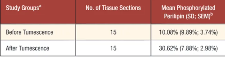
Table 1. Percentages of Phosphorylated Perilipin in Adipocytes Harvested Before and After the Tumescent Technique
Study Groupsa No. of Tissue Sections Mean Phosphorylated Perilipin (SD; SEM)b
Before Tumescence 15 10.08% (9.89%; 3.74%)
After Tumescence 15 30.62% (7.88%; 2.98%)
SD, standard deviation; SEM, standard error of the mean.aLipoaspirates were harvested from
patients before and after tumescence was achieved with a solution containing lidocaine and epinephrine.bThe percentage of phosphorylated perilipin was calculated in terms of pixel
intensity by dividing the mean phospho-perilipin pixel intensity by the sum of the mean phospho-perilipin pixel intensity and the mean perilipin pixel intensity, then multiplying by 100.
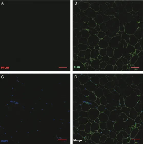
Figure 2. Confocal micrographs of immunostained adipocytes harvested before tumescence was induced (original magnification × 10; bar = 10 µm). (A) PPLIN; phosphorylated perilipin (red) staining is very limited. (B) PLIN; unphosphorylated perilipin (green) is more pronounced. (C) DAPI, nuclear stain (blue). (D) Merged image.
1:400,000) had negligible effects on adipocyte survival. The results of an animal study by Livaoğlu et al14also indicated
that lidocaine plus epinephrine or prilocaine did not affect graft survival with respect to angiogenesis or graft volume.
Several investigators have evaluated fat tissue in terms of adipocyte morphology.3,15,16However, standard stains such
as hematoxylin and eosin are insufficient to discern viable and nonviable adipocytes. Trypan blue is excluded by intact cell membranes and absorbed by permeable membranes. With this stain, dead or dying cells appear blue under a mi-croscope. However, trypan blue does not differentially stain necrotic and apoptotic cells and involves some subjective analysis. In contrast, perilipin immunostaining offers a robust and precise marker of viable adipocytes.21,22Perilipin
detection also can be applied to infer adipocyte differentia-tion.22 Therefore, we did not consider it necessary to
perform an additional test of adipocyte survival.
A lipid droplet comprises a core of neutral lipids that is enveloped in a phospholipid monolayer and coated with specific proteins.23Perilipins are a family of surface proteins
that coat lipid droplets and constitute 0.25% to 0.50% of total cell protein.19Knowledge regarding the biology of lipid
droplets and perilipin has increased in recent years, but the mechanisms by which perilipin functions remain poorly un-derstood. Perilipins appear to function as a physical barrier, protecting stored triglycerides from hydrolysis by hormone-sensitive lipase (HSL).24Depending on the energy status of
the organism, perilipin restricts or enables access of lipases to stored triglycerides.25
Epinephrine is a catecholamine that binds α1-, α2-, β1-, and β2-adrenergic receptors. When it binds α-adrenergic receptors, epinephrine induces vasoconstrictive and hemostatic effects that enable prolonged lidocaine analge-sia. When it binds β-adrenergic receptors, epinephrine
Figure 3. Confocal micrographs of immunostained adipocytes harvested after tumescent solution, containing lidocaine and epi-nephrine, was infiltrated at the donor site (original magnification × 10; bar = 10 µm). (A) PPLIN; phosphorylated perilipin (red) staining is readily visible, albeit less pronounced than in panel B. (B) PLIN; unphosphorylated perilipin (green). (C) DAPI, nuclear stain (blue). (D) Merged image.
nude mice. In this study, fat was harvested by suction-assisted lipectomy, and the donor site was injected either with lido-caine and epinephrine or with normal saline (control).8
These authors found no significant between-group differenc-es in terms of weight or volume of the fat grafts; they conclud-ed that lidocaine and epinephrine did not alter the success of fat grafts or the viability of adipocytes.8
In 2009, Keck et al9compared the effects of the following
incubation environments on the viability of preadipocytes: 1% lidocaine, 1% articaine with epinephrine (1:200,000), 1% prilocaine, 0.75% ropivacaine, and tumescent solution. These authors found that incubation of preadipocytes with li-docaine, ropivacaine, or prilocaine plus epinephrine signifi-cantly reduced preadipocyte viability.9 In 2010, Keck et al10
found that several local anesthetics significantly impaired adi-pocyte differentiation. In subsequent in vitro11and in vivo12
studies, Girard et al demonstrated that clinical concentrations of lidocaine were toxic to adipose-derived stem cells. However, in a 2011 study, Kim et al13determined that various
concentrations of epinephrine (1:100,000, 1:200,000, and
Table 1. Percentages of Phosphorylated Perilipin in Adipocytes Harvested Before and After the Tumescent Technique
Study Groupsa No. of Tissue Sections Mean Phosphorylated Perilipin (SD; SEM)b
Before Tumescence 15 10.08% (9.89%; 3.74%)
After Tumescence 15 30.62% (7.88%; 2.98%)
SD, standard deviation; SEM, standard error of the mean.aLipoaspirates were harvested from
patients before and after tumescence was achieved with a solution containing lidocaine and epinephrine.bThe percentage of phosphorylated perilipin was calculated in terms of pixel
intensity by dividing the mean phospho-perilipin pixel intensity by the sum of the mean phospho-perilipin pixel intensity and the mean perilipin pixel intensity, then multiplying by 100.
Figure 2. Confocal micrographs of immunostained adipocytes harvested before tumescence was induced (original magnification × 10; bar = 10 µm). (A) PPLIN; phosphorylated perilipin (red) staining is very limited. (B) PLIN; unphosphorylated perilipin (green) is more pronounced. (C) DAPI, nuclear stain (blue). (D) Merged image.
1:400,000) had negligible effects on adipocyte survival. The results of an animal study by Livaoğlu et al14also indicated
that lidocaine plus epinephrine or prilocaine did not affect graft survival with respect to angiogenesis or graft volume.
Several investigators have evaluated fat tissue in terms of adipocyte morphology.3,15,16However, standard stains such
as hematoxylin and eosin are insufficient to discern viable and nonviable adipocytes. Trypan blue is excluded by intact cell membranes and absorbed by permeable membranes. With this stain, dead or dying cells appear blue under a mi-croscope. However, trypan blue does not differentially stain necrotic and apoptotic cells and involves some subjective analysis. In contrast, perilipin immunostaining offers a robust and precise marker of viable adipocytes.21,22Perilipin
detection also can be applied to infer adipocyte differentia-tion.22 Therefore, we did not consider it necessary to
perform an additional test of adipocyte survival.
A lipid droplet comprises a core of neutral lipids that is enveloped in a phospholipid monolayer and coated with specific proteins.23Perilipins are a family of surface proteins
that coat lipid droplets and constitute 0.25% to 0.50% of total cell protein.19Knowledge regarding the biology of lipid
droplets and perilipin has increased in recent years, but the mechanisms by which perilipin functions remain poorly un-derstood. Perilipins appear to function as a physical barrier, protecting stored triglycerides from hydrolysis by hormone-sensitive lipase (HSL).24Depending on the energy status of
the organism, perilipin restricts or enables access of lipases to stored triglycerides.25
Epinephrine is a catecholamine that binds α1-, α2-, β1-, and β2-adrenergic receptors. When it binds α-adrenergic receptors, epinephrine induces vasoconstrictive and hemostatic effects that enable prolonged lidocaine analge-sia. When it binds β-adrenergic receptors, epinephrine
Figure 3. Confocal micrographs of immunostained adipocytes harvested after tumescent solution, containing lidocaine and epi-nephrine, was infiltrated at the donor site (original magnification × 10; bar = 10 µm). (A) PPLIN; phosphorylated perilipin (red) staining is readily visible, albeit less pronounced than in panel B. (B) PLIN; unphosphorylated perilipin (green). (C) DAPI, nuclear stain (blue). (D) Merged image.
stimulates lipolysis by activating PKA, which phosphorylates perilipin.23 Specifically, epinephrine activates β-adrenergic
receptors to signal heterotrimeric G-proteins to activate adenyl cyclase, which in turn elevates levels of cyclic adeno-sine monophosphate and activates PKA. In vivo, this signal-ing pathway proceeds within seconds. PKA subsequently phosphorylates perilipin A and HSL. Maximal phosphoryla-tion of perilipin occurs in less than 2 minutes, and transloca-tion of HSL from the cytosol to lipid droplets peaks within 5 minutes.18
Phosphorylation of perilipin has not been studied in great detail, but Brasaemle26proposed an intriguing model
whereby phosphorylation of perilipin facilitates access of HSL to the droplet surface, thus stimulating lipolysis in a hormone-dependent manner. HSL requires phosphorylated perilipin for optimal activity.23-29Phosphorylated perilipin
provides a docking site for phosphorylated HSL on lipid droplets, but the mechanism for this is not known. Murine perilipin A is phosphorylated by PKA at≤6 potential sites; it is unknown whether additional kinases contribute. Perilipin 1 is potentially phosphorylated by PKA at several sites, in-cluding conserved C-terminal residues, serine 497 (PKA-site 5), and serine 522 (PKA-site 6).18,30Phosphorylation of
peril-ipin is essential for maximal PKA-stimulated lipolysis.31 A
decrease in perilipin expression also may promote lipolysis, thereby reducing intracellular triacylglycerol and adipose mass.28Knockout of perilipin 1 in mice confers resistance to
obesity, induced by a high-fat diet, and is associated with in-creased basal lipolysis.26 The adipose mass of perilipin 1
knockout mice was reduced by 65% to 75%.
Although epinephrine is well known to reduce bleeding intraoperatively and intensify anesthesia, the effects of epi-nephrine on adipocyte viability have not been established. We sought to infer adipocyte viability in terms of perilipin phosphorylation status as a means to inform clinical prac-tice, given that the number of viable adipocytes in a graft correlates with the volume of persisting fat.3 We
demon-strated that tumescent solution containing epinephrine and lidocaine increased the phosphorylation of perilipin in adi-pocytes, which may make these cells vulnerable to lipoly-sis. Although the phosphorylation status of perilipin is only 1 component in a multimolecular process, our findings suggest that adipocytes may be more prone to lipolysis after they are exposed to epinephrine and lidocaine and harvest-ed from tumescent tissues. Additional research is neharvest-edharvest-ed to elucidate the molecular mechanisms leading to perilipin phosphorylation in this context. We recommend harvesting fat immediately after injection of local anesthetics, thereby limiting exposure of adipocytes.
Study Limitations
It was not possible to evaluate the effects of epinephrine or lidocaine alone because fat tissues for this study were
collected from patients, and ethical approval would have been denied if nonstandard tumescent solutions were to be applied. Therefore, we cannot discern whether 1 or both re-agents were responsible for increased phosphorylation of perilipin. Similarly, our collection of patient tissues pre-cluded any modification to saline content of the tumescent solution to ascertain whether phosphorylation could be stimulated osmotically. This study also was limited by its relatively small sample size (15 patients).
CONCLUSIONS
The effectiveness and predictability of autologous fat trans-plantation depends on multiple factors, including adipocyte viability. Tumescent solution containing lidocaine and epi-nephrine significantly increases the phosphorylation status of perilipin in adipocytes, potentially subjecting these cells to lipolysis. These findings suggest that the tumescent tech-nique may decrease adipocyte viability and increase resorp-tion of the fat graft. We recommend minimizing the concentrations of lidocaine and epinephrine in tumescent solution when fat is to be harvested for transplantation. More research is warranted to elucidate the mechanism of tumescence-associated perilipin phosphorylation and to confirm the clinical relevance of our findings.
Disclosures
The authors declared no potential conflicts of interest with respect to the research, authorship, and publication of this article.
Funding
The authors received no financial support for the research, authorship, or publication of this article.
REFERENCES
1. Carpaneda CA, Ribeiro MT. Percentage of graft viability versus injected volume in adipose autotransplants. Aesthetic Plast Surg. 1994;18(1):17-19.
2. Mikus JL, Koufman JA, Kilpatrick SE. Fate of liposuc-tioned and purified autologous fat injections in the canine vocal fold. Laryngoscope. 1995;105(1):17-22. 3. Sommer B, Sattler G. Current concepts of fat graft
sur-vival: histology of aspirated adipose tissue and review of the literature. Dermatologic Surg. 2000;26(12):1159-1166. 4. Smith P, Adams WP, Lipschitz AH, et al. Autologous human fat grafting: effect of harvesting and preparation techniques on adipocyte graft survival. Plast Reconstr Surg. 2006;117(6):1836-1844.
5. Monreal J. Fat tissue as permanent implant: new instru-ments and refineinstru-ments. Aesthet Surg J. 2003;23(3): 213-216.
6. Lin JY, Wang C, Pu LL. Can we standardize the tech-niques for fat grafting? Clin Plast Surg. 2015;42(2): 199-208.
7. Moore JH, Kolaczynski JW, Morales LM, et al. Viability of fat obtained by syringe suction lipectomy: effects of local anesthesia with lidocaine. Aesthetic Plast Surg. 1995;19 (4):335-339.
8. Shoshani O, Berger J, Fodor L, et al. The effect of lido-caine and adrenaline on the viability of injected adipose tissue--an experimental study in nude mice. J Drugs Dermatol. 2005;4(3):311-316.
9. Keck M, Janke J, Ueberreiter K. Viability of preadipocytes in vitro: the influence of local anesthetics and pH. Dermatologic Surg. 2009;35(8):1251-1257.
10. Keck M, Zeyda M, Gollinger K, et al. Local anesthetics have a major impact on viability of preadipocytes and their differentiation into adipocytes. Plast Reconstr Surg. 2010;126(5):1500-1505.
11. Girard A-C, Atlan M, Bencharif K, et al. New insights into lidocaine and adrenaline effects on human adipose stem cells. Aesthetic Plast Surg. 2013;37(1):144-152.
12. Girard A-C, Mirbeau S, Gence L, et al. Effect of washes and centrifugation on the efficacy of lipofilling with or without local anesthetic. Plast Reconstr Surg Glob Open. 2015;3(8):e496.
13. Kim IH, Yang JD, Lee DG, Chung HY, Cho BC. Evaluation of centrifugation technique and effect of epinephrine on fat cell viability in autologous fat injection. Aesthet Surg J. 2011;29(1):35-39.
14. Livaoğlu M, Buruk CK, Uraloğlu M, et al. Effects of lido-caine plus epinephrine and prilolido-caine on autologous fat graft survival. J Craniofac Surg. 2012;23(4):1015-1018. 15. Niechajev I, Oleg S. Long-term results of fat
transplanta-tion: clinical and histologic studies. Plast Reconstr Surg. 1994;94(3):496-506.
16. Pu LL, Yoshimura K, Coleman SR. Future perspectives of fat grafting. Clin Plast Surg. 2015;42(3):389-394, ix-x. 17. Cinti S, Mitchell G, Barbatelli G, et al. Adipocyte death
defines macrophage localization and function in adipose tissue of obese mice and humans. J Lipid Res. 2005;46 (11):2347-2355.
18. Mcdonough PM, Maciejewski-Lenoir D, Hartig SM, et al. Differential phosphorylation of perilipin 1a at the initia-tion of lipolysis revealed by novel monoclonal antibodies and high content analysis. PLoS One. 2013;8(2):e55511. 19. Brown DA. Lipid droplets: proteins floating on a pool of
fat. Curr Biol. 2001;11(11):446-449.
20. Bartynski J, Marion MS, Wang TD. Histopathologic eval-uation of adipose autografts in a rabbit ear model. Otolaryngol Head Neck Surg. 1990;102(4):314-321. 21. Ataru S, Yasushi S. The fate of nonvascularized fat grafts:
histological and bioluminescent study. Plast Reconstr Surg Glob Open. 2013;1(6):e41.
22. Eto H, Kato H, Suga H, et al. The fate of adipocytes after non-vascularized fat grafting: evidence of early death and replacement of adipocytes. Plast Reconstr Surg. 2012;129: 1081-1092.
23. Bickel PE, Tansey JT, Welte MA. PAT proteins, an ancient family of lipid droplet proteins that regulate cellular lipid stores. Biochim Biophys Acta. 2009;1791(6):419-440. 24. Moore HP, Silver RB, Mottillo EP, Bernlohr DA,
Granneman JG. Perilipin targets a novel pool of lipid droplets for lipolytic attack by hormone-sensitive lipase. J Biol Chem. 2005;280(52):43109-20.
25. Aon MA, Bhatt N, Cortassa SC. Mitochondrial and cellu-lar mechanisms for managing lipid excess. Front Physiol. 2014;5:1-13.
26. Brasaemle DL. Thematic review series: adipocyte biology. The perilipin family of structural lipid droplet proteins: stabilization of lipid droplets and control of li-polysis. J Lipid Res. 2007;48(12):2547-2559.
27. Tansey JT, Sztalryd C, Gruia-Gray J, et al. Perilipin ablation results in a lean mouse with aberrant adipocyte lipolysis, enhanced leptin production, and resistance to diet-induced obesity. Proc Natl Acad Sci. 2001;98(11):6494-6499. 28. Mcdonough PM, Ingermanson RS, Loy PA, et al.
Quantification of hormone sensitive lipase phosphoryla-tion and colocalizaphosphoryla-tion with lipid droplets in murine 3t3l1 and human subcutaneous adipocytes via automated digital microscopy and high-content analysis. Assay Drug Dev Technol. 2011;9(3):262-280.
29. Greenberg AS, Egan JJ, Wek SA, et al. Perilipin, a major hormonally regulated adipocyte-specific phosphoprotein associated with the periphery of lipid storage droplets. J Biol Chem. 1991;266(17):11341-11346.
30. Yeaman SJ. Hormone-sensitive lipase--new roles for an old enzyme. Biochem J. 2004;379(Pt 1):11-22.
31. Granneman JG, Moore HP, Granneman RL, Greenberg AS, Obin MS, Zhu Z. Analysis of lipolytic protein traffick-ing and interactions in adipocytes. J Biol Chem. 2007;282 (8):5726-35.
stimulates lipolysis by activating PKA, which phosphorylates perilipin.23 Specifically, epinephrine activates β-adrenergic
receptors to signal heterotrimeric G-proteins to activate adenyl cyclase, which in turn elevates levels of cyclic adeno-sine monophosphate and activates PKA. In vivo, this signal-ing pathway proceeds within seconds. PKA subsequently phosphorylates perilipin A and HSL. Maximal phosphoryla-tion of perilipin occurs in less than 2 minutes, and transloca-tion of HSL from the cytosol to lipid droplets peaks within 5 minutes.18
Phosphorylation of perilipin has not been studied in great detail, but Brasaemle26proposed an intriguing model
whereby phosphorylation of perilipin facilitates access of HSL to the droplet surface, thus stimulating lipolysis in a hormone-dependent manner. HSL requires phosphorylated perilipin for optimal activity.23-29 Phosphorylated perilipin
provides a docking site for phosphorylated HSL on lipid droplets, but the mechanism for this is not known. Murine perilipin A is phosphorylated by PKA at≤6 potential sites; it is unknown whether additional kinases contribute. Perilipin 1 is potentially phosphorylated by PKA at several sites, in-cluding conserved C-terminal residues, serine 497 (PKA-site 5), and serine 522 (PKA-site 6).18,30Phosphorylation of
peril-ipin is essential for maximal PKA-stimulated lipolysis.31A
decrease in perilipin expression also may promote lipolysis, thereby reducing intracellular triacylglycerol and adipose mass.28Knockout of perilipin 1 in mice confers resistance to
obesity, induced by a high-fat diet, and is associated with in-creased basal lipolysis.26 The adipose mass of perilipin 1
knockout mice was reduced by 65% to 75%.
Although epinephrine is well known to reduce bleeding intraoperatively and intensify anesthesia, the effects of epi-nephrine on adipocyte viability have not been established. We sought to infer adipocyte viability in terms of perilipin phosphorylation status as a means to inform clinical prac-tice, given that the number of viable adipocytes in a graft correlates with the volume of persisting fat.3 We
demon-strated that tumescent solution containing epinephrine and lidocaine increased the phosphorylation of perilipin in adi-pocytes, which may make these cells vulnerable to lipoly-sis. Although the phosphorylation status of perilipin is only 1 component in a multimolecular process, our findings suggest that adipocytes may be more prone to lipolysis after they are exposed to epinephrine and lidocaine and harvest-ed from tumescent tissues. Additional research is neharvest-edharvest-ed to elucidate the molecular mechanisms leading to perilipin phosphorylation in this context. We recommend harvesting fat immediately after injection of local anesthetics, thereby limiting exposure of adipocytes.
Study Limitations
It was not possible to evaluate the effects of epinephrine or lidocaine alone because fat tissues for this study were
collected from patients, and ethical approval would have been denied if nonstandard tumescent solutions were to be applied. Therefore, we cannot discern whether 1 or both re-agents were responsible for increased phosphorylation of perilipin. Similarly, our collection of patient tissues pre-cluded any modification to saline content of the tumescent solution to ascertain whether phosphorylation could be stimulated osmotically. This study also was limited by its relatively small sample size (15 patients).
CONCLUSIONS
The effectiveness and predictability of autologous fat trans-plantation depends on multiple factors, including adipocyte viability. Tumescent solution containing lidocaine and epi-nephrine significantly increases the phosphorylation status of perilipin in adipocytes, potentially subjecting these cells to lipolysis. These findings suggest that the tumescent tech-nique may decrease adipocyte viability and increase resorp-tion of the fat graft. We recommend minimizing the concentrations of lidocaine and epinephrine in tumescent solution when fat is to be harvested for transplantation. More research is warranted to elucidate the mechanism of tumescence-associated perilipin phosphorylation and to confirm the clinical relevance of our findings.
Disclosures
The authors declared no potential conflicts of interest with respect to the research, authorship, and publication of this article.
Funding
The authors received no financial support for the research, authorship, or publication of this article.
REFERENCES
1. Carpaneda CA, Ribeiro MT. Percentage of graft viability versus injected volume in adipose autotransplants. Aesthetic Plast Surg. 1994;18(1):17-19.
2. Mikus JL, Koufman JA, Kilpatrick SE. Fate of liposuc-tioned and purified autologous fat injections in the canine vocal fold. Laryngoscope. 1995;105(1):17-22. 3. Sommer B, Sattler G. Current concepts of fat graft
sur-vival: histology of aspirated adipose tissue and review of the literature. Dermatologic Surg. 2000;26(12):1159-1166. 4. Smith P, Adams WP, Lipschitz AH, et al. Autologous human fat grafting: effect of harvesting and preparation techniques on adipocyte graft survival. Plast Reconstr Surg. 2006;117(6):1836-1844.
5. Monreal J. Fat tissue as permanent implant: new instru-ments and refineinstru-ments. Aesthet Surg J. 2003;23(3): 213-216.
6. Lin JY, Wang C, Pu LL. Can we standardize the tech-niques for fat grafting? Clin Plast Surg. 2015;42(2): 199-208.
7. Moore JH, Kolaczynski JW, Morales LM, et al. Viability of fat obtained by syringe suction lipectomy: effects of local anesthesia with lidocaine. Aesthetic Plast Surg. 1995;19 (4):335-339.
8. Shoshani O, Berger J, Fodor L, et al. The effect of lido-caine and adrenaline on the viability of injected adipose tissue--an experimental study in nude mice. J Drugs Dermatol. 2005;4(3):311-316.
9. Keck M, Janke J, Ueberreiter K. Viability of preadipocytes in vitro: the influence of local anesthetics and pH. Dermatologic Surg. 2009;35(8):1251-1257.
10. Keck M, Zeyda M, Gollinger K, et al. Local anesthetics have a major impact on viability of preadipocytes and their differentiation into adipocytes. Plast Reconstr Surg. 2010;126(5):1500-1505.
11. Girard A-C, Atlan M, Bencharif K, et al. New insights into lidocaine and adrenaline effects on human adipose stem cells. Aesthetic Plast Surg. 2013;37(1):144-152.
12. Girard A-C, Mirbeau S, Gence L, et al. Effect of washes and centrifugation on the efficacy of lipofilling with or without local anesthetic. Plast Reconstr Surg Glob Open. 2015;3(8):e496.
13. Kim IH, Yang JD, Lee DG, Chung HY, Cho BC. Evaluation of centrifugation technique and effect of epinephrine on fat cell viability in autologous fat injection. Aesthet Surg J. 2011;29(1):35-39.
14. Livaoğlu M, Buruk CK, Uraloğlu M, et al. Effects of lido-caine plus epinephrine and prilolido-caine on autologous fat graft survival. J Craniofac Surg. 2012;23(4):1015-1018. 15. Niechajev I, Oleg S. Long-term results of fat
transplanta-tion: clinical and histologic studies. Plast Reconstr Surg. 1994;94(3):496-506.
16. Pu LL, Yoshimura K, Coleman SR. Future perspectives of fat grafting. Clin Plast Surg. 2015;42(3):389-394, ix-x. 17. Cinti S, Mitchell G, Barbatelli G, et al. Adipocyte death
defines macrophage localization and function in adipose tissue of obese mice and humans. J Lipid Res. 2005;46 (11):2347-2355.
18. Mcdonough PM, Maciejewski-Lenoir D, Hartig SM, et al. Differential phosphorylation of perilipin 1a at the initia-tion of lipolysis revealed by novel monoclonal antibodies and high content analysis. PLoS One. 2013;8(2):e55511. 19. Brown DA. Lipid droplets: proteins floating on a pool of
fat. Curr Biol. 2001;11(11):446-449.
20. Bartynski J, Marion MS, Wang TD. Histopathologic eval-uation of adipose autografts in a rabbit ear model. Otolaryngol Head Neck Surg. 1990;102(4):314-321. 21. Ataru S, Yasushi S. The fate of nonvascularized fat grafts:
histological and bioluminescent study. Plast Reconstr Surg Glob Open. 2013;1(6):e41.
22. Eto H, Kato H, Suga H, et al. The fate of adipocytes after non-vascularized fat grafting: evidence of early death and replacement of adipocytes. Plast Reconstr Surg. 2012;129: 1081-1092.
23. Bickel PE, Tansey JT, Welte MA. PAT proteins, an ancient family of lipid droplet proteins that regulate cellular lipid stores. Biochim Biophys Acta. 2009;1791(6):419-440. 24. Moore HP, Silver RB, Mottillo EP, Bernlohr DA,
Granneman JG. Perilipin targets a novel pool of lipid droplets for lipolytic attack by hormone-sensitive lipase. J Biol Chem. 2005;280(52):43109-20.
25. Aon MA, Bhatt N, Cortassa SC. Mitochondrial and cellu-lar mechanisms for managing lipid excess. Front Physiol. 2014;5:1-13.
26. Brasaemle DL. Thematic review series: adipocyte biology. The perilipin family of structural lipid droplet proteins: stabilization of lipid droplets and control of li-polysis. J Lipid Res. 2007;48(12):2547-2559.
27. Tansey JT, Sztalryd C, Gruia-Gray J, et al. Perilipin ablation results in a lean mouse with aberrant adipocyte lipolysis, enhanced leptin production, and resistance to diet-induced obesity. Proc Natl Acad Sci. 2001;98(11):6494-6499. 28. Mcdonough PM, Ingermanson RS, Loy PA, et al.
Quantification of hormone sensitive lipase phosphoryla-tion and colocalizaphosphoryla-tion with lipid droplets in murine 3t3l1 and human subcutaneous adipocytes via automated digital microscopy and high-content analysis. Assay Drug Dev Technol. 2011;9(3):262-280.
29. Greenberg AS, Egan JJ, Wek SA, et al. Perilipin, a major hormonally regulated adipocyte-specific phosphoprotein associated with the periphery of lipid storage droplets. J Biol Chem. 1991;266(17):11341-11346.
30. Yeaman SJ. Hormone-sensitive lipase--new roles for an old enzyme. Biochem J. 2004;379(Pt 1):11-22.
31. Granneman JG, Moore HP, Granneman RL, Greenberg AS, Obin MS, Zhu Z. Analysis of lipolytic protein traffick-ing and interactions in adipocytes. J Biol Chem. 2007;282 (8):5726-35.