62
However, the prognostic value of this unusual plasma cell phenotype remains unclear.
Keywords: Light chain myeloma, Plasma cells, Bone marrow
aspirate
Anahtar Sözcükler: Hafif zincir myeloma, Plazma hücreleri,
Kemik iliği aspirasyonu
Informed Consent: Was obtained from the patient.
Conflict of Interest: The authors of this paper have no conflicts
of interest, including specific financial interests, relationships, and/or affiliations relevant to the subject matter or materials included.
References
1. Castoldi G, Piva N, Tomasi P. Multiple myeloma with Auer-rod-like inclusions. Haematologica 1999;84:859-860.
2. Metzgeroth G, Back W, Maywald O, Schatz M, Willer A, Hehlmann R, Hastka J. Auer rod-like inclusions in multiple myeloma. Ann Hematol 2003;82:57-60.
3. Abdulsalam AH, Al-Yassin FM. Myeloma cells with Auer rod-like inclusions. Turk J Hematol 2012;29:206.
4. Noujaim JC, D’Angelo G. Auer rod-like inclusions in ĸ light chain myeloma. Blood 2013;122:2932.
5. Ho WK, Zantomio D. Auer rod-like inclusions in plasma cells in multiple myeloma. J Clin Pathol 2014;67:547-548.
LETTERS TO THE EDITOR Turk J Hematol 2019;36:52-71
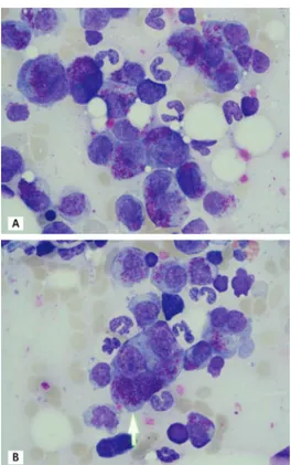
Figure 1. Bone marrow aspirate smear of a 73-year-old patient
with κ-type light chain myeloma (A and B). The arrow marks a binuclear plasmablastic cell containing numerous Auer rod-like inclusions.
Address for Correspondence/Yazışma Adresi: Gernot KRIEGSHÄUSER, M.D.,
General Hospital Steyr, Institute of Clinical Chemistry and Laboratory Medicine, Steyr, Austria
Phone : +43 50554 66 25308
E-mail : [email protected] ORCID-ID: orcid.org/0000-0003-2768-795X
Received/Geliş tarihi: June 05, 2018 Accepted/Kabul tarihi: July 09, 2018
DOI: 10.4274/tjh.galenos.2018.2018.0197
©Copyright 2019 by Turkish Society of Hematology
Turkish Journal of Hematology, Published by Galenos Publishing House
Aggressive Clinicopathological Course of Myeloma with t(3;16)
(q21;q22) Cytogenetic Abnormality
t(3;16)(q21;q22) Anomalili Myeloma Olgusunda Agresif Klinikopatolojik Seyir
Süreyya Bozkurt1, Müfide Okay2, İbrahim Haznedaroğlu2
1İstinye University Faculty of Medicine, Department of Medical Biology, İstanbul, Turkey
2Hacettepe University Faculty of Medicine, Department of Internal Medicine, Division of Hematology, Ankara, Turkey
To the Editor,
Multiple myeloma (MM) is a heterogeneous disease and patients present with a wide variety of cytogenetic anomalies reflecting the nature of the disease [1]. The aim of this letter is to report a rare karyotypic abnormality with an aggressive clinical course of MM.
A 56-year-old male patient was admitted to the neurosurgery clinic with dorsal shoulder pain and inability to walk in April 2011. He underwent thoracic and lumbar spinal magnetic resonance imaging. Laminectomy was performed on the patient upon detecting masses at the levels of the first and seventh thoracic vertebrae. The patient was referred to our center when he was determined to have “lymphoma” based on the first evaluation
63 of his biopsy material. The specimen was then reevaluated in
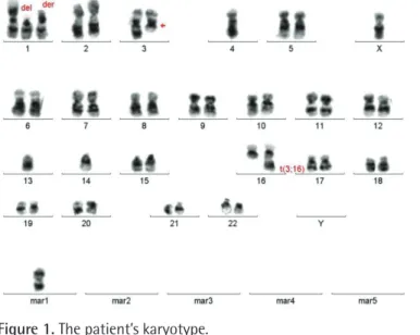
our center. A high-grade hematopoietic neoplasia was detected. Immunophenotypic findings suggested neoplasia with plasma cell origin. Immunohistochemically, neoplastic cells were positive for CD38, MUM-1, and kappa and negative for lambda. The karyotype of the patient was identified as 44,X,-Y,del(1) (p13p35),+der(1),t(3;16)(q21;q22),-4,-13,-14,+mar[8]/46,XY[42] (Figure 1).
Upon detection of newly developed lesions in the tenth thoracic vertebra in the control imaging obtained after the radiotherapy of the patient, treatment with vincristine, adriamycin, and dexamethasone (VAD) was initiated. The paraprotein levels in the patient’s serum decreased after four cycles of chemotherapy. However, the treatment was planned to be continued with only bortezomib (Velcade) and dexamethasone due to severe infection after chemotherapy and the development of decubitus infection during the follow-up of the patient. After three cycles of treatment, nodular lesions compatible with plasmacytomas appeared on his skin. The treatment strategy was changed since the patient was considered to have disease progression and had responded better to VAD chemotherapy. The patient was hospitalized due to pneumonia after chemotherapy and died due to severe sepsis.
The t(3;16)(q21;q22) anomaly reported in this case is a quite rare cytogenetic anomaly and, according to the databases that we have investigated [2,3], it was reported only in three adults and one child to date [4]. One of these three adult cases was a male
patient with the diagnosis of myelodysplastic syndrome. In this patient, the translocation of t(3;16)(q21;q22) was found in the complex karyotype, as in our case [5]. The other two cases were female patients and they were diagnosed with acute myeloid leukemia that developed after acute myelomonocytic leukemia and primary tumor mantle cell lymphoma, respectively. None of the reported patients were diagnosed with MM. Therefore, we report the t(3;16)(q21;q22) anomaly, which is a quite rare translocation, in a patient with MM for the first time in this case. The prognostic effect of t(3;16)(q21;q22) is unknown. In the presented case, the anomaly of t(3;16)(q21;q22) coexisted with the anomalies of 1p deletion and monosomy 13, which are markers of progressive disease and short survival, or in other words, poor prognostic markers [6]. The clinical course of our case progressed in a manner that was consistent with poor prognosis. We believe that our case contributes to the literature since it is the first MM case with a very rarely encountered translocation.
Keywords: Multiple myeloma, Rare translocations, Cytogenetic
abnormality
Anahtar Sözcükler: Multiple myelom, Nadir translokasyonlar,
Sitogenetik anomali
Informed Consent: Recevied.
Conflict of Interest: No benefits in any form have been received
or will be received from a commercial party related directly or indirectly to the subject of this article.
References
1. Sawyer JR. The prognostic significance of cytogenetics and molecular profiling in multiple myeloma. Cancer Genet 2011;204:3-12.
2. Huret JL. Atlas of Genetics and Cytogenetics in Oncology and Haematology. 2017. Available at http://atlasgeneticsoncology.org/.
3. Mitelman F, Johansson B, Mertens F. Mitelman Database of Chromosome Aberrations and Gene Fusions in Cancer, 2017. Available at http://cgap.nci. nih.gov/Chromosomes/Mitelman.
4. Huret JL. t(3;16)(q21;q22). Atlas Genet Cytogenet Oncol Haematol 2009;13:747.
5. Neri G, Daniel A, Hammond N. Chromosome 16q, eosinophilia, and leukemia. Cancer Genet Cytogenet 1985;14:371-372.
6. Fonseca R, Bergsagel PL, Drach J, Shaughnessy J, Gutierrez N, Stewart AK, Morgan G, Van Ness B, Chesi M, Minvielle S, Neri A, Barlogie B, Kuehl WM, Liebisch P, Davies F, Chen-Kiang S, Durie BG, Carrasco R, Sezer O, Reiman T, Pilarski L, Avet-Loiseau H; International Myeloma Working Group. International Myeloma Working Group molecular classification of multiple myeloma: spotlight review. Leukemia 2009;23:2210-2221.
LETTERS TO THE EDITOR
Turk J Hematol 2019;36:52-71
Figure 1. The patient’s karyotype.
Address for Correspondence/Yazışma Adresi: Süreyya BOZKURT, M.D.,
İstinye University Faculty of Medicine, Department of Medical Biology, İstanbul, Turkey
Phone : +90 850 283 60 00
E-mail : [email protected] ORCID-ID: orcid.org/0000-0002-1765-9894
Received/Geliş tarihi: January 30, 2018 Accepted/Kabul tarihi: May 02, 2018
DOI: 10.4274/tjh.galenos.2018.2018.0049
©Copyright 2019 by Turkish Society of Hematology