Antioxidant SMe1EC2 modulates
pentose phosphate pathway and
glutathione-dependent enzyme activities
in tissues of aged diabetic rats
Nuray Nuriye ULUSU 1, Müslüm GÖK 2, Arzu Ayşe SAYIN ŞAKUL 3, Nuray ARI 4, Milan STEFEK 5, Çimen KARASU 6, “The ADIC (Antioxidants in Diabetes-Induced Complications) Study Group”
1 Department of Medical Biochemistry, School of Medicine, Koc University, Istanbul, Turkey 2 Department of Biochemistry, Faculty of Medicine, Hacettepe University, Ankara, Turkey 3 Department of Pharmacology, Faculty of Medicine, Medipol University, Istanbul, Turkey 4 Department of Pharmacology, Faculty of Pharmacy, Ankara University, Ankara, Turkey 5 Department of experimental Pharmacology, Slovak Academy of Sciences, Bratislava, Slovakia
6 Cellular Stress Response & Signal Transduction Research Laboratory, Department of Medical Pharmacology, Faculty of Medicine, Gazi University, Ankara, Turkey
ITX100417A03 • Received: 27 November 2017 • Accepted: 10 December 2017 ABSTRACT
The pentose phosphate pathway and glutathione-associated metabolism are the main antioxidant cellular defense systems. This study investigated the effects of the powerful antioxidant SMe1EC2 (2-ethoxycarbonyl-8-methoxy-2,3,4,4a,5,9b-hexahydro-1H-pyrido[4,3-b] indolinium dichloride) on pentose phosphate pathway (PPP) and glutathione-dependent enzyme activities in aged diabetic and aged matched control rats. Diabetes was induced by streptozotocin injection in rats aged 13–15 months. Diabetic and control rats were divided into two subgroups, one untreated and one treated with SMe1EC2 (10 mg/kg/day, orally) for 4 months. SMe1EC2 ameliorated body weight loss, but not hyperglycemia of aged diabetic rats. Diabetes resulted in decreased glucose-6-phosphate dehydrogenase (G6PD), 6-phosphogluconate dehydrogenase (6PGD) and glutathione-S-transferase (GST), yet in unchanged glutathione reductase (GR) in the liver of aged diabetic rats. In the liver of the aged control rats, SMe1EC2 did not affect G6PDH, 6PGDH and GR, but it inhibited GST. SMe1EC2 also failed to affect diabetes-induced decline in 6PGDH, it ameliorated G6PDH but produced further decline in GST in the liver of aged diabetic rats. In the kidney of aged rats, G6PDH and GST were found to be comparable among the groups, but diabetes up-regulated 6PGDH and GR; these alterations were prevented by SMe1EC2. In the heart of aged diabetic rats, while GST remained unchanged, the recorded increase in G6PD, 6PGD, GR was prevented by SMe1EC2. Furthermore, an unchanged GR and remarkable increases in G6PD, 6PGD and GST were found in the lung of the aged diabetic group. These alterations were completely prevented by SMe1EC2. The results suggest that in aged rats SMe1EC2 can ameliorate the response of the kidney, heart and lung but not that of the liver against diabetes-induced glucotoxicity by interfering with the activity of redox network enzymes.
KEY WORDS: SMe1EC2; antioxidant; diabetes; aging; rat; pentose phosphate pathway; glutathione-dependent enzymes
Correspondence address: Prof. Çimen Karasu
Cellular Stress Response & Signal Transduction Research Laboratory Faculty of Medicine, Gazi University, Turkey
TEL.: +90 312 2026921 • FAX +90 312 2124647 E-MAIL: [email protected], [email protected]
Introduction
Increases in the intracellular levels of reactive oxygen species (ROS), frequently referred to as oxidative stress, represent a potentially toxic insult. If not counteracted, it will attack lipids, sugars and proteins and oxidize them. Chronic oxidative stress, resulting in the accumulation of dysfunctional and damaged biomolecules, has numerous pathological consequences, including diabetes (Karasu, 2010; Stefek & Karasu, 2011; Şakul et al., 2013). On the other hand, aging is caused by the accumulation of ran-dom molecular damage due to ROS (Ergin et al., 2013; Cencioni et al., 2013). The degenerative complications and deficits in organ performance as a result of enhanced susceptibility to the long-term effects of increased Copyright © 2017 SETOX & IEPT, SASc.
This is an Open Access article distributed under the terms of the Creative Commons Attri-bution-NonCommercial-NoDerivatives 4.0 License (https://creativecommons.org/licenses/ by-nc-nd/4.0).
ORIGINAL ARTICLE
ABBREVIATIONS:
SMe1EC2: 2-ethoxycarbonyl-8-methoxy-2,3,4,4a,5,9b-hexahydro-1H-pyrido[4,3-
b]indolinium dichloride; G6PD: glucose-6-phosphate dehydrogenase; 6PGD: 6-phosphogluconate dehydrogenase; GST: glutathione-S-transferase; GR: glu-tathione reductase; GSSG: oxidized gluglu-tathione; ROS: reactive oxygen species;
PPP: pentose phosphate pathway; NADPH: reduced nicotinamide adenine
dinucleotide phosphate; Nrf2: the nuclear factor erythroid 2-related factor 2;
oxidative stress and inflammation are increased by aging and diabetes. A decrease in endogenous antioxidant mechanisms with aging or diabetes increases the vulner-ability of the tissues to oxidative damage (Shakeel, 2015; Panigrahy et al., 2017). Because of the potential impact of oxidative stress and inflammatory vulnerability in aging and diabetes, research has focused on the use of synthetic anti-oxidant and anti-inflammatory agents that protect against stress-related phenomena (Karasu 2010, Aldini et
al., 2013). Many synthetic compounds have been
evalu-ated as inhibitors of ROS formation, but none have yet been approved for clinical use.
In extensive preclinical studies, dietary supplemen-tation with a potent antioxidant 2-ethoxycarbonyl-8-methoxy-2,3,4,4a,5,9b-hexahydro-1H-pyrido[4,3-b] indolinium dichloride (SMe1EC2), which is a hexahydro-pyridoindole derivative, revealed significant anti-oxidant, anti-inflammatory, vasculoprotective, antidysrhythmic and neuroprotective effects (Drimal et al., 2008; Stefek
et al., 2013; Broskova et al., 2013; Gasparova et al., 2014).
However, the regulating role of SMe1EC2 on pentose phosphate pathway (PPP) and glutathione-dependent enzymatic activities is not yet known, while PPP is one of the major sources of reduction equivalents for the glutathione peroxidase (GPx)/glutathione reductase (GR) antioxidant system, which plays a key role in preventing oxidative stress (Stanton, 2012; Ulusu, 2015). The pres-ent investigation was undertaken to assess the effects of SMe1EC2 treatment on the activities of glucose-6-phosphate dehydrogenase (G6PD), 6-phosphogluconate dehydrogenase (6PGD), glutathione reductase (GR) and glutathione-S-transferase (GST) in the liver, kidney, heart and lung of aged STZ diabetic rats.
Materials and methods
Animals and TreatmentThirteen- to fifteen-month-old adult male Wistar rats were obtained from the “Laboratory Animal Unit of Ankara University, Faculty of Pharmacy”. They were caged in groups of four with free access to food and water and were maintained on a 12-h light–dark cycle (7:00–19:00 h), at a temperature-controlled colony room (23±1 °C). These conditions were maintained constant throughout the experiments. All experimental procedures were performed in accordance with the National Institute of Health Guide for the Care and Use of Laboratory Animals (NIH publication number 80–23 revised 1996). Our research protocol was approved by the Ethical Committee for Animal Experimentation of the Ankara University (No: 2010-56-280).
Diabetes was induced in 13–15-month-old rats by two intravenous injections within an interval of two days of 2 × 20 mg/kg, i.p mg/kg Streptozotocin (STZ) in a 0.05 mol/l citrate buffer solution (Zúrová-Nedelcevová
et al., 2006). Ten days after treatment with STZ, tail
vein blood glucose samples were measured with (Accu-check go®) to ensure induction of diabetes. The animals
with a blood glucose level ≥250 mg/dl were accepted to be diabetic. Rats were given either SMe1EC2 (2-eth- oxycarbonyl-8-methoxy-2,3,4,4a,5,9b-hexahydro-1H-pyrido[4,3-b]indolinium dichloride) (10 mg/kg/day) or vehicle (0.15 M saline) daily for 4 months by oral gavage (Sakul et al., 2013). An initial 10-week period without treatment was introduced to avoid β-cell regeneration and alleviation of hyperglycemia, which is known to occur when antioxidants are administered together with streptozotocin or shortly after induction of diabetes (Koçak et al., 2000).
Tissue homogenates
The tissues derived from the liver, kidney, heart and lung were kept in deep freeze (–85 °C) until the experiment date. Samples were homogenized in 10 mM Tris/HCl buffer (pH 7.6) containing 1 mM 2-Merkaptoetanol by using Ultraturrax IKA T18 homogenization (22 000 rpm/ minute). After homogenization, samples were
cen-trifuged in Eppendorf centrifuge 5417 R device,
(20 800 rcf/25 min/4 °C). All of the steps mentioned above were completed on ice in order not to diminish the cata-lytic activity of the enzymes.
Glucose-6-phosphate dehydrogenase activity (G6PD)
All enzyme activities were determined spectrophoto-metrically using an ultraspec 2100 pro spectrophotom-eter at 340 nm and at 37 °C. G6PD was dspectrophotom-etermined by monitoring the NADPH production at 340 nm and at 37 °C. The 500 μl assay mixture contained 50 μl of 2 mM NADP+ and 50 μl of 6 mM glucose-6-phosphate, 100 μl of 100 mM Tris/HCl buffer, pH 8.0 (containing 10 mM MgCl2), 280 μl distilled water and 20 μl supernatant. Assays were carried out in duplicate and the activities were followed for 40 s. The reaction was linear during this period. One unit (U) of activity is the amount of enzyme required to reduce one μmol of NADP+ per min under the assay conditions. Specific activity was defined as units per mg of protein (Betke et al., 1967).
6-phosphategluconate dehydrogenase activity (6PGDH)
6PGDH was measured by substituting 50 μl 0.6 mM 6-phosphogluconate as substrate in the assay mixture given above for G6PD measurement. The activity mixture contained 0.2 mM NADP+, 6 mM D-6-phosphateglucone-lactone (6PGA) as substrate in a
100 mM Tris/HCl+MgCl2 (10 mM) buffer, pH 8.0. The
enzyme activities were followed for 60 seconds (Pearse & Rosemeyer, 1975).
Glutathione-S-transferase activity (GST)
The tissue GST was measured by a previously described method using 1-choloro-2-4 dinitrobenzene as substrate. GST was determined by using 1 mM of glutathione (GSH), 1 mM of 1-chloro-2, 4-dinitrobenzene, 0.1 M of potassium phosphate buffer, pH 6.5, and tissue supernatants. The linear increase in absorbance at 340 nm was monitored at 37 °C. The enzyme activities were followed for 60 seconds (Habig et al., 1981).
Glutathione reductase activity (GR)
GR was performed in the cytosolic fraction of tissue homogenates by monitoring the oxidation of NADPH in the presence of oxidized glutathione according to the previous method (Acan & Tezcan, 1989). The incubation mixture contained 100 mM sodium phosphate buffer, pH 7.4; 1 mM GSSG; 0.2 mM NADPH. Decrease in the absorbance of NADPH at 340 nm was monitored spec-trophotometrically at 37 °C. A unit of activity (U) was defined as the amount of enzyme that catalyzes the oxida-tion of 1 μmol of NADPH in 1 min under these condioxida-tions (Tandogan et al., 2011).
Protein determination
Protein content of the tissues was determined spectropho-tometrically according to Bradford Coomassie brilliant blue G-250 assay using BSA as standard (Bradford, 1976). Chemicals
SMe1EC2 was synthesized at the Institute of Experimental Pharmacology and Toxicology, Slovak Academy of Sciences, and was available as hydrochlorides. Tris [Tris (hydroxymethyl) aminomethane], Glucose-6-Phosphate (G6P), NADP+, 6-phosphogluconate (6PGA) were obtained from Sigma-Aldrich Chemical Co., MO, USA.
BSA was obtained from Amersco Chemical Co., USA. All other chemicals were obtained from Sigma, USA.
Statistical analysis
Data were expressed as mean ± SD. Statistical com-parisons were carried out by one-way analysis of variance (ANOVA) and the Newman-Keuls test. A p-value <0.05 was considered statistically significant.
Results
Body weights and blood glucose levels of animals
The body weights and blood glucose levels of the animals before sacrifice are described in Figure 1. While diabetes resulted in loss of weight gain in aged rats, SMe1EC2 treatment produced a less but significant amelioration in the final body weight of aged diabetic rats. Diabetes also caused hyperglycemia that was not significantly affected by SMe1EC2, and the blood glucose levels of SMe1EC2-treated diabetic aged rats were found to be increased compared to aged control rats (Figure 1B).
Enzyme activities
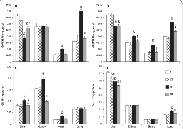
Glucose-6-phosphate dehydrogenase activity (G6PD) significantly increased in the heart (p<0.01) and lung (p<0.001), was unchanged in the kidney (p>0.05), and decreased in the liver of aged diabetic rats compared to control rats treated or untreated with SMe1EC2. In the liver, heart and lung, SMe1EC2 treatment significantly prevented diabetes-induced alterations in G6PD in comparison with the aged diabetic untreated animals (p<0.001) (Figure 2A).
Diabetes resulted in a significant increase in kidney, heart and lung 6-phosphogluconate dehydrogenase activ-ity (6PGD) (p<0.001) and a significant decrease in liver 6PGD (p<0.001). Treatment of aged diabetic animals with SME1EC2 significantly inhibited the increase of G6PD (p<0.001) but had no effect on liver G6PD in comparison with the aged diabetic untreated rats (p>0.05). SMe1EC2 treatment did not change liver, kidney, heart and lung G6PD in aged control rats (p>0.05) (Figure 2B).
Glutathione reductase activity (GR) did not change in the lung (p>0.05). It was elevated in the liver, kidney and heart of aged diabetic rats as compared to aged control rats (p<0.001). The treatment with SMe1EC2 led to a significant decrease in the liver, kidney and heart GR of diabetic rats compared to aged diabetic untreated rats (p<0.001). The treatment with SMe1EC2 did not signifi-cantly change GR in aged control animals (Figure 2C).
In the kidney and heart of aged rats, glutathione-S-transferase activity (GST) was not affected by diabetes (p>0.05). However, GST was significantly inhibited in the liver (p<0.001) and increased in the lung (p<0.001) of aged diabetic rats compared to aged control rats. SMe1EC2 treatment normalized lung GST (p<0.001) but not liver GST (Figure 2D). Liver GST was significantly decreased by SMe1EC2 treatment when compared to that of aged control and of aged diabetic group (p<0.001).
0 50 100 150 200 250 300 350 400 450 Fin a l b o d y we igh t, g 0 50 100 150 200 250 300 350 400 450 C CT D DT Final blood glucose, mg/dl & & # ƒ A B
Figure 1. Eff ects of STZ-diabetes and SMe1EC2 treatment on fi nal
body weight (g; A) and fi nal blood glucose (mg/dl; B) of rats. The rats were grouped as control (C), control treated (CT), diabetic (D), diabetic treated (DT). All data are expressed as the means ± SD. #p<0.01, &p<0.001 vs. control; ƒp<0.05 vs. diabetic (ANOVA fol-lowed by Newman-Keuls test).
Discussion
An accelerated formation of protein glyco-lipo-oxidation occurs in diabetes due to higher blood glucose. The long-term oxidation causes an inability of the proteins to do their function, which plays a key role in the pathogenesis of diabetic complications. Besides, oxidative stress is also the primary cause that links the loss of physiological functions with senescence. Thus the suppression of exac-erbated oxidation is supposed to play an important role in the prevention and/or delay of diabetic complications and in the aging process. In this context, our group has undertaken a research program to assess the mechanisms of antioxidative, antiglycative and tissue protecting activ-ity of synthetic pyridoindolic compounds for a long time. The present work is the first to investigate SMe1EC2 effects on PPP and glutathione-dependent enzyme activi-ties, acting as the key modulators in the control of ROS, in aged diabetic rats.
We have already demonstrated that G6PD is increased in the brain, lung, pancreas and eye-lens of diabetic animals (Ulusu et al., 2003; Ulusu et al., 2005; Gök et
al., 2016). Accordingly, the present study showed an
increased activation of heart and lung G6PD even in the presence of aging and diabetes. PPP generates NADPH and ribonucleotides, with enzymatic regulation by glucose-6-phosphate dehydrogenase (G6PDH), 6-phos-phogluconolactonase (6PGL), and 6-phosphogluconate dehydrogenase (6PGDH). In fact, PPP is a main source for NADPH, which can be used as a cofactor of GR for the regeneration of GSH or as a substrate for NADPH oxidase for the release of free radicals. NADPH oxidases generally act as pro-oxidant enzymes, playing a crucial role in the pathogenesis of diabetes complications and the aging pro-cess. In our study, diabetes in aged rats led to a decrease in G6PDH, 6PGDH and GST only in the liver and no other tissues studied that displayed increased or unchanged G6PDH, 6PGDH or GST, suggesting the cell type-specific roles of PPP in the regulation of ROS balance, as indicated before (Park et al., 2017). The decline in liver G6PDH, 6PGDH and GST may be due to excessive oxidative stress as indicated previously, showing that different types of
& & & & &ƒ ƒ * & & & & &‡ &‡ ‡ ‡ ‡ ‡ ‡ † † ‡ & & & & A B 0 0.005 0.01 0.015 0.02 0.025 0.03 0.035 0.04 0.045 G6P D H , U /mg protei n C 0 0.05 0.1 0.15 0.2 0.25 GR, U /mg protei n
Liver Kidney Heart Lung
D 0 0.2 0.1 0.3 0.5 0.4 0.6 0.7 0.8 GST, U/mg pr otein
Liver Kidney Heart Lung
C CT D DT 0 0.005 0.01 0.015 0.02 0.025 0.03 0.035 0.04 0.045 6PGDH, U/mg protein
Figure 2. Eff ects of STZ-diabetes and SMe1EC2 treatment on glucose-6-phosphate dehydrogenase (G6PD), 6-phosphogluconate
dehydroge-nase (6PGD), glutathione reductase (GR) and glutathione-S-transferase activities in liver, kidney, heart and lung of rats. The rats were grouped as control (C), control treated (CT), diabetic (D), diabetic treated (DT). The enzyme activities were measured spectrophotometrically. All data are expressed as the means ± SD.*p<0.05, #p<0.01, &p<0.001 vs. control; ƒp<0.05, †p<0.01, ‡p<0.001 vs. diabetic (ANOVA followed by Newman-Keuls test).
tissue increase the utilization of G6PDH and 6PGDH under oxidative stress conditions to allow efficient regeneration of NADPH and the reduction of glutathione disulfide (GSSG) to GSH via GR, which enhances the antioxidant defense system (Bolaños and Almeida, 2010). In accordance with this, we showed an up-regulated GR in the aged diabetic liver. Since G6PD is a key regulator responsible for intracellular redox homeostasis, in long-term conditions G6PD deficiency can alter redox balance leading to many abnormal cellular effects, such as the cellular inflammatory and immune response against viral infection (Yang et al., 2016). Decreased G6PD activity and, as a result, decreased NADPH level may associate with liver disease in aged diabetics (Gupte et al., 2009) since NADPH is required not only for the antioxidant system and NADPH oxidase but also for many essential cellular processes such as nitric oxide synthase and cytochrome p450 enzymes (Sahoo et al., 2016).
Conversely, our study showed that G6PDH and 6PGDH were up-regulated along with increased or unchanged GR in the heart and lung of aged diabetic rats. This finding confirms our previous reports (Gök et al., 2016) and is consistent with reports of others showing that high glucose concentrations leads to overexpres-sion of G6PD and augmented PPP activity in different cell types (Peiró et al., 2012). The overactivation of PPP may be linked with increased depletion of NADPH in these tissues due to chronic hyperglycemia via increased polyol pathway activity and upregulated aldose reductase (Kyselova et al., 2005). However, the long-term overacti-vation of PPP would favor the utilization of NADPH by NADPH oxidase and the excess of free radical generation would further contribute to exhausting GSH (Peiró et
al., 2016). NADPH oxidase requires higher
concentra-tions of NADPH to be active, as the Michaelis constant for this enzyme is five times higher than for glutathione reductase (Matsui et al., 2005). It has been known for many years that diabetic patients have a decreased tissue concentration of GSH and the defects in GSH-dependent antioxidant enzymatic activity have been related to diabetes-associated tissue complications (Özdemir et
al., 2009; Raza et al., 2012). In this context, we recall
the early reports indicating that some tissues are highly sensitive even to low levels of hyperglycemia and NADPH than other tissues (Chandrasena et al., 2008; Zhang et
al., 2010). It may be reasonable to explain the differences
in enzymatic activities as the tissue’s responses to aged diabetes.
It has been demonstrated that exacerbated tissue dysfunctions observed in diabetes were prevented both by NADPH oxidase inhibition or PPP blockade (Peiró et
al., 2016; Gök et al., 2016). This is the first study showing
that alterations in G6PD or 6PGDH due to aging and dia-betes are prevented by a pyridoindol compound SMe1EC2 in the liver, kidney, heart and lung of rats. This is also consistent with our previous findings which showed that diabetes-induced regulations on PPP and glutathione-dependent enzymes were prevented by vitamin E and the antioxidant stobadine (Ulusu et al., 2003).
In fact, there is but limited report indicating effects of aging on PPP and GSH-dependent enzymes. This stresses the importance of our work as the first study to evaluate PPP enzyme activities in different tissues of aged diabetic animals. Only one recent study reported that aging affected PPP and produced an age-dependent decrease in erythrocytes G6PDH, which was correlated with decreased GSH and increased oxidative stress (Maurya
et al., 2016). On the other hand, G6PD-Tg mice were
shown to exhibit higher levels of NADPH, lower levels of ROS-derived damage, and better protection from aging-associated functional decline, including extended median lifespan in females. Nevertheless, the authors conclude that a modest increase in G6PD activity is beneficial for healthspan through increased NADPH levels and protec-tion from the deleterious effects of ROS (Nóbrega-Pereira
et al., 2016).
By using a proteomic approach, it has been found that the regulatory role of G6PD in xenobiotic metabo-lism occurs possibly via NOX and the redox-sensitive Nrf2-signaling pathway to modulate the expression of xenobiotic-metabolizing enzymes (Yang et al., 2016). In addition, GST, an important enzyme in detoxification, catalyzes the reaction of glutathione conjugation with many electrophilic xenobiotics (e.g. drugs, toxins, envi-ronmental pollutants, products of oxidative stress, and carcinogens) and their reactive metabolites formed via the cytochrome P-450 monooxygenase system. This enzyme was reported to be induced as a protective mechanism under conditions of increased oxidative stress, including diabetes (Pahwa et al., 2017) and aging (Vyskočilová et al., 2013) and favorable effects were achieved by treatment with antioxidants (Gök et al., 2016; Chandran et al., 2016). GST increased only in the lung, was unchanged in the kidney and heart and attenuated in the liver of aged dia-betic rats. Except the liver, kidney and heart, only the lung showed an unaffected GR by diabetes and aging, while G6PDH and 6PGDH were up-regulated in the lung of aged diabetic rats. GST helps in lowering oxidative stress, thus the increment of GST in aged diabetic rats might be due to its utilization to alleviate the oxidative stress in the lung of aged diabetic rats. SMe1EC2 treatment also reduced GR in the kidney of aged diabetic rats below its normal level, implicating SMe1EC2-mediated dysregulation of reduc-tion of the oxidized glutathione (GSSG).
GST activity depends on a steady quantity of GSH. If GSH is oxidized, it is reduced back by GR, which requires NADPH. Another interesting finding are the effects of SMe1EC2 treatment on liver GST. SMe1EC2 treatment lowered GST in the liver of aged rats in the presence or absence of diabetes, while it restored G6PDH, the rate-limiting enzyme of the pentose phosphate pathway, and GR to its normal levels in aged diabetic rats. With increas-ing age, a decline of biotransformation capacity was observed in laboratory animals and in man (Warrington
et al., 2004) and a marked fall in cytochrome p450 (CYP)
activities. Oxidative biotransformation has been repeat-edly described in old animals and humans (Yun et al., 2010). Thus SMe1EC2-mediated increase in age- and
diabetes-related decline in liver GST may have important clinical consequences by affecting both drug efficacy and toxicity. Nevertheless, we have already demonstrated that SMe1EC2 decreases age- and diabetes-induced up-regulation of oxidative protein modification in rat brain and peripheral tissues (Şakul et al., 2013).
Acknowledgements
This work was supported by VEGA (2/0041/15), SAS – TÜBİTAK Joint Project (No. JRP 2015/7), Ankara University BAP (10B336002) and Gazi University BAP (01/2010-126). and Gazi University BAP (01/2010-126).
Declaration of interest:
The authors report no conflict of interest. The authors alone are responsible for the content and writing of this paper.
Habig WH, Jakoby WB. (1981). Glutathione transferase (human placenta). Methods Enzymol 77: 218–231.
Karasu C. (2010). Glycoxidative stress and cardiovascular complications in ex-perimentally-induced diabetes: eff ects of antioxidant treatment. Open Car-diovasc Med J 4: 240–256.
Koçak G, Aktan F, Canbolat O, Ozoğul C, Elbeğ S, Yildizoglu-Ari N, Karasu C; ADIC Study Group-Antioxidants in Diabetes-Induced Complications. (2000). Alpha-lipoic acid treatment ameliorates metabolic parameters, blood pressure, vascular reactivity and morphology of vessels already damaged by streptozotocin-diabetes. Diabetes Nutr Metab 13: 308–318. Kyselova Z, Gajdosik A, Gajdosikova A, Ulicna O, Mihalova D, Karasu C,
Ste-fek M. (2005). Eff ect of the pyridoindole antioxidant stobadine on devel-opment of experimental diabetic cataract and on lens protein oxidation in rats: comparison with vitamin E and BHT. Mol Vis 11: 56–65.
Matsui R, Xu S, Maitland KA, Hayes A, Leopold JA, Handy DE, Loscalzo J, Co-hen RA. (2005). Glucose-6 phosphate dehydrogenase defi ciency decreases the vascular response to angiotensin II. Circulation 112: 257–263.
Maurya PK, Kumar P, Chandra P. (2016). Age-dependent detection of erythro-cytes glucose-6-phosphate dehydrogenase and its correlation with oxida-tive stress. Arch Physiol Biochem 122: 61–66.
Nóbrega-Pereira S, Fernandez-Marcos PJ, Brioche T, Gomez-Cabrera MC, Sal-vador-Pascual A, Flores JM, Viña J, Serrano M. (2016). G6PD protects from oxidative damage and improves healthspan in mice. Nat Commun 7: 10894. Ozdemir S, Tandogan B, Ulusu NN, Turan B. (2009). Angiotensin II receptor blockage prevents diabetes-induced oxidative damage in rat heart. Folia Biol (Praha) 55: 11–16.
Pahwa S, Sharma R, Singh B. (2017). Role of Glutathione S-Transferase in Cor-onary Artery Disease Patients with and Without Type 2 Diabetes Mellitus. J Clin Diagn Res 11: BC05–BC08.
Panigrahy SK, Bhatt R, Kumar A. (2017). Reactive oxygen species: sources, con-sequences and targeted therapy in type 2 diabetes. J Drug Target 25: 93–101. Park YJ, Choe SS, Sohn JH, Kim JB. (2017). The role of glucose-6-phosphate
dehydrogenase in adipose tissue infl ammation in obesity. Adipocyte 8: 1–7. Pearse BMF, Rosemeyer MA. (1975). 6-Phosphogluconate dehydrogenase
from human erythrocytes. Methods Enzymol 41: 220–226.
Peiró C, Romacho T, Azcutia V, Villalobos L, Fernández E, Bolaños JP, Moncada S, Sánchez-Ferrer CF. (2016). Infl ammation, glucose, and vascular cell dam-age: the role of the pentose phosphate pathway. Cardiovasc Diabetol 15: 82. Raza H, John A, Howarth FC. (2012). Alterations in glutathione redox metabo-lism, oxidative stress, and mitochondrial function in the left ventricle of el-derly Zucker diabetic fatty rat heart. Int J Mol Sci. 13: 16241–16254. Sahoo S, Meijles DN, Pagano PJ. (2016). NADPH oxidases: key modulators in
aging and age-related cardiovascular diseases? Clin Sci 130: 317–335. Sakul A, Cumaoğlu A, Aydin E, Ari N, Dilsiz N, Karasu C. (2013) Age- and
dia-betes-induced regulation of oxidative protein modifi cation in rat brain and peripheral tissues: consequences of treatment with antioxidant pyridoin-dole. Exp Gerontol 48: 476–484.
Shakeel M. (2015). Recent advances in understanding the role of oxidative stress in diabetic neuropathy. Diabetes Metab Syndr 9: 373–378.
Stanton RC. (2012). Glucose-6-phosphate dehydrogenase, NADPH, and cell survival. IUBMB Life 64: 362–369.
Stefek M, Karasu C. (2011). Eye lens in aging and diabetes: eff ect of quercetin. Rejuvenation Res 14: 525–534.
Stefek M, Milackova I, Juskova-Karasova M, Snirc V. (2013). Antioxidant action of the hexahydropyridoindole SMe1EC2 in the cellular system of isolated red blood cells in vitro. Redox Rep 18: 71–75.
Tandogan B, Güvenç A, Çalış İ, Ulusu NN. (2011). In vitro eff ects of compounds isolated from Sideritis brevibracteata on bovine kidney cortex glutathione reductase. Acta Biochim Pol 58: 471–475.
Ulusu NN, Ozbey G, Tandogan B, Gunes A, Durakoglugil DB, Karasu C, Uluo-glu C, Zengil H. (2005). Circadian variations in the activities of 6-phospho-gluconate dehydrogenase and glucose-6-phosphate dehydrogenase in the liver of control and streptozotocin-induced diabetic rats. Chronobiol Int
22: 667–677.
Ulusu NN, Sahilli M, Avci A, Canbolat O, Ozansoy G, Ari N, Bali M, Stefek M, Stolc S, Gajdosik A, Karasu C. (2003). Pentose phosphate pathway, glutathi-one-dependent enzymes and antioxidant defense during oxidative stress in diabetic rodent brain and peripheral organs: eff ects of stobadine and vi-tamin E. Neurochem Res 28: 815–823.
REFERENCES
Acan NL, Tezcan EF. (1989). Sheep brain glutathione reductase: Purifi cation and general properties. FEBS Lett 250: 72–74.
Aldini G, Vistoli G, Stefek M, Chondrogianni N, Grune T, Sereikaite J, Sad-owska-Bartosz I, Bartosz G. (2013). Molecular strategies to prevent, inhibit, and degrade advanced glycoxidation and advanced lipoxidation end products. Free Radic Res 47 Suppl 1: 93–137.
Betke K, Brewer GJ, Kirkman HN, Luzzato L, Motulsky AG, Ramot B, Siniscalco M. (1967). Standardized method for G-6-PD assay of haemolysates. WHO Tech Rep Ser 366: 30–32.
Bolaños JP, Almeida A. (2010). The pentose-phosphate pathway in neuronal survival against nitrosative stress. IUBMB Life 62: 14–18.
Bradford MM. (1976). A rapid and sensitive method for the quantitation of microgram quantities of protein utilizing the principle of protein dye bind-ing. Anal Biochem 72: 248–254.
Broskova Z, Sotnikova R, Nedelcevova J, Bagi Z. (2013). Eff ect of a novel sto-badine derivative on isolated rat arteries. Interdiscip Toxicol 6: 63–66. Cencioni C, Spallotta F, Martelli F, Valente S, Mai A, Zeiher AM, Gaetano C.
(2013). Oxidative stress and epigenetic regulation in ageing and age-re-lated diseases. Int J Mol Sci 14: 17643–17663.
Chandran R, Parimelazhagan T, Shanmugam S, Thankarajan S. (2016). Anti-diabetic activity of Syzygium calophyllifolium in Streptozotocin-Nicotin-amide induced Type-2 diabetic rats. Biomed Pharmacother 82: 547–554. Chandrasena LG, De Silva LD, De Silva KI, Dissanayaka P, Peiris H. (2008).
Changes in erythrocyte glucose-6-phosphate dehydrogenase (G6PD) and reduced glutathione (GSH) activities in the development of senile and dia-betic cataracts. Southeast Asian J Trop Med Public Health 39: 731–736. Drimal J, Knezl V, Navarova J, Nedelcevova J, Paulovicova E, Sotnikova R,
Snirc V, Drimal D. (2008). Role of infl ammatory cytokines and chemoattrac-tants in the rat model of streptozotocin-induced diabetic heart failure. En-docr Regul 42: 129–135.
Gasparova Z, Stara V, Janega P, Navarova J, Sedlackova N, Mach M, Ujhazy E. (2014). Pyridoindole antioxidant-induced preservation of rat hippocampal pyramidal cell number linked with reduction of oxidative stress yet with-out infl uence on cognitive deterioration in Alzheimer-like neurodegenera-tion. Neuro Endocrinol Lett 35: 454–462.
Gök M, Ulusu NN, Tarhan N, Tufan C, Ozansoy G, Arı N, Karasu Ç. (2016). Flax-seed Protects Against Diabetes-Induced Glucotoxicity by Modulating Pen-tose Phosphate Pathway and Glutathione-Dependent Enzyme Activities in Rats. J Diet Suppl 13: 339–351.
Gupte RS, Floyd BC, Kozicky M, George S, Ungvari ZI, Neito V, Wolin MS, Gupte SA. (2009). Synergistic activation of glucose-6-phosphate dehydro-genase and NAD(P)H oxidase by Src kinase elevates superoxide in type 2 diabetic, Zucker fa/fa, rat liver. Free Radic Biol Med 47: 219–228.
Ulusu NN. (2015). Glucose-6-phosphate dehydrogenase defi ciency and Al-zheimer’s disease: Partners in crime? The hypothesis. Med Hypotheses 85: 219–223.
Vyskočilová E, Szotáková B, Skálová L, Bártíková H, Hlaváčová J, Boušová I. (2013). Agerelated changes in hepatic activity and expression of detoxifi -cation enzymes in male rats. Biomed Res Int 2013: 408573.
Warrington JS, Greenblatt DJ, von Moltke LL. (2004). Age-related diff erences in CYP3A expression and activity in the rat liver, intestine, and kidney. J Pharmacol Exp Ther 309: 720–729.
Yang HC, Wu YH, Liu HY, Stern A, Chiu DT. (2016). What has passed is prolog: new cellular and physiological roles of G6PD. Free Radic Res 50: 1047–1064.
Yun KU, Oh SJ, Oh JM, Kang KW, Myung CS, Song GY, Kim BH, Kim SK. (2010). Age-related changes in hepatic expression and activity of cytochrome P450 in male rats. Arch Toxicol 84: 939–946.
Zhang Z, Liew CW, Handy DE, Zhang Y, Leopold JA, Hu J, Guo L, Kulkarni RN, Loscalzo J, Stanton RC. (2010). High glucose inhibits glucose-6-phosphate dehydrogenase, leading to increased oxidative stress and beta-cell apop-tosis. FASEB J 24: 1497–1505.
Zúrová-Nedelcevová J, Navarová J, Drábiková K, Jancinová V, Petríková M, Bernátová I, Kristová V, Snirc V, Nosál’ová V, Sotníková R. (2006). Participa-tion of reactive oxygen species in diabetes-induced endothelial dysfunc-tion. Neuro Endocrinol Lett 27 Suppl 2:168–171.