Turk Kardiyol Dern Ars 2015;43(6):565–567 doi: 10.5543/tkda.2015.49060
Non-surgical treatment of a right ventricle puncture
during diagnostic pericardiocentesis
Tanısal perikardiyosentez sırasında gelişen sağ ventrikül ponksiyonunun
ameliyatsız tedavisi
Department of Cardiology, Istanbul Medipol University Faculty of Medicine, Istanbul
Ekrem Güler, M.D., Gamze Babur Güler, M.D., Gültekin G. Demir, M.D., Filiz Kızılırmak, M.D.
Özet– Perikardiyosentez, enfektif, enflamatuvar veya ma-lignite nedenli kalp tamponadı veya kalp cihazı yerleştiril-mesi, perkütan koroner girişim, kateter ablasyonu benzeri işlemler sonrası uygulanan hayat kurtarıcı bir işlemdir. Ar-tan görüntüleme yöntemlerine rağmen komplikasyon riski az değildir. Özellikle kateterin kalp boşluklarına ilerletilmesi veya duvar rüptürü sonrası acil cerrahi müdahale gerekli olabilmektedir. Ancak bu hastaların mevcut komorbiditeleri sebebiyle acil cerrahi riski yüksektir. Bu yazıda sunulan ol-guda, tüberküloz perikardit ön tanısı ile takip edilen bir has-tada tanı amaçlı perikardiyosentez sırasında sağ ventriküle yerleştirilen kateteri, perikart boşluğuna yerleştirilen ikinci bir kateter yardımı ile geri aldık. Spontan kanama kontrolü sağlandığı için perikart sıvısında artış izlenmedi ve cerrahi girişim gerekmedi. Bu yöntem perikardiyosentez gibi kal-be dışardan müdahale veya intrakardiyak manipülasyon-lar sebebiyle görülebilecek kardiyak hasarmanipülasyon-lar sonrasında uygulanabilir ve hasta cerrahinin olası yüksek riskinden korunabilir.
Summary– Pericardiocentesis is a life-saving procedure performed in cardiac tamponade cases occurring in infec-tive, inflammatory or malignancy conditions, or following percutaneous coronary intervention, cardiac device implan-tation or catheter ablation. In spite of advanced imaging methods, a substantial risk of complication persists. Emer-gent surgical intervention may be required, in particular dur-ing advancement of the catheter into the heart chambers or in cases of wall rupture. Furthermore, in all these cases, patients have a high risk of surgery because of existing comorbidities. This case presents a patient suspected of tuberculous pericarditis who underwent diagnostic pericar-diocentesis complicated by right ventricular puncture. The catheter in the right ventricle was withdrawn via a second catheter placed in the pericardial cavity. Spontaneous blood control was established, and with no increase in pericardial effusion surgical intervention was not required. This method can be applied in certain conditions, including cardiac injury caused by pericardiocentesis or intracardiac manipulations, thus eliminating the need for high-risk surgical intervention.
565
P
ericardiocentesis is a commonly-used procedure for diagnostic or therapeutic purposes, especially in cardiac tamponade.[1] While most procedures arecur-rently performed under the guidance of advanced im-aging methods,[2] there still exists a complication risk.
Inadvertent cardiac puncture during introduction of the needle or sheath is a serious complication and, unless appropriately managed, is associated with high mortal-ity and surgical risk due to patient comorbidites.[3]
This case report presents a patient with a prelimi-nary diagnosis of tuberculosis pericarditis in whom
cardiac puncture through the right ventricle occurred during diagnostic pericardiocentesis, and who was successfully treated by withdrawal of the catheter by a second catheter placed in the pericardial cavity.
CASE REPORT
A 52-year-old female patient with a progressive cough and shortness of breath was admitted to the pulmonary disease outpatient clinic. She was sched-uled for echocardiography for increased cardiac size on chest X-ray. Echocardiography revealed normal Received:January 23, 2015 Accepted:May 11, 2015
Correspondence: Dr. Ekrem Güler. İstanbul Medipol Üniversitesi Hastanesi, Tem Avrupa Otoyolu Göztepe Çıkışı, No: 1, Bağcılar, İstanbul, Turkey.
Tel: +90 212 - 460 77 77 e-mail: [email protected] © 2015 Turkish Society of Cardiology
left ventricular systolic function, mild mitral regurgi-tation and pericardial effusion (1.8 cm in posterior, 1.2 cm in right ventricle adjacency, 1.3 cm in apex and 2.7 cm in lateral segments). The patient had a his-tory of tuberculosis, and diagnostic pericardiocente-sis for sampling was planned. Informed consent was obtained and preparation for subxiphoid access was made.[4,5] Following sedation and local anesthesia,
the needle was gently advanced under echocardiog-raphy guidance and hemorrhagic fluid was aspirated. Agitated saline was infused for confirmation, but no intramyocardial bubble was observed. A 6 Fr pigtail-catheter was advanced over a 0.035” guiding wire. In order to confirm appropriate location of the catheter, an insecure method with indefinite results was used, in which hemorrhagic fluid was tested several times on gauze-pad for coagulation control. The results were coagulation in some, but failure to coagulate in others, which caused doubt about appropriate catheter location. The agitated saline test was repeated and showed bubbles in the right ventricle. The patient was asymptomatic, with blood pressure 132/75 mmHg, heart rate 98/min and oxygen saturation 96%. Cardio-vascular surgery was consulted and operative prepa-rations were initiated. It was decided to withdraw the first catheter immediately after placement of a second catheter in the pericardial cavity and perform pericar-diocentesis via this second catheter to avoid cardiac tamponade in the event of any increase in pericardial effusion during follow-up. In comparison to the first
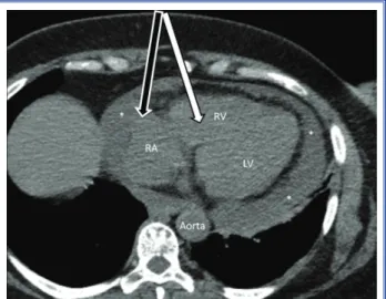
puncture, the second was performed more medially towards the right atrium, which was contiguous with a greater amount of fluid. When the second catheter was advanced into the pericardial cavity, serous fluid was aspirated. After obtaining samples for LDH, pro-tein, albumin, cytology and culture, 550 mL fluid was drained. Following completion of emergency surgery preparations, the first catheter was gently withdrawn under guide wire control, and the second catheter left in place for back-up purposes in the pericardial cavity (Figure 1). Follow-up echocardiography revealed no increase in effusion and the patient had stable hemo-dynamic parameters. No fluid drainage was observed through the second catheter during 24-hour follow-up. The catheter was removed after follow-up and the patient discharged.
DISCUSSION
This case demonstrates the possibility of treating in-advertent right ventricle puncture during diagnostic pericardiocentesis without cardiac surgery by place-ment of a second catheter support in the pericardial cavity.
Cardiac tamponade may sometimes occur follow-ing infections, malignancy or cardiac interventions, and pericardiocentesis is a life-saving procedure in its occurrence.[6] Major and minor complication rates for
pericardiocentesis under echocardiography guidance are 1.2% and 3.5% respectively.[7] Complications
in-clude right atrium or ventricle laceration, coronary artery injury, injury to the mammary or intercostal arteries, hypotension, arrhythmia, pneumothorax, pericardial decompression and death.[8,9] A larger
amount of fluid in cardiac tamponade facilitates the procedure and decreases complication rates. How-ever, diagnostic pericardiocentesis is associated with increased complication rates. In the present case, de-spite larger amounts of regional fluid accumulation, there was a smaller amount at the access site, and the right heart was in a closer position to the pericardial border. Catheter introduction following needle access into the right ventricle prevented bleeding into the pericardial space, so hemodynamics parameters were stable. Absence of bleeding into the pericardial space following withdrawal of the catheter may be related to both fibrillary structures in the pericardium and lack of anticoagulation in the patient. The gelatinous characteristics of the fibrillary structures on the heart Turk Kardiyol Dern Ars 566
Figure 1. Computed tomography image showing pericardial effusion and catheters placed in right ventricle (white arrow) and pericardial cavity (black arow). RA: Right atrium; RV: Right ventricle; LV: Left ventricle; (*) Pericardial effusion.
Non-surgical treatment of a right ventricle puncture during diagnostic pericardiocentesis 567 surface may have acted as a size-limiting and
anti-coagulative factor.
In our opinion, even though echocardiographic guidance was used in this case, the main cause of complication was the smaller amount of fluid at the puncture site. In contrast to the more commonly per-formed subxiphoid approach, large pericardial ef-fusions should be drained through an apical or left ventricle lateral wall puncture when appropriate. In a study including 32 patients undergoing apical pericar-diocentesis with echocardiographic guidance, the pro-cedural success rate was reported as 96%, with only 4 patients having serious complications (hemopneu-mothorax requiring tube drainage, vasovagal reaction, nonsustained ventricular tachycardia, and frequent ventricular extrasystoles). As a result, the authors of the study suggested apical pericardiocentesis, particu-larly in cases of anterior fluid accumulation.[10]
Inadvertent cardiac puncture during pericardio-centesis is conventionally treated with surgery. Ap-propriate selection of puncture site under echocar-diographic guidance may reduce complication risks. However, withdrawal of the catheter with the support of a second catheter placed in the pericardial space and observation of spontaneous bleeding control may be an alternative method.
Conflict-of-interest issues regarding the authorship or article: None declared.
REFERENCES
1. Hsu LF, Scavée C, Jaïs P, Hocini M, Haïssaguerre M. Trans-cardiac pericardiocentesis: an emergency life-saving
tech-nique for cardiac tamponade. J Cardiovasc Electrophysiol 2003;14:1001–3.
2. Salem K, Mulji A, Lonn E. Echocardiographically guided pericardiocentesis - the gold standard for the management of pericardial effusion and cardiac tamponade. Can J Cardiol 1999;15:1251–5.
3. Imazio M, Adler Y. Management of pericardial effusion. Eur Heart J 2013;34:1186–97.
4. Tsang TS, Freeman WK, Sinak LJ, Seward JB. Echocardio-graphically guided pericardiocentesis: evolution and state-of-the-art technique. Mayo Clin Proc 1998;73:647–52.
5. Gring C, Griffin BP. Pericardiocentesis. In: Griffin BP, Topol EJ, editors. Manual of cardiovascular medicine. 2nd ed. Phila-delphia: Lippincott Williams&Wilkins; 2004. p. 709–13. 6. Soler-Soler J, Sagristà-Sauleda J, Permanyer-Miralda G.
Management of pericardial effusion. Heart 2001;86:235–40. 7. Tsang TS, Enriquez-Sarano M, Freeman WK, Barnes ME,
Sinak LJ, Gersh BJ, et al. Consecutive 1127 therapeutic echo-cardiographically guided pericardiocenteses: clinical profile, practice patterns, and outcomes spanning 21 years. Mayo Clin Proc 2002;77:429–36.
8. Wong B, Murphy J, Chang CJ, Hassenein K, Dunn M. The risk of pericardiocentesis. Am J Cardiol. 1979;44:1110–4. 9. Imazio M. Pericardial decompression syndrome: A rare but
potentially fatal complication of pericardial drainage to be recognized and prevented. Eur Heart J Acute Cardiovasc Care 2015;4:121–3.
10. Ozer HO, Davutoğlu V, Cakici M, Doğan A, Sari I, Oylumlu M, et al. Echocardiography-guided pericardiocentesis with the apical approach. [Article in Turkish] Turk Kardiyol Dern Ars 2009;37:177–81.
Keywords: Cardiac tamponade; pericardiocentesis; rupture. Anahtar sözcükler: Kalp tamponadı; perikardiyosentez; yırtık.