Protective effect of lycopene on diethylnitrosamine-induced oxidative
stress and catalase expression in rats
*Emre KAYA
1, Seval YILMAZ
1, Ali Osman ÇERİBAŞI
2, Selda TELO
31Fırat University, Faculty of Veterinary Medicine, Department of Biochemistry; 2Fırat University, Faculty of Veterinary Medicine,
Department of Pathology, 3Fırat University, Faculty of Dentistry, Department of Basic Science, Elazığ, Turkey.
Summary: The purpose of this study is to investigate the protective role of lycopene on diethylnitrosamine (DEN)-induced
hepatotoxicity using biochemical and histopathological approaches. The rats were divided into 5 groups as control, lycopene, DEN, lycopene+DEN and DEN+lycopene. DEN was administered to rats at 200 mg/kg, a single dose intraperitoneally for 30 days. Lycopene was administered to rats every other day at 10 mg/kg, gavage for 10 days. Lycopene administration was started 10 days before the DEN administration in lycopene+DEN group and together with the DEN administration in DEN+lycopene group. In this study, malondialdehyde (MDA), reduced glutathione (GSH) levels, catalase (CAT), glutathione peroxidase (GSH-Px), glutathione-S-transferase (GST), superoxide dismutase (SOD) activities and the expression levels of the CAT enzyme were measured in blood and liver tissues. DEN caused the oxidative stress by the increased MDA level and the reduced GSH level, antioxidant enzyme activities in tissues. Lycopene administration produced amelioration in biochemical indices of hepatotoxicity in both blood and liver tissues when compared to DEN group; simultaneous-administration with DEN has been more effective. It was determined to increase expression levels of the CAT enzyme in the DEN group in RT-PCR. The improvement in expression levels in DEN+lycopene group was observed to be better than the lycopene+DEN group. Histopathologically, many different histopathological changes were observed in liver tissues of DEN and lycopene+DEN groups, it was determined that these changes reduced in DEN+lycopene group. The results from the present study indicate that lycopene exhibits good hepatoprotective and antioxidant potential against DEN-induced hepatocellular damage in rats.
Keywords: Antioxidant, CAT expression, diethylnitrosamine, lycopene, oxidative stress.
Ratlarda likopenin dietilnitrozamin kaynaklı oksidatif stres ve katalaz ekspresyonu üzerine koruyucu
etkisi
Özet: Çalışmanın amacı, likopenin dietilnitrozamin (DEN) kaynaklı hepatotoksisite üzerindeki koruyucu rolünü biyokimyasal
ve histopatolojik yaklaşımları kullanarak araştırmaktır. Ratlar kontrol, likopen, DEN, likopen+DEN ve DEN+likopen olmak üzere 5 gruba ayrılmıştır. DEN ratlara 200 mg/kg dozunda tek doz 30 gün süreyle intraperitoneal olarak uygulanmıştır. Likopen ratlara 10 mg/kg dozunda gün aşırı olarak 10 gün boyunca gavajla uygulanmıştır. Likopen uygulamasına, likopen+DEN grubunda DEN uygulanmasından 10 gün önce, DEN+likopen grubunda ise DEN uygulaması ile birlikte başlanmıştır. Çalışmada kan ve karaciğer dokularında malondialdehit (MDA), redükte glutatyon (GSH) düzeyleri, katalaz (CAT), glutatyon peroksidaz (GSH-Px), glutatyon-S-transferaz (GST), süperoksit dismutaz (SOD) aktiviteleri ve CAT enziminin ekspreson düzeyleri ölçülmüştür. DEN dokularda MDA düzeyinde artış, GSH düzeyi ve antoksidan enzim aktivitelerinde düşüşe neden olarak oksidatif strese sebep olmuştur. Likopen uygulaması hem kan hem de karaciğer dokusunda DEN grubuna kıyasla hepatotoksisitenin biyokimyasal indekslerinde iyileşme sağlamıştır; DEN ile eşzamanlı uygulanması daha etkili olmuştur. RT-PCR analizlerinde CAT enziminin ekspresyon düzeylerinin DEN grubunda arttığı belirlenmiştir. DEN+likopen grubundaki ekspresyon seviyelerindeki düzelmenin likopen+DEN grubundan daha iyi olduğu da gözlenmiştir. Histopatolojik olarak, DEN ve likopen+DEN gruplarının karaciğer dokularında birçok farklı histopatolojik değişiklikler gözlenmiştir ve bu değişikliklerin DEN+likopen grubunda azaldığı belirlenmiştir. Bu çalışmanın sonuçları, ratlarda DEN ile oluşturulan hepatoselüler hasara karşı likopenin iyi bir hepatoprotektif ve antioksidan potansiyel taşıdığını göstermektedir.
Anahtar sözcükler: Antioksidan, CAT ekspresyonu, dietilnitrosamin, likopen, oksidatif stres.
* This article is a part of the first author's doctoral thesis named ’’The Effect of Lycopene on Oxidative Stress and DNA Damage in
Diethylnitrosamine Administered Rats’’. The abstract of this research was published in the proceedings of the 8th National Veterinary
Introduction
Nitroso compounds are divided into two groups as nitrosamides and nitrosamines. Nitrosamides are derivatives of substances such as amides, guanidines, carbamates, carboxamides. Nitrosamines are a group of hepatotoxic and carcinogenic, alkylating chemical compounds formed by the combining of nitrites and amines in the digestive tract. Nitrosamines can be formed endogenously in vivo, as well as nitrite taken from various foods, especially as a result of the reaction with secondary amines. The first occurrence of nitrosamines in vivo is the mouth, which is the beginning of the digestive system. The salivary secretion contains abundant amounts of nitrate and this nitrate is reduced to nitrite by the nitrate reductase enzyme. Thus, formed nitrite causes the formation of nitrosamines (11).
Nitrosamines, besides being substances with strong carcinogenic effect, also show mutagenic and teratogenic effects. The dose, exposure frequency, and shape can lead to the differentiation of the affected organ (such as lung, kidney or liver) and the alteration of the target cell where the tumor is formed (26).
Diethylnitrosamine (DEN) is a nitrosamine compound with potent hepatocarcinogenic initiator effects. It is reported that DEN, a carcinogenic substance, can be formed from chemicals used in agriculture, from insecticides and nitrate, as well as being present in the cigarette smoke, and nitrates found in foodstuffs combined with secondary and tertiary amines. In addition, DEN is found in workplaces such as the rubber industry, in alcoholic beverages and processed meat products, and can also occur during the metabolism of certain therapeutic drugs in the liver. DEN, an environmental carcinogen, and hepatotoxin are widely used in experimental animal models. DEN is found in many types of products, which humans are exposed to, such as cigarette smoke, meat, and whiskey, and cause degenerative, proliferative, and neoplastic lesions in the liver (3, 17).
DEN is hydroxylated by cytochrome P-450 isoenzymes in the liver and becomes bioactive by the mechanism of alkylation. As a result of activation, the radical character metabolites, mainly the ethyl radical occurs (26).
There have been many studies on the effect of carotenoids on oxidative stress. Tomato and the products obtained from tomato contain lycopene and other similar carotenoids. Lycopene is an aliphatic hydrocarbon and one of about 600 naturally occurring carotenoids (31).
Lycopene exhibits antioxidant activity due to its extremely hydrophobic nature, acyclic in long chain form and its conjugated double bond. Lycopene shows antioxidant property by destroying oxygen radicals like retinol, α-tocopherol and carotenoids (34). Lycopene is
effective in eliminating singlet oxygen (1O
2), which is
responsible for the formation of other free radicals (5). In addition, lycopene is an excellent superoxide radical (O·̄2)
cleanser that resembles liposomes in biological membrane models. While lycopene exhibits a strong antioxidant property outside the cell, it has a protective effect against the oxidation of DNA, proteins and lipids in the cell. Lycopene, which contains 11 conjugated double bonds in the structure, can retain active oxygen species much more effectively than β-carotene containing 9 conjugated double bonds (30). Furthermore, in a number of clinical trials, consumption of tomatoes has been shown to prevent oxidative DNA damage in human leukocytes (35). Therefore, in this study, it is aimed to reveal the possible protective or healing effects of lycopene on oxidative damage and catalase expression which may be caused by DEN in blood and liver in rats.
Materials and Methods
Chemicals: DEN was purchased from Sigma (St.
Louis, MO, USA, Code: N0258, CAS No: 55-18-5), Lycopene 10% FS (RedivivoTM) from DSM Nutritional
Products (Heerlen, Netherlands), RNA isolation and Real-Time Polymerase Chain Reaction (RT-PCR) chemicals from Qiagen (Hilden, Germany), glutathione, glutathione reductase (GR), thiobarbituric acid (TBA), hydrogen peroxide (H2O2), nicotinamide adenine dinucleotide
phosphate (NADPH) and other reagents were supplied from Sigma (St. Louis, MO, USA).
Experimental Groups: Thirty-five healthy male
Wistar-Albino rats (250-300 g body weight) were used in this study. The protocol for the use of animals was approved by the Firat University Animal Experiments Local Ethics Committee (Protocol No: 2014/18). DEN was administered the animals a single intraperitoneal (i.p.) dose at the dose of 200 mg/kg body weight dissolved in serum physiological (0.9% NaCl) for 30 days. Lycopene was suspended in corn oil and administered to the animals by gavage every other day at the dose of 10 mg⁄kg body weight for 10 days. The doses of DEN (15) and lycopene (25) used in this study was selected according to previous studies. The animals were randomly divided into five experimental groups, including seven rats in each. These groups were arranged as follows: control group, lycopene group (lycopene was administered), DEN group (a single dose of DEN was administered), lycopene+DEN (lycopene was started 10 days before the DEN administration), DEN+lycopene group (DEN and lycopene were administered together).
Sample Collection and Biochemical Analysis: At the
end of the 30 days experiment, under ether anesthesia, blood samples were withdrawn by injector from the heart of the animals and collected into tubes containing EDTA.
Then, the rats in control and experimental groups were sacrificed by decapitation under ether anesthesia.
Plasma was used to measure malondialdehyde (MDA) level as a marker of lipid peroxidation (LPO) and to determine aspartate transaminase (AST), alanine transaminase (ALT), alkaline phosphatase (ALP), lactate dehydrogenase (LDH) activities and cholesterol levels. Tissue samples were quickly removed and perfused with ice-cold saline for biochemical and histopathological evaluation. The tissues were homogenized in distilled water by using a Potter-elvehjem Homogenizer. The homogenate was centrifuged (at 3500 rpm for 15 min to MDA reduced glutathione (GSH) levels, catalase (CAT), glutathione-S-transferase (GST), superoxide dismutase (SOD) analyzes and at 14000 rpm for 55 min to glutathione peroxidase (GSH-Px) analyze at +4°C).
MDA and GSH levels, CAT, GSH-Px, GST and SOD activities were analyzed in blood and liver tissues. The MDA level was measured according to the method of Placer et al. (32). This method was based on the reaction of TBA and MDA, one of the aldehyde products of LPO. GSH level was determined by the method of Ellman et al. (13). This method was a spectrophotometric method based on the formation of highly stable yellow color of sulfhydryl groups when 5,5'dithiobis-2-nitrobenzoic acid (DTNB) was added. CAT activity was carried out by using the Aebi’s method (2). CAT activity was determined by measuring the resolution of H2O2 at 240 nm. GSH-Px
activity was measured by the Beutler method (7). GSH-Px catalyzes the oxidation of GSH to oxide glutathione (GSSG) using H2O2. The rate of formation of GSSG was
measured by the GR reaction. GST activity was measured by the method of Habig et al. (19). Enzyme activity was determined by measuring the amount of enzyme catalyzing 1 μmol of 1- (S-glutathionyl) -2,4 dinitrobenzene formed at 340 nm at 37°C per minute using GSH and 1-chloro-2,4-dinitrobenzene (CDNB). SOD activity was measured by using xanthine and xanthine oxidases to generate O·̄2 reacting with nitroblue
tetrazolium (40). The protein concentration determination was based on the method of Lowry et al. (27). The plasma AST, ALT, ALP, LDH activities and cholesterol levels were measured by using an AutoAnalyzer (Olympus AU 600, Tokyo, Japan).
Total RNA Isolation and Gene Expression Analysis:
Total RNA was isolated from blood and liver tissue with the RNeasy Plus Mini Kit according to the manufacturer's protocol. Total RNA concentration was determined using a nanodrop spectrophotometer at 260 nm. Total RNA was reverse transcribed to cDNA in 20 µl reactions composed with the First-Strand Kit. The reaction was incubated for 15 minutes at 42°C and at 95°C for 5 minutes according to the manufacturer's protocol. Specific rat CAT primers (NM_012520) for RT-PCR were obtained from Qiagen.
Gene-specific PCR products were quantitated with RT-PCR Instrument (Applied Biosystem, 7500 Fast Real-Time PCR System). Reactions were prepared in 25 µl volumes using the SYBR Green Master Mix. The cycling conditions were performed according to the manufacturer’s protocol as follows: 15 minute at 95oC; 15
seconds at 95oC, 1 minute at 60oC (repeated for 40 cycles).
The RT-PCR analysis results were normalized against the expression level of the internal control β-Actin (NM_031144). The values of the all samples were expressed as percentages with respect to the control.
Histopathological Examination: Necropsy of the rats
was performed and liver tissue samples were fixed at 10% neutral buffered formalin. Paraffin-embedded blocks were routinely processed. 5-μm thick sections were stained with hematoxylin-eosin (H-E) and examined under a microscope (28). Then random 10 microscopic fields were examined in x40 magnification.
Statistical Analysis: The results are expressed as a
mean ± standard error (S.E.). Statistical significance between the different groups was determined by using one-way analysis of variance (ANOVA) in the SPSS 21 software package. Post hoc test was performed for between-group comparisons by using the Tukey multiple comparison test. All data are expressed as mean ± S.E. The level of significance was set at p<0.05 and p<0.001.
Results
MDA, GSH Levels, and Antioxidant Enzymes Activities: Tables 1 and 2 shows the MDA, GSH levels
and the activities of antioxidant enzymes such as CAT, GSH-Px, GST and SOD in the control and experimental groups in the blood and liver tissues. The data indicated that the DEN group had a significantly higher MDA level than the control group in plasma and liver tissue. Lycopene group had a significantly lower MDA level than DEN group. Treatment with DEN and lycopene provided an apparent normalization in MDA level compared to DEN group. GSH level significantly decreased in the DEN group compared to the control group. Upon supplementation of lycopene to DEN group, a significant increase was observed in the GSH level compared to DEN group. The GSH level restored in DEN+lycopene groups. A significant reduction was found in CAT, GSH-Px, GST and SOD activities in the DEN group compared to the control group. Upon simultaneous supplementation of lycopene to DEN group, significant increases were observed in antioxidant enzyme activities compared to DEN group (p<0.05, p<0.001).
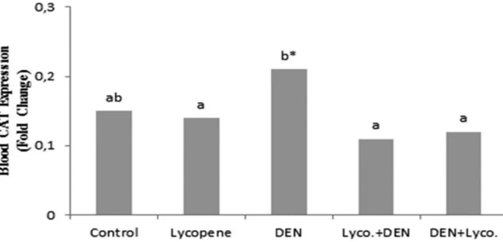
Effect of Lycopene on CAT Expression: Figure 1 and
2 show CAT expression levels in the blood and liver tissue of the control and experimental groups. While the data indicated that DEN group had a significantly higher CAT expression level than the control group in liver tissue, not statistically significant higher CAT expression level than
the control group in blood. Lycopene group had a significantly lower CAT expression levels than DEN group. Treatment with DEN and lycopene provided an apparent normalization in CAT expression levels
compared to the DEN group. Upon supplementation of lycopene to DEN group, a significant decrease was observed in the CAT expression levels compared to the DEN group.
Figure 1. The effect of lycopene supplementation on blood CAT expression level (*P < 0.05) Şekil 1. Likopen ilavesinin kan CAT ekspresyon düzeyi üzerine etkisi
Figure 2. The effect of lycopene supplementation on liver CAT expression level (**P < 0.001). Şekil 2. Likopen ilavesinin karaciğer CAT ekspresyon düzeyi üzerine etkisi
Table 1. The effect of lycopene supplementation on plasma MDA level and erythrocyte GSH level, CAT, GSH-Px, SOD activities Tablo 1. Likopen ilavesinin plazma MDA ve eritrosit GSH düzeyi, CAT, GSH-Px, SOD aktivitelerine etkisi
Parameters Control (1. Group) Lycopene (2. Group) DEN (3. Group) Lyco.+DEN (4. Group) DEN+Lyco. (5. Group) MDA (nmol/ml) 8.34±0.04ab 8.00±0.10a 11.41±0.22c** 8.50±0.14b 7.93±0.13a GSH (µmol/ml) 47.21±0.22a 47.01±0.40a 38.02±0.53c** 43.25±0.50b 46.16±0.49a CAT (k/g Hb) 65.33±0.78a 65.22±0.52a 48.63±0.74c** 56.20±1.21b 65.08±1.90a GSH-Px (U/g Hb) 86.07±1.74a 82.41±2.11ab 62.52±1.54c** 78.47±2.48b 80.79±2.62ab SOD (U/g Hb) 72.93±0.49a 72.01±0.44a 66.71±0.46b** 71.77±0.28a 72.61±0.32a
The data are expressed in mean±S.E. for seven animals per group. Within rows, means with different letters (a, b and c) are significantly different (**P < 0.001).
Table 2. The effect of lycopene supplementation on liver MDA, GSH levels and CAT, GSH-Px, GST, SOD activities Tablo 2. Likopen ilavesinin karaciğer MDA, GSH düzeyleri ve CAT, GSH-Px, SOD aktivitelerine etkisi
Parameters Control (1. Group) Lycopene (2. Group) DEN (3. Group) Lyco.+DEN (4. Group) DEN+Lyco. (5. Group) MDA (nmol/g tissue) 0.63±0.01ab 0.61±0.01a 0.97±0.02c** 0.67±0.01b 0.62±0.01a GSH (µmol/ml) 17.12±0.23a 17.08±0.10a 15.03±0.07c** 16.51±0.07b 17.01±0.08a CAT (k/mg protein) 0.378±0.01a 0.381±0.01a 0.181±0.01c** 0.240±0.01b 0.353±0.01a GSH-Px (U/g protein) 34.19±1.31a 35.07±0.88a 26.29±0.57c** 32.52±0.84b 33.25±0.54a GST (U/mg protein) 20.92±0.28a 20.56±0.46a 16.51±0.22c** 19.43±0.10b 20.21±0.26ab SOD (U/mg protein) 0.646±0.01a 0.645±0.01a 0.628±0.01c** 0.635±0.01b 0.644±0.01a
The data are expressed in mean±S.E. for seven animals per group. Within rows, means with different letters (a, b and c) are significantly different (**P < 0.001).
Table 3. The effect of lycopene supplementation on plasma AST, ALT, ALP, LDH activities and cholesterol level Tablo 3. Likopen ilavesinin plazma AST, ALT, ALP, LDH aktiviteleri ve kolesterol düzeyi üzerine etkisi
Parameters Control (1. Group) Lycopene (2. Group) DEN (3. Group) Lyco.+DEN (4. Group) DEN+Lyco. (5. Group) AST (U/L) 171.00±5.77a 146.33±2.87a 230.00±9.81c** 200.00±11.11b 165.80±7.77a ALT (U/L) 78.25±6.10a 83.75±2.32ab 111.75±5.05d** 97.25±2.74c 93.00±3.90bc ALP (U/L) 6.50±0.22a 7.00±0.28a 14.60±0.69d** 11.33±0.33b 15.00±1.34c LDH (U/L) 794.00±49.07a 724.57±63.34a 1235.75±93.85b** 895.00±61.37a 744.50±49.84a Cholesterol (mg/dl) 38.00±2.08a 39.66±0.42a 49.66±3.41b* 39.33±1.52a 38.20±1.98a
The data are expressed in mean±S.E. for seven animals per group. Within rows, means with different letters (a, b, c and d) are significantly different (*P <0.05 and **P <0.001).
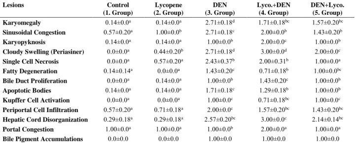
Table 4. The effect of lycopene supplementation on liver histopathological changes Table 4. Likopen ilavesinin karaciğer histopatolojik değişiklikleri üzerine etkisi
Lesions Control (1. Group) Lycopene (2. Group) DEN (3. Group) Lyco.+DEN (4. Group) DEN+Lyco. (5. Group) Karyomegaly 0.14±0.0a 0.14±0.0a 2.71±0.18d 1.71±0.18bc 1.57±0.20bc Sinusoidal Congestion 0.57±0.20a 1.00±0.0b 2.71±0.18c 2.00±0.0b 1.43±0.20b Karyopyknosis 0.14±0.0a 0.14±0.0a 1.00±0.0b 2.00±0.0c 1.00±0.0b
Cloudy Swelling (Periasiner) 0.0±0.0a 0.44±0.20b 2.71±0.18d 3.00±0.0d 2.00±0.0c
Single Cell Necrosis 0.0±0.0a 0.57±0.20a 2.43±0.37b 2.00±0.31b 1.00±0.0a
Fatty Degeneration 0.14±0.14a 0.0±0.0a 1.43±0.20c 0.71±0.18b 1.00±0.0bc
Bile Duct Proliferation 0.0±0.0a 0.14±0.0a 1.00±0.0b 1.43±0.20c 1.00±0.0b
Apoptotic Bodies 0.14±0.0a 0.14±0.0a 1.71±0.18c 1.29±0.18b 1.00±0.0b
Kupffer Cell Activation 0.0±0.0a 0.0±0.0a 1.00±0.0c 0.71±0.18bc 1.00±0.0c
Periportal Cell Infiltration 0.57±0.20a 0.71±0.18a 2.00±0.0c 1.57±0.20bc 1.43±0.20bc
Hepatic Cord Disorganization 0.29±0.18a 0.29±0.18a 2.57±0.20bc 3.00±0.0c 2.14±0.14bc
Portal Congestion 1.00±0.0a 1.00±0.0a 1.00±0.0b 2.00±0.0a 1.00±0.0a
Bile Pigment Accumulations 0.0±0.0 0.0±0.0 1.00±0.0 1.00±0.0 1.00±0.0
The data are expressed in mean±S.E. for seven animals per group. Within rows, means with different letters (a, b, c, and d) are significantly different (P <0.001).
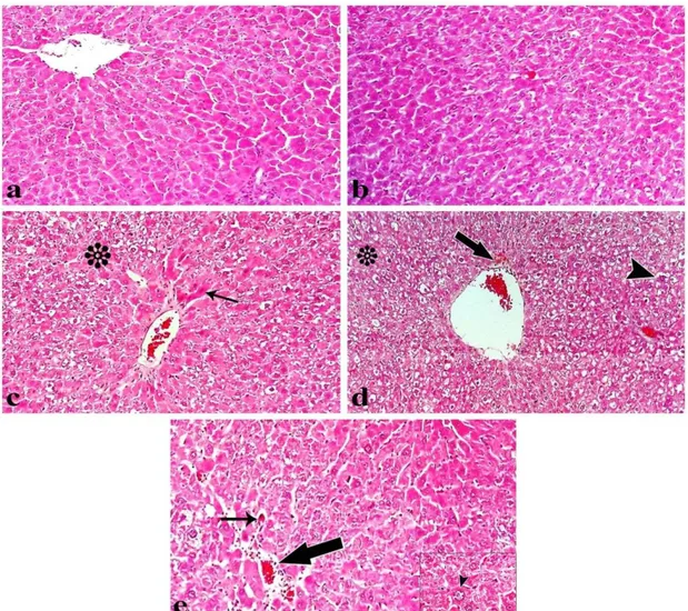
Figure 3. Light microscopy of liver tissue from rats injecting (a) control group, (b) lycopene treated group, (c) DEN treated group, (d) lycopene+DEN treated group, (e) DEN+lycopene treated group. Normal appearanced liver structure (a and b). DEN treated group (c) shows acute cellular swelling (*) and karyopyknosis (arrow) in hepatocytes. Lycopene+DEN treated group (d) shows acute cellular swelling (*), bile pigment accumulations (arrow) and karyomegaly (arrow head). DEN+lycopene treated group (e) shows karyomegaly (arrowhead), apoptosis (thin arrow) and periportal cell infiltration (thick arrow) (H-E staining, magnificationx x40).
Şekil 3. (a) Kontrol, (b) likopen, (c) DEN, (d) likopen+DEN, (e) DEN+likopen uygulanan ratların karaciğerinin ışık mikroskobik görüntüleri. Normal görünümlü karaciğer yapısı (a ve b). DEN uygulanan grupta (c), hepatositlerde bulanık şişkinlik (*) ve karyopiknoz (ok) görülmektedir. Likopen+DEN uygulanan grupta (d), bulanık şişkinlik (*), safra pigmenti birikimleri (ok) ve karyomegali (ok başı) görülmektedir. DEN+likopen uygulanan grupta (e), karyomegali (ok başı), apoptozis (ince ok) ve periportal hücre infiltrasyonu (kalın ok) görülmektedir. (H-E boyama, büyütme x40).
Biochemical Parameters: Table 3 shows the
biochemical parameters such as AST, ALT, ALP, LDH activities and cholesterol levels in the control and experimental groups.
There was no statistical difference between control and lycopene group in this biochemical parameters. Plasma AST, ALT, ALP, LDH activities and cholesterol levels were significantly higher after administration of DEN compared to the control group (p<0.05 and p<0.001). Plasma AST, ALT, ALP, LDH activities and cholesterol levels in lycopene+DEN and DEN+lycopene groups were lower than those determined in the DEN group.
Histopathological Examination: The histological
changes in the tissues were assessed as defined in Figure
3. Control and lycopene groups had a normal histological appearance in liver tissue. As histopathological changes, a statistically significant difference was found in DEN and lycopene+DEN groups compared to the control group (p<0.05).
While severe necrotic changes to the character with karyopyknosis, acute cellular swelling in hepatocytes, karyomegaly, apoptosis, sinusoidal and portal congestion, periportal cell infiltration, intra- and extracellular bile pigment accumulations and Kupffer cell hyperplasia were observed in liver tissues of DEN and lycopene+DEN groups. The severity of degenerative changes in the liver of DEN+lycopene treated group attracted attention decreased to the DEN treated group.
Discussion and Conclusion
DEN is a potent nitrosamine compound that forms tumors in the liver, lung, kidney, gallbladder, esophagus and trachea and nasal cavity. DEN is usually used as a tumor inducer in cancer research. Nitrosamines, primarily DEN, form toxic effects primarily in the blood and especially in the liver. At the same time, other organs, such as the kidneys, where the blood flow is high, are also affected if the toxic effects of nitrosamines are lower than in liver (41).
DEN is an environmental carcinogen that causes free radical formation by affecting membrane lipids. Nitrosamines such as DEN and dimethylnitrosamine have been reported to be effective as they are not much mutagenic themselves, but are metabolized to strong mutagenic intermediates by mammalian enzyme systems. DEN causes an increase in reactive oxygen species (ROS) and resulting in oxidative stress and cell damage (29). High levels of ROS result in many diseases, including cancer in humans, due to causing mitochondrial damage, DNA modification, and LPO (22).
MDA is one of the parameters showing LPO. In many experimental studies, it has been reported that DEN causes LPO, characterized by elevated levels of MDA in tissue damage, especially in the liver (38, 41). In this study, the data indicated that the DEN group had a significantly higher MDA level than the control group in plasma and liver tissue. It was demonstrated that DEN causes the formation of high levels of free radicals that can not be tolerated by the cellular antioxidant defense system. But lycopene treatment reversed the increased MDA contents to the control level, which is suggestive of that lycopene may be successful in quenching the free radicals, inhibiting LPO and protecting membrane lipids from oxidative damage in plasma and liver of rats.
GSH and GST play an important role in the protection of tissues from the deleterious effects of DEN. GSH can act direct interaction of -SH group with ROS, as a cofactor or coenzyme. It helps to protect biological membranes, which are susceptible to peroxidation (39). DEN has been found to conjugate readily with GSH. The observed decrease in the GSH level and GST activity in the present study may reflect an increased demand for GSH by the cell possibly to combat ROS generation during DEN metabolism. Thus, significantly lower GSH level would further aggravate the toxic effects of DEN.
Free radical scavenging enzymes like CAT and SOD protect the biological systems from oxidative stress. SOD, CAT, and GSH-Px act mutually and constitute the enzymic antioxidative defense mechanism against reactive oxygen species (16, 39). With regard to the antioxidant state of the animal in this study, results indicate that the changes in LPO were also accompanied by a concomitant decrease in the activities of the enzymes
involved in the disposal of O·̄2 anions and peroxides,
namely SOD and CAT, as well as the levels of GSH and its related enzymes (GST and GSH-Px) in the DEN administration and this is because of generation of ROS by DEN. From these findings, it appears that the initial changes induced by DEN are due to the formation of LPO and toxicity is mediated through antioxidant enzymes as well as GSH metabolism. The decline in the activities of these enzymes may either be due to the inhibitory effect of DEN on these enzymes or due to consumption during the breakdown of the high level of H2O2, which forms inside
the cells during DEN metabolism (39).
Sahin et al. (37) found in the study of the underlying mechanisms in the development of hepatocarcinogenesis induced by DEN application that the activities of antioxidant enzymes such as CAT, SOD, GSH-Px decreased serum ALT, AST, total bilirubin and MDA levels in liver tissues of rats. Sayed-Ahmed et al. (38) have examined effect of thymoquinone against hepatotoxicity caused by DEN, Bishayee et al. (9) have examined effect of pomegranate emulsion, Bishayee et al. (10) have examined effects of the black grape extract on liver in another study and observed that antioxidants had positive effects on hepatotoxicity of DEN. Some researchers have investigated the effects of ursolic acid on DEN-induced hepatocarcinogenesis and found that LPO increases with the application DEN, and applications of ursolic acid have decreased the LPO with respect to DEN applied group (17). Karahan and Yilmaz (23) have investigated MDA and GSH levels and GSH-Px and CAT activities, which are indicative of LPO in blood, liver, and kidney, following the long-term administration of some nitrosamines in rats. Similarly, many researchers have found significant increases in MDA levels, and significant decreases in GSH levels, and antioxidant enzyme activities after DEN administration (36). Banakar et al. (6) have found that the levels of MDA, a product of LPO, formed as a result of radical formation in liver tissue cells of rats subjected to DEN and phenobarbital have increased and some antioxidant enzyme activities have decreased. They have shown that these enzymes play a role in the defense system and are very important for the defense system.
The statistical result being significant in gene expression can be expressed by genetic modification with RT-PCR. Low expressions indicate that protein formation that is encoded by the gene is suppressed, whereas high expression indicates the formation of more proteins (24). It has been determined by RT-PCR that DEN not only affects the CAT enzyme activity but also expression level. CAT gene expression levels, especially among antioxidant enzymes. These changes are thought to result from oxidative DNA damage due to DEN in the rat liver. Metabolites formed during the metabolism of DEN via
cytochrome P-450 cause oxidative, mutagenic and genotoxic damage to tissues. In a study conducted, the researchers observed a significant decrease in gene expression levels of GST, CAT and GSH-Px enzymes in the DEN-administered group in parallel with the decrease in antioxidant enzyme activities. Decrease in antioxidant enzyme activities and CAT gene expression after DEN administration was explained by an increase in free radical production during DEN metabolism (38). Bingül et al. (8) also found in their study a decrease in CAT activity and expression levels in the rats which were administered DEN. However, in our study, an increase in CAT enzyme expression levels was found in the DEN-applied groups, contrary to the results of the researchers. The difference between CAT activity and expression level may be due to post-translational transformations.
Enzymes are, at the same time, a protein. Proteins formed as a result of gene expression of CAT form the CAT enzyme (24). In this context, there should be a relationship between enzyme activity and gene expressions. CAT enzyme activity in blood and liver tissues decreased significantly compared to the control group and the expression level of the same enzyme was increased in the groups treated with DEN. It is contemplated that the difference between CAT activity and gene expression level may be due to variation in protein and mRNA conversion. In addition, the relationship between genotype and mRNA/protein levels can be influenced by various factors (such as environment, transcriptional regulation). It has been determined that the level of CAT expression decreases with lycopene administration, and this is thought to be due to inhibition of radical-related oxidation of transcriptional factors of lycopene. The difference between CAT activity and expression level may be due to the oxidation of transcriptional factors responsible for the initiation mechanism of the CAT enzyme in the transcription process. Furthermore, the change detected in the expression of the enzyme may be related to the half-lives of the mRNAs. It is also known that increased oxidative stress may have triggered mRNA's instability.
There are researches available reporting that lycopene has significantly increased antioxidant enzyme activities (20, 34). Giovannucci (18) reported that lipoproteins of lycopene can protect nucleic acids and proteins from oxidative stress that could lead to cancer. Astorg et al. (4) have observed that lycopene decreased liver preneoplastic foci in DEN-treated rats. Abdallah et al. (1) have demonstrated that in DEN-induced experimental hepatocarcinogenesis, lycopene inhibits the development of hepatocellular carcinoma. In the study, the effect of lycopene on oxidative stress and DNA damage in the blood and liver tissues of rats was investigated. It was determined that antioxidant parameters were closer to the
control group when lycopene, which has an antioxidant effect on rats, is given with DEN. This can be explained by the fact that lycopene has the ability to inhibit oxidative stress and free radicals by limiting the production of free radicals. Lycopene is known to exert a protective effect by inhibiting proliferation by interrupting intercellular communication by affecting cell surface receptors, especially in cancer cells. There are studies showing that pretreatment with lycopene is effective (31, 33). There are also studies showing that lycopene is effective when administered in combination with toxic substances (12, 42). In our study, it was observed that lycopene against DEN-induced hepatotoxicity was more effective in the group that was started lycopene administration together with DEN.
The altered activities of some liver-specific enzymes reflect the effects of cell proliferation and their metabolic transformations in tumor cells are quite different from normal cells (21). Many researchers have shown that DEN administration causes an increase in some liver-specific enzyme activities (14, 21). In this study, DEN administration to rats lead to a marked elevation in the levels of plasma AST, ALT, ALP, LDH activities and cholesterol level which is indicative of hepatocellular damage. This might be due to the possible release of these enzymes from the cytoplasm, into the blood circulation rapidly after rupture of the plasma membrane and cellular damage.
In conclusion, DEN-induced hepatic damage includes oxidative stress triggered by ROS generation. Toxicity induced by DEN ameliorates by the oral administration of lycopene which is indicated by a decrease in MDA levels with a concomitant increase in non-enzymatic and enzymatic antioxidants levels. In conclusion, lycopene showed a comparable antioxidant property against DEN-induced LPO and its related enzymes in rats. These results suggest that the protective effect of lycopene is possibly due to its antioxidants and ROS scavenging properties against DEN-induced hepatotoxicity.
Acknowledgment
I would like to thank FUBAP (VF.14.19) for financially supporting this study and this work was produced from a doctoral thesis.
References
1. Abdallah IZ, Khattab HA (2004): Protective role of
lycopene against diethylnitrosamine induced experimental hepatocarcinogenesis. Egypt J Hosp Med, 16, 1-13.
2. Aebi H (1974): Catalase. 673-8. In: Bergmeyer HU, editor. Methods of enzymatic analysis. 2nd English ed. Weinheim:
Verlag Chemie.
3. Akyuz F, Inal M, Baycu C, et al, (2001): Changes in
tissues of the rats that were given diethylnitrosamine. Ann
Med Sci, 10(2), 50-54.
4. Astorg P, Gradelet S, Bergès R, et al. (1997): Dietary lycopene decreases the initiation of liver preneoplastic foci by diethylnitrosamine in the rat. Nutr. Cancer. 29(1), 60-68. 5. Atessahin A, Yilmaz S, Karahan I, et al. (2005): Effects
of lycopene against cisplatin-induced nephrotoxicity and oxidative stress in rats. Toxicology, 212(2-3), 116-123.
6. Banakar MC, Paramasivan SK, Chattopadhyay MB, et al. (2004): 1,25-Dihydroxyvitamin D3 prevents DNA
damage and restores antioxidant enzymes in rat hepatocarcinogenesis induced by diethylnitrosamine and promoted by phenobarbital. World J Gastroenterol, 10(9),
1268-1275.
7. Beutler E (1984): Red cell metabolism. 160. A manual of biochemical methods. Grune and Starton (Editors) 2nd
Edition, New York.
8. Bingul I, Basaran-Kucukgergin C, Aydin AF, et al. (2016): Blueberry treatment attenuated cirrhotic and
preneoplastic lesions and oxidative stress in the liver of diethylnitrosamine-treated rats. Int J Immunopathol Pharm,
29(3), 426-437.
9. Bishayee A, Bhatia D, Thoppil RJ, et al. (2011):
Pomegranate-mediated chemoprevention of experimental hepatocarcinogenesis involves Nrf2-regulated antioxidant mechanisms. Carcinogenesis, 32(6), 888-896.
10. Bishayee A, Mbimba T, Thoppil RJ, et al. (2011):
Anthocyanin-rich black currant (Ribes nigrum L.) extract affords chemoprevention against diethylnitrosamine-induced hepatocellular carcinogenesis in rats. J Nutr
Biochem, 22(11), 1035-1046.
11. Dalling JW, Pachen DMFA, Lousberg AHPJ, et al. (1998:) Volatile N-nitrosamines in gastric juice of patients
with various conditions of the gastrointestinal tract determined by gas chromatography–mass spectrometry and related to intragastric pH and nitrate and nitrite levels.
Cancer Lett, 124(2), 119-125.
12. El-Gerbed MS (2014): Protective effect of lycopene on
deltamethrin-induced histological and ultrastructural changes in kidney tissue of rats. Toxicol and Health, 30(2),
160-173
13. Ellman GL, Courtney KD, Andres V, et al. (1961): A new
and rapid colorimetric determination of acetylcholinesterase activity. Biochem Pharmacol, 7, 88-95.
14. El-Mesallamy HO, Metwally NS, Soliman MS, et al. (2011): The chemopreventive effect of Ginkgo biloba and
Silybum marianum extracts on hepatocarcinogenesis in rats. Cancer Cell Int, 11(1), 38.
15. El-Shahat M, El-Abd S, Alkafafy M, et al. (2012):
Potential chemoprevention of diethylnitrosamine-induced hepatocarcinogenesis in rats: myrrh (Commiphora molmol) vs. turmeric (Curcuma longa). Acta Histochem, 114(5),
421-428.
16. Fridovich I (1986): Superoxide dismutases. Adv Enzymol, 58, 61-97.
17. Gayathri R, Priya DKD, Gunassekaran GR, et al. (2009): Ursolic acid attenuates oxidative stress-mediated
hepatocellular carcinoma induction by diethylnitrosamine in male Wistar rats. Asian Pacific J Cancer Prev, 10,
933-938.
18. Giovannucci E (1999): Tomatoes, tomato-based products,
lycopene, and cancer: review of the epidemiologic literature. J National Cancer Ins, 91(4), 317-331.
19. Habig WH, Pabst MJ, Jakoby WB (1974): Glutathione
S-transferases. The first enzymatic step in mercapturic acid formation. J Biol Chem, 249, 130-139.
20. Ip BC, Hu KQ, Liu C, et al. (2013): Lycopene metabolite,
apo-10′-lycopenoic acid, ınhibits diethylnitrosamine-ınitiated, high fat diet–promoted hepatic ınflammation and tumorigenesis in mice. Cancer Pre Res, 6(12), 1304-1316.
21. Jahan MS, Vani G, Shyamaladevi CS (2007): Effect of
Solanum trilobatum on hepatic drug metabolising enzymes during diethylnitrosamine-induced hepatocarcinogenesis promoted by Phenobarbital in rat. Hepatol Res, 37(1),
35-49.
22. Kang JS, Wanibuchi H, Morimura K (2007): Role of
CYP2E1 in Diethylnitrosamine-induced hepatocarcinogenesis in vivo. Cancer Res, 67(23), 11141-11146.
23. Karahan I, Yılmaz S (2006): Effects of prolonged low
amounts some nitrosoamines administrations on oxidative stress in blood, liver and kidney of rats. Firat University
Veterinary Journal of Health Sciences, 20(1), 73-78. 24. Kubista M, Andrade JM, Bangtsson M, et al. (2006): The
real-time polymerase chain reaction. Mol Aspects Med,
27(2-3), 95-125.
25. Kumar P, Kumar A (2009): Effect of lycopene and
epigallocatechin-3-gallate against 3-nitropropionic acid induced cognitive dysfunction and glutathione depletion in rat: a novel nitric oxide mechanism. Food and Chem
Toxicol, 47(10), 2522-2530.
26. Lijinsky W (1992): Chemistry and Biology of N-nitroso
Compounds. Cambridge, UK: Cambridge Univ. Pres, 1992.
27. Lowry OH, Rosenbrough NJ, Farr A, et al. (1951):
Protein measurement with the folin-phenol reagent. J Biol
Chem, 193, 265-257.
28. Luna LG (1968): Manuel of histologic staining methods of
Armed Forces Institute of Pathology. 1-36, McGraw-Hill
Book Co, New York.
29. Man S, Fan W, Gao W, et al. (2014): Anti-fibrosis and
anti-cirrhosis effects of Rhizoma paridis saponins on diethylnitrosamine induced rats. J Ethnopharmacol, 151(1),
407-412.
30. Matos HR, Capelozzi VL, Gomes OF, et al. (2001):
Lycopene inhibits DNA damage and liver necrosis in rats treated with ferric nitrilotriacetate. Arch Biochem Biophys,
396, 171-177.
31. Ozkan E, Akyuz C, Dulundu E, et al. (2012): Protective
effects of lycopene on cerulein-induced experimental acute pancreatitis in rats. J Surg Res, 176(1), 232-238.
32. Placer ZA, Cushman L, Johnson BC (1966): Estimation
of products of lipid peroxidation in biological fluids. Anal Biochem, 16, 359-364.
33. Qu M, Nan X, Gao Z, et al. (2013): Protective effects of
lycopene against methylmercury-induced neurotoxicity in cultured rat cerebellar granule neurons. Brain Res, 1540,
92-102.
34. Rao AV, Agarwal S (1999): Role of lycopene as
antioxidant carotenoid in the prevention of chronic diseases. Nutr Res, 19, 199-203.
35. Rehman A, Bourne LC, Halliwell B, et al. (1999): Tomato
consumption modulates oxidative DNA damage in humans.
Biochem Biophys Res Commun, 262, 828-831.
36. Rezaie A, Fazlara A, Karamolah MH, et al. (2013):
Effects of Echinacea purpurea on hepatic and renal toxicity induced by diethylnitrosamine in rats. Jundishapur J Nat
Pharm Prod, 8(2), 60-64.
37. Sahin K, Orhan C, Tuzcu M, et al. (2014): Orally
administered lycopene attenuates diethylnitrosamine-induced hepatocarcinogenesis in rats by modulating Nrf-2/HO-1 and Akt/mTOR pathways. Nutrition and Cancer,
66(4), 590-598.
38. Sayed-Ahmed MM, Aleisa AM, Al-Rejaie S, et al. (2010):
Thymoquinone attenuates diethylnitrosamine induction of hepatic carcinogenesis through antioxidant signaling. Oxid
Med Cell Longev, 3, 254-261.
39. Sies H (1991): Oxidative Stress: Oxidants and Antioxidants. Academic Press, San Diego, California.
40. Sun Y, Oberly LW, Ying LA (1988): Simple method for
clinical assay of superoxide dismutase. Clin Chem, 34,
497-500.
41. Yamada K, Yamamiya I, Utsumi H (2006): In vivo
detection of free radicals induced by diethylnitrosamine in rat liver tissue. Free Radic Biol Med, 40, 2040-2046.
42. Yilmaz S, Atessahin A, Sahna E, et al. (2006): Protective
effect of lycopene on adriamycin-induced cardiotoxicity and nephrotoxicity. Toxicology, 218(2), 164-171.
Geliş tarihi : 01.12.2017 / Kabul tarihi : 19.03.2018
Address for correspondence:
Emre KAYA, PhD, DVM
Firat University, Faculty of Veterinary Medicine, Department of Biochemistry, Elazig, Turkey e-mail: [email protected]