Q - Z
,ZOZ
'VS'S
MOLECULAR ANALYSES OF HEMATOLOGICAL MALIGNANCIES
^'WE:8IS SUBMITTED TO
THE DEPARTMENT OF MOLECULAR BIOLCGY AND GENETICS
AND THE INSTITUTE OF ENGINEERING AND SCIENCE OF
BILKENT UNIVERSITY
IN PARTIAL FULFILLMENT OF THE REQUIREMENTS FOR
MOLECULAR ANALYSES OF HEMATOLOGICAL MALIGNANCIES
A THESIS SUBMITTED TO
THE DEPARTM ENT OF M OLECULAR BIOLOGY AND GENETICS AND THE INSTITUTE OF ENGINEERING AND SCIENCE OF
BiLKENT UNIVERSITY
IN PARTIAL FULFILLMENT OF THE REQUIREM ENTS FOR THE DEGREE OF M ASTER OF SCIENCE
BY
Z. BUKET YILMAZ AUGUST 1998
о г
л о і
I certify that I have read this thesis and that in my opinion it is fully adequate, in scope and quality, as a dissertation for the degree o f M aster o f Science.
P ro f Dr. Ay Ogii§
I certify that I have read this thesis and that in my opinion it is fiilly adequate, in scope and quality, as a dissertation for the degree o f M aster o f Science.
Assoc. P ro f Dr. Tayfiin Özçelik
I certify that I have read this thesis and that in my opinion it is fully adequate, in scope and quality, as a dissertation for the degree o f M aster o f Science.
Asst. P ro f Dr. Işık G. Yuluğ
Approved for the Institute o f Engineering and Science.
P ro f Dr. Mehmet Baray D irector o f Institute o f Engineering and Science
ABSTRACT
Molecular Analyses o f Hematological Malignancies
Z. Buket YILMAZ
M.Sc. in Molecular Biology and Genetics Supervisor: Assoc. P ro f Dr. Tayfim Oz9elik
August 1998, 140 pages
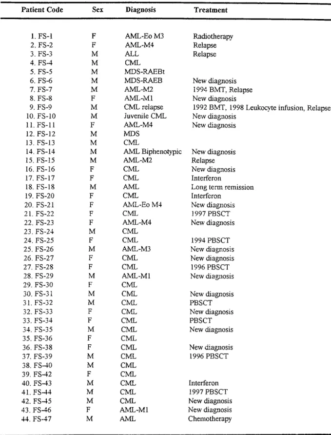
Normal hematopoiesis is known to be disrupted in hematological malignancies. As research advances, many new treatment techniques are introduced, among which allogeneic transplantation is the most effective. In patients who undergo such treatment, different hematopoietic chimeric states may result which can be detected by indirect analyses using DNA polymorphisms. MRD (minimal residual disease) describes leukemia cells present at a level below that is detectable by conventional means. The detection o f MRD has gained special significance in transplantation patients. The detection o f fusion transcripts that are generated by chromosomal rearrangements in various hematological malignancies is the direct and the most powerful approach for the detection o f MRD. It not only has a prognostic significance but also has a diagnostic importance for patients who have not taken any treatment. The aim o f this thesis is the development o f PCR-based tests for the diagnosis and monitoring o f patients with different hematological malignancies. Therefore the chimerism status o f 22 recipient-donor pairs have been evaluated by PCR amplification o f STR and VNTR polymorphisms followed by polyacrylamide gel electrophoresis and silver staining. In addition, 44 patients with acute and chronic leukemias have been analyzed for the detection o f fusion transcripts generated by t (9;22) (q 3 4 ;q ll), t (8;21) (q22;q22), t (15;17) (q22;q21), inv 16 (pl3;q22), t (4;11) (q21;q23), and t (1;19) (q23;p23) chromosomal rearrangements with RT-PCR.
ÖZET
Hematolojik Kanserlerde Moleküler Analiz
Z. Buket YILMAZ
Yüksek Lisans Tezi, Moleküler Biyoloji ve Genetik Bölümü Tez Damşmam: Doç. Dr. Tayfun Özçelik
Ağustos 1998, 140 sayfa
Hematolojik kanserlerde normal hematopoezin zarara uğradığı bilinmektedir. Araştırmalardaki gelişmelere paralel olarak aralannda allojenik kemik iliği transplantasyonunun en etkili olduğu, birçok tedavi tekniği uygulamaya girmiştir. Bu tür tedavi alan hastalarda, DNA polimorfizmlerinin kullamidığı dolaylı analiz yöntemleriyle tespit edilebilecek değişik kimerizm durumları görülmektedir. MRD (minimal residüel hastalık), standart analiz yöntemleriyle tespit edilebilecek seviyenin altındaki lösemi hücrelerinin varlığım ifade etmektedir. MRD analizi, transplantasyon hastalarında özel bir önem kazanmıştır. Değişik kan kanserlerinde kromozomlar arası parça değişimleriyle ortaya çıkan füzyon transkriptlerinin analizi, MRD tespitindeki doğrudan ve en etkili yöntemdir. Bu yöntem, prognostik özelliğinin yam sıra, herhangi bir tedavi almamış hastalar için diagnostik açıdan da önem taşımaktadır.
Bu tezin amacı, hematolojik kanser hastalannda diagnostik ve prognostik amaçlı olarak PCR (polimeraz zincir reaksiyonu) yöntemine dayalı testlerin geliştirilmesidir. Bu amaca yönelik olarak, STR ve VNTR polimorfizmlerinin amplifikasyonu, bunu takiben poliakrilamid jel elektroforezi ve gümüş boyama yöntemleriyle 22 ahcı-verici çiftindeki kimerizm durumu değerlendirildi. Buna ek olarak, RT-PCR yöntemiyle t (9;22) (q 3 4 ;q ll), t (8;21) (q22;q22), t (15;17) (q22;q21), inv 16 (pl3;q22), t (4;11) (q21;q23) ve t (1;19) (q23;p23) kromözomlar arası parça değişimleri sonucu ortaya çıkan füzyon transkriptlerinin analizi için, 44 akut ve kronik lösemi hastası incelendi.
ACKNOWLEDGEMENTS
First, and foremost I would like to express my gratitude to
Assoa Prof. Dr. Tayfun
özçelik
for Ms support, friendly and kind-hearted policy, and for teaching that one is alone in life to overcome obstacles.Special thanks go to
Prof. Dr. Mehmet öztürk
for his invaluable intellectual contributions and advice whenever one is in need o f I also would like to thankAsst
Prof. Dr. Işık G. Yulug
for her support, friendly approach and advice, andProf. Dr.
Ay Öğüş
for her contributions.I would like to thank
Prof. Dr. Meral Beksaç
andDr. Celalettin Üstün
for their contributions in the. generation o f this work.Özlem Karaca
is the one who deserves the most special thanks for just being herselfand for being with me during the past thirteen years. I also thank my dear friend
Dr.
Tutku Soyer
for enabling the unconditioned reliance and for showing that truefriendship is forever. I thank
Füsun Türesin, Y. Senem Ceylan,
andSeval Pehlivan
for being real friends for the last six years and for making me feel very special.
I w ould like to thank
Burçak Vural
for her love, care and for being my elder sister;Alper Romano
for all those scientific and other (!) discussions and deepunderstanding;
Birsen Cevher
for her valuable friendship;Hilal Özdağ
andLütfîye
Mesci
for sharing their experience;Cemaliye Akyerli
andArzu Atalay
for making mefeel in a family;
Tuba Dinger
for making our house “Л о те” full o f joy and happiness and for sharing my problems patiently;Tolga Çağatay
for his thoughtful, kind behaviours and support;Necati Fındıklı, Çağla Eroğlu, Korkut Vata,
andEmre
Öktem
for being my sweet friends; andDr. Gökçe Törüner
for his always available,special thanks to
Emre Sayan
for making all impossible things possible for me, andto
Berna Özgelik
for her great support especially during the time o f writing the thesis.I w ould also like to thank
Dr. Gürol Tunçman
for all the joy we shared; andFüsun
Elvan
for providing my instant requests.I would like to express my very special thanks to dear
Reşat Ünal,
who made many things here worthwhile. Thanks for everything but particularly for being yourself, for never disappointing me and for your deep belief, love, care, and support I always felt.I would also like to thank my other friends, and my family, whom I could not mention here one by one. I love you all.
Finally I would like to express my deepest gratitude to my precious mother
Halime
Çapkı,
and my dear fatherH. Orhan Yılmaz
for their invaluable, unconditionedsupport, understanding, and endless love. You are the center o f my life.
Thank you all from the heart for making everything w orthw hile...
TABLE OF CONTENTS
T IT L E S IG N A T U R E PA G E A B S T R A C T 111 O Z E T IV A C K N O W L E D G E M E N T S V T A B L E O F C O N T EN T S Vll L IS T O F TA B LES XU L IS T O F FIG U R E S Xlll A B B R E V L 4T IO N S XV I. IN T R O D U C T IO N I. 1. H E M A T O P O IE T IC SY STEM AND H E M A T O L O G IC A L M A L IG N A N C IE S1.1 Hematopoiesis : Genesis and Differentiation o f Blood Cells 1.2 Description and Function o f Bone M arrow
1.3 Hematological Malignancies
1
1 4
1.3.1 Leukemias
1.3.1.1 Acute Myeloblastic Leukemia (AML) 1.3.1.2 Acute Lymphoblastic Leukemia (ALL) 1.3.1.3 Chronic Myelogenous Leukemia (CML) 1.3.2 Lymphomas
1.3.3 Myelodysplastic Syndromes (M DS)
1.4 Chromosomal Rearrangements in Hematological Malignancies 1 .4 .1 1 (9; 22) ( q 3 4 ;q ll) 1 .4 .2 t( 8 ;2 1 ) ( q 2 2 ; q22) 1.4.3 t (15; 17) (q22; q21) 1.4.4 inv 16 (p l3 ; q22) 1.4.5 t (4; H )(q 2 1 ;q 2 3 ) 1.4.6 t ( l ; 19) (q23;p23)
12
15 16 17 19 20 22 25 28 31 33 35 8I. 2. MOLECULAR DIAGNOSIS OF HEMATOLOGICAL MALIGNANCIES
2.1 Cytogenetics
2.2 Immunological Methods 2.3 Fusion Transcript Detection
37 39 39
I. 3. TREATMENT OF HEMATOLOGICAL MALIGNANCIES
3.1 Chemotherapy and Biologic Agents 3.2 Transplantation o f Hematopoietic Cells
41
41
44
I. 4. MONITORING OF MINIMAL RESIDUAL DISEASE IN
HEMATOLOGICAL MALIGNANCIES
4.1 The Concept o f Chimerism 4.2 Minimal Residual Disease
4.3 Direct M onitoring o f MRD Using Chromosomal Rearrangements 4.4 Indirect M onitoring o f MRD Using D NA Polymorphisms
50 50 51 52
50
I. 5. AIM
56H. MATERIALS AND METHODS
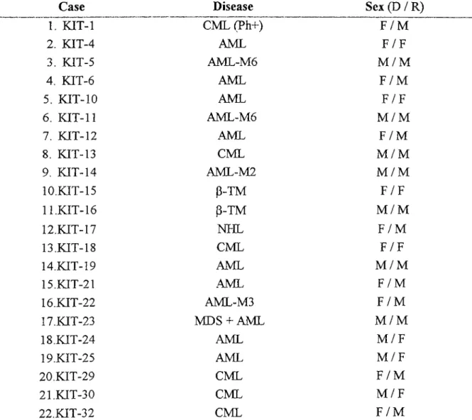
571. MATERIALS
1.1 Patient Samples 1.2 Chemicals 1.3 Equipment 1.4 Others 1.5 Oligonucleotides 1.6 Solutions1.6.1 General Stock Solutions 1.6.2 Buffers 1.6.3 D NA Isolation Solutions 57 57 61 62 63 65 67 67 67 68
1.6.4 Agarose Gel Electrophoresis Solutions 1.6.5 Polyacrylamide Gel Electrophoresis Solutions 1.6.6 Silver Staining Solutions
1.6.7 RNA Isolation Solutions
70 71 72 73
2. METHODS
2.1 D N A Isolation from Whole Blood 2.2 D N A Isolation from Buccal Wash 2.3 D NA Isolation from Skin Biopsy 2.4 Polymerase Chain Reaction (PCR) 2.5 Agarose Gel Electrophoresis
2.5.1 M etaphor Agarose Gel Electrophoresis 2.5.2 Formaldehyde-Agarose Gel Electrophoresis 2.6 Polyacrylamide Gel Electrophoresis (PAGE)
2.7 Silver Staining
2.8 White Blood Cell Separation by Ficoll Centrifugation 2.9 RNA Isolation Using TRI Reagent
2.10 First Strand cDNA Synthesis
2.11 Reverse-Transcriptase Polymerase Chain Reaction (RT-PCR)
74 74 75 76 76 78 79 80 81 82 83 83 86 88
m . RESULTS
921. Evaluation of Chimerism with DNA Polymorphisms After Allogeneic
Transplantation
921.1 Visualization o f DNA Samples 96
1.2 Polymerase Chain Reaction (PCR) 96
1.3 Polyacrylamide Gel Electrophoresis 98
2. Detection of Fusion Transcripts by RT-PCR Analysis
2.1 Visualization o f RNA Samples 2.2 cDNA Synthesis
2.3 Reverse-Transcriptase Polymerase Chain Reaction (RT-PCR)
2.3.1
1 (9:22) (q 3 4 ;q ll) 2.3.2 t (8;21) (q22;q22)2.3.3
t(1 5 ;1 7 ) (q22;q21) 2 .3 .4 t( 4 ;ll) ( q 2 1 ;q 2 3 )2.3.5
t( l ;1 9 ) (q23;p23)2.3.6
inv 16(pl3;q22) 102 106 107 108 109112
114 116 118120
IV. DISCUSSION
122V. REFERENCES
131LIST OF TABLES
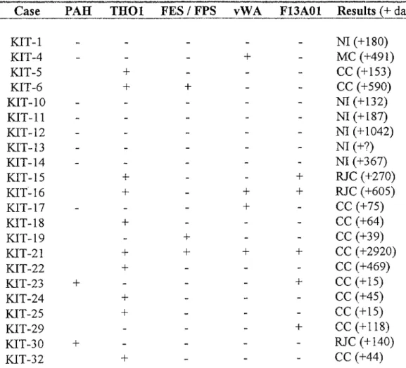
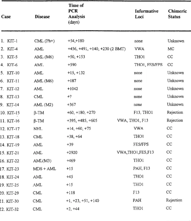
Table 1
Hematopoietic Growth Factors and Some o f Their Characteristics 6Table 2
FAB Classification o f Acute Myeloblastic (Myelocytic) Leukemias 13Table 3
Chromosomal Abnormalities in AML and Their Significance 14Table 4
Classification o f Acute Lymphoblastic Leukemia (ALL) 15
Table 5
A Classification o f CML 17Table
6 FAB Classification and Leukemic Transformation in MDS 20Table
7 Chromosomal Rearrangements Analyzed in the Scope o f This Study 21Table 8
Locus Specific Information o f the STR Loci Studied55
Table 9
Patient Samples for Evaluation o f Chimerism Status After AllogeneicTransplantation 59
Table 10
Patient Samples for Detection o f Fusion Transcripts 60Table 11
List o f the Chemicals Used in This Study 61Table 12
Primers Used in This Study 66Table 13
The Components o f PCR Reaction M ixture 77Table 14
Agents Used to Preserve RNA Integrity During Extraction 84Table 15
Panel for RT-PCR Analysis 89Table 16
The Components o f RT-PCR Reaction M ixture 90Table 17
Analyzed Polymorphic Loci for Chimerism Evaluation 94Table 18
STR and PAH VNTR Analyses for Chimerism Evaluation 95Table 19
RT-PCR Analysis for Detection o f Fusion Transcripts in Acute LeukemiaPatients 104
Table 20
RT-PCR Analysis for Detection o f Fusion Transcripts in ChronicMyelogenous Leukemia (CML) Patients 105
Figure 1
Differentiation o f Hematopoietic Cells 2Figure 2
Clonality and Transformation in Acute Leukemia 8Figures
Fusion in t (9;22) (q34;q 11) 24Figure 4
t (8;21) (q22;q22) Rearrangement 27Figures
t (15;17) (q22;q21)Rearrangem ent 30Figu re
6 inv 16 (p 13 ;q22) Rearrangement 31Figure 7
t (16;16) (p 13 ;q22) Rearrangement 32Figure 8
t (4;11) (q21;q23) Rearrangement 34Figure 9
t (1; 19) (q23;p23) Rearrangement 36Figure 10
Bone M arrow Transplantation Centers in Turkey 49Figure 11
Patient DNA Samples 96Figure 12
PCR Amplification o f TH O l and FES / FPS Loci 97Figure 13
PCR Amplification o f PAH VNTR Locus for KIT-2 and KIT-23 97Figure 14
M etaPhor Agarose Gel Electrophoresis Showing CompleteChimerism at vWA Locus for KIT -17 98
Figure 15
Rejection Based on vW A and TH O l Genotype in K IT -16 99Figure 16
Complete Chimerism Based on FES / FPS Genotype in K IT -19 100Figure 17
Complete Chimerism Based on TH O l Genotype in KIT-25 100Figure 18
Non-Informative Results at vW A ,TH 01, F13A 0F and FES / FPSLoci in KIT-21 101
Figure 19
Patient RNA Samples 106Figure 20
Amplification o f cDNA Samples 107Figure 21
Schematic Representation o f the Amplified Region for t (9;22) 109Figure 22
RT-PCR Amplification for t (9;22) (q34;q 11) 110Figure 23
Schematic Representation o f the Amplified Region for t (8 ;21) 112Figure 24
RT-PCR Amplification for t (8;21) (q22;q22) 113Figure 25
Schematic Representation o f the Amplified Region for t (15; 17) 114Figure 26
RT-PCR Amplification for t (15;17) (q22;q21) 115Figure 27
Schematic Representation o f the Amplified Region for t (4;11) 116Figure 28
RT-PCR Amplification for t (4;11) (q21;q23) 117Figure 29
Schematic Representation o f the Amplified Region for t (1; 19) 118Figure 30
RT-PCR Amplification for t (1;19) (q21;p23) 119Figure 31
Schematic Representation o f the Amplified Region for inv 16 120Figure 32
RT-PCR Amplification for inv 16 (pl3;q22) 121ABBREVIATIONS
ALL Acute lymphocytic leukemia AML Acute myelogenous leukemia APL Acute promyelocytic leukemia APS Ammonium persulphate ATRA : All-trans retinoic acid BFU Burst forming unit
BM Bone marrow
BMT Bone marrow tranplantation
bp Base pair
ß-TM ; Beta-thalassemia
cc
Complete chimeric CPU Colony forming unitCLL Chronic lymphocytic leukemia CML Chronic myelogenous leukemia CNS Central nervous system
CR Complete remission CSF Cerebrospinal fluid DEPC : Diethylpyrocarbonate DFS Disease fi"ee survival
EDTA : Ethylenediaminetetraacetate FAB French-American-British
FISH Fluorescence
in situ
hybridization GVHD: Graft versus-host diseaseGVL Graft versus-leukemia HLA Human leukocyte antigen IFN Interferon
Ig Immunoglobulin IL Interleukin kb iCilobase kD Kilodalton MC Mixed chimeric MDS Myelodysplastic syndrome
MIC Morphological immunological and cytogenetic MOPS ; 3-(N-morpholino) propanesulfonic acid
M M ) Minimal residual disease NHL N on-Hodgkin’s lymphoma p Short arm o f the chromosome PAGE : Polyacrylamide gel electrophoresis PAH Phenylalanine hydroxylase gene
PBSCT; Peripheral blood stem cell transplantation PCR Polymerase chain reaction
Ph Philadelphia
q Long arm o f the chromosome
R .E .A L : Revised European-American Classification o f Lymphoid Neoplasms RA Retinoic acid
RAEB : Refractory anemia with excess blasts
R A E B -t; Refractory anemia with excess blasts in transformation RAR Retinoic acid receptor
RFLP : Restriction length polymorphism
RT-PCR; Reverse transcriptase - polymerase chain reaction SDS Sodium dodecyl sulphate
STR Short tandem repeat t Translocation
TEMED: N,N,N,N-tetram ethyl-l,2 diaminoethane VNTR : Variable number o f tandem repeats
I. INTRODUCTION
I. 1. HEMATOPOIETIC SYSTEM AND HEMATOLOGICAL
MALIGNANCIES
1.1 Hematopoiesis : Genesis and Differentiation of Blood Cells
Hematopoiesis
is the formation o f blood cells (Fauciet a i,
1998). The bone marrow, lymph nodes, and spleen are all involved in hematopoiesis. These organs and tissues have traditionally been divided intomyeloid tissue
including the bone marrow and the cells derived from it- erythrocytes, platelets, granulocytes, and monocytes, andlymphoid tissue
consisting o f thymus, lymph nodes, and spleen (Cotranet al.,
1989).Maximow, in 1924, postulated that blood cells were derived from a single class o f progenitors. In 1938, Downey added the concept o f hierarchies o f pluripotent cells. The demonstration that single cells were capable o f establishing nodules o f hematopoietic grow th in the spleen o f irradiated mice and that such colonies displayed
midtilineage
orpluripotent
differentiation (erythroid, myeloid, megakaryocytic) came in 1961 by Till and McCulloch. These landmark experiments established that astem
cell
existed for hematopoiesis. A stem cell is a cell with the ability o f self renewal and the production o f progeny destined to differentiate (Leeet al.,
1993).The common pluripotent hematopoietic stem cell gives rise to lymphoid stem cells and the trilineage myeloid stem cells respectively (Figure 1).
ui·· ' ‘ io
The lymphoid stem cell is the origin o f precursors o f T cells (pro-T cells) and B cells (pro-B cells). The former differentiates into mature T cells under the inductive influence o f the thymus and the latter to mature B cells under the influence o f bursa- equivalent tissue. An important difference between lymphoid and myeloid differentiation is that, there are no distinctive, morphologically recognizable stages in lymphoid differentiation. From the multipotent myeloid stem cell, three types o f
committed stem cells
arise which differentiate along the erythroid / megakaryocytic, eosinophilic, and granulocyte / macrophage pathways (Cotranet al.,
1989).Pluznik and Sachs and Bradley and M etcalf reported that hematopoietic colonies could be grow n in semisolid medium (Lee
et al.,
1993). Thus, the committed stem cells have been called the colony - forming units (CFU). As it is indicated in Fig. 1, granulocytes and macrophages have a common precursor, CFU-G / M, which gives rise to colonies having a mixture o f neutrophils and macrophages. In the erythroid pathway, tw o distinct committed stem cells can be recognized. Based on the morphology o f the colonies, BFU-E (burst-forming unit-eiythroid) is the more primitive one. The later stage is CFU-E. From all these different committed stem cells, intermediate stages are derived, and finally the morphologically recognizable precursors o f the differentiated cell lines are formed. These are, proerythroblasts, myeloblasts, megakaryoblasts, monoblasts, and eosinophiloblasts which in turn give rise to mature progeny . The mature blood elements have a finite life span and their numbers must be constantly replenished. Thus, self renewal is an important propertyo f stem cells. The pluripotent stem cells have the greatest capacity o f self renewal, but normally most o f them are not in cycle. Self renewal ability declines as commitment proceeds, but a greater fraction o f the stem cells are found in cycle (Cotran
et al.,
1989).
During the development o f systems for the grow th o f hematopoietic colonies in vitro, it was understood that, in the absence o f either serum or conditioned medium, hematopoietic colonies did not grow. It was then demonstrated that glycoproteins termed
colony-stimulating factors
(CSF) were necessary for granulocyte-macrophage colony growth (Leeet al.,
1993). Some hematopoietic growth factors and their characteristics are summarized in Table 1.1.2 Description and Function of Bone Marrow
The bone m arrow is a reservoir o f stem cells which also provides a unique microenvironment for the proliferation and the differentiation o f precursor cells. In addition, it regulates the release o f fully differentiated cells into the circulation. Both structural (stromal) and humoral components o f the bone marrow are involved in support o f hematopoiesis (Cotran
et al.,
1989).The m arrow is a highly organized and complex organ. It is a gelatinous material interspersed within trabecular bone, which is supported by the cylindric medullary bone. A complex network o f vessels organized into repeating units supplies the m arrow cavity. Venous sinusoids, which are surrounded by hematopoietic tissue in which blood cell formation takes place, is formed by the combination o f small vessels that reenter the marrow cavity. The production o f the various marrow cells takes place by a ‘colonial’ proliferation o f islands o f erythroid or granulocytic cells, rather than by random distribution. Sinusoids are formed before myeloid hematopoiesis begins in the fetus. The venous sinusoids which eventually flow into a central vein, thus seem to be essential for hematopoiesis. The stromal elements o f the marrow have been proposed as major factors in. the regulation o f hematopoiesis mainly through the mechanism o f various cytokines that they export (Lee
et al.,
1993).Table 1 : Hematopoietic Growth Factors and Some of Their Characteristics
Factor*
Cells Stimulated
Production Sources
CSF-1 (M -CSF)
Monocytes Endothelial cells,
monocytes, fibroblasts GM-CSF All granulocytes, megakaryocytes,
erythrocytes, stem cells, leukemic blasts
T cells, endothelial cells, fibroblasts
G-CSF Granulocytes, macrophages, endothelial cells, fibroblasts, leukemic blasts
Endothelial cells, placenta, monoc34es IL-3 Granulocytes, erythroid cells, multipotential
progenitors, leukemic blasts
T cells
IL-4 B, T cells T cells
E.-5 B cells, CFU-Eo T cells
IL-6 B, T cells, CFU-GEMM, CFU-GM, BFU-E, macrophages, neural cells, hepatocytes
Fibroblasts leukocytes, epithelial cells
IL-7 B cells Leukocytes
IL-8 T cells, neutrophils Leukocytes
E.-9 BFU-E, CFU-GEMM Lymphocytes
IL-11 B, T cells, CFU-GEMM, macrophages Macrophages
Erythropoietin CFU-E, BFU-E Kidney, liver
*GM, granulocyte-macrophage; IL, interleukin; GEMM, granulocytes, eiythroid cells, macrophages, megakaryocytes (L eei?/a/., 1993).
1.3 Hematological Malignancies
Hematological malignancies arise as a clonal proliferation o f one o f the hematopoietic progenitor cells. The clinical manifestations o f a particular malignancy depend on the stage o f differentiation and lineage o f the affected cell. The specific nature o f the initial mutation and o f subsequent mutations that may take place during clonal evolution is also critical. The abnormal clone o f cells must possess either a growth advantage or a block in apoptosis and / or differentiation over the normal cells. In lymphocyte proliferation, a causative mechanism has been shown to be a block in
fas-
mediated apoptosis. Clonal analysis o f mature blood cells indicates that many myeloproliferative and / or myelodysplastic disorders generally arise in the pluripotent stem cell (Fauciet al.,
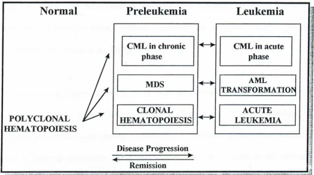
1998). The relationship between clonality and transformation into acute leukemia is summarized in Figure 2.Figure 2 : Clonality and Transformation in Acute Leukemia
(Adapted from Lee
et al.,
1993)1.3.1 Leukemias
The term
leukemia
was derived from the Greek word meaning ‘white blood’ and was first described by John Hughes Bennett and R udolf Vichow in 1845 (Guinn B.A., and Padua R.A., 1997). The leukemias are a heterogeneous group o f neoplasms that arise from the malignant transformation o f hematopoietic cells (Wilsonet al.,
1991). This group o f cancers arise in immature hematopoietic cells and leukemias are constantly characterized by the disturbance o f normal hematopoiesis and some degree o f failure in production o f normally functioning cells. The associated clinical features appear toreflect the level and the lineage in the stem cell hierarchy at which malignant transformation has taken place. Leukemia represents approximately 5% o f all cancers (Guinn B.A., and Padua R.A., 1997).
Leukemic cells proliferate primarily in the bone marrow and lymphoid tissues where they interfere with normal hematopoiesis and immunity. The accumulation o f leukemic cells in the bone marrow is both due to excessive proliferation and to a defect in terminal maturation. These cells further enter the circulation and infiltrate into other tissues such as lymph nodes, liver, spleen, skin, viscera and the central nervous system (Wilson
etal.,
1991).Environmental toxins (e.g. benzene or other industrial chemicals), cancer chemotherapy, radiation, and viruses such as HTLV-I (human T-cell lymphotropic virus type-I in adult T-cell leukemia / lymphoma) are among the well-established leukemogenic factors. Several hereditary factors have also been implicated as significant risk factors in leukemia, especially in childhood. M ost o f them appear to be related to either some form o f immune deficiency (agammaglobulinaemia, severe combined immunodeficiency, adenosinedeaminase deficiency), or a syndrome o f chromosomal instability (Down syndrome) (Wilson
et al.,
1991).Disruption o f Hematopoiesis in Leukemia
A limited number o f hematopoietic stem cells sequentially enter into cell cycle. These then differentiate into multiple lineages in the peripheral blood and lymphoid organs. According to the traditional concepts o f hematopoietic development, progenitor cells differentiate into a single phenotype without the ability to switch lineage. In general, leukemic transformation involves a particular lineage. However, some adult acute leukemias are “biphenotypic” in nature with the expression o f both lymphoid and myeloid lineage cell surface antigens. In addition, in a subset o f acute leukemias, lymphoid or myeloid lineage markers fail to be expressed and these are known as
acute undifferentiated leukemias.
Such leukemic cells may represent leukemic expansion o f the stem cell itself with no lineage markers (Sawyerset a l,
1991).Classification o f Leukemias
The cell type involved and the state o f maturity o f the leukemic cells are the traditional bases on which leukemias are classified. Thus,
acute leukemias
are characterized by the presence o f veiy immature cells, blasts, and by a rapid fatal course in untreated patients. On the other hand,chronic leukemias
are at least initially associated with well-differentiated, mature, leukocytes and with a relatively slow course. There are two major variants o f acute and chronic leukemias:lymphocytic
andmyelocytic
(myelogenous). Therefore a simple classification yields four patterns o fleukemia: acute lymphocytic leukemia (ALL), chronic lymphocytic leukemia (CLL), acute myelocytic (myeloblastic) leukemia (AML), and chronic myelocytic leukemia (CML) (Cotran
et al.,
1989).FAB and MIC Classifications
In 1976, a uniform classification system for acute leukemias and myelodysplastic syndromes was developed by an international group o f investigators. This system, known as the FAB (French-American-British) classification, is based on the morphologic appearance o f bone marrow and blood leukemic blasts. This classification system has been adopted to allow comparison o f treatment results and to take advantage o f biologic differences between morphologic subtypes (Lee
et al.,
1993).
A classification system based solely on morphological criteria proved to be inadequate. Therefore an additional system was introduced. Morphological, immunological and cytogenetic methods were integrated into this new classification by the M IC (Morphological, Immunological and Cytogenetic) Cooperative Study Group, but not all acute myeloid leukemias could be categorized by this classification as well (Segeren C. M., and van’t Veer M. B., 1996).
Nevertheless, immunophenotyping can be helpful in confirmation o f the diagnosis o f AML and is necessary in differentiation o f AML-Mo and AML-M7 from ALL (Table 2). However, it makes no contribution in distinguishing between AML and MDS. In this case, cytogenetic analysis will add important information as in MDS, numerical chromosomal abnormalities are more frequently observed while in
de novo
AML translocations are common. Immunophenotyping also is indispensable for the classification o f acute undifferentiated leukemia which is not described in FAB classification. In the FAB classification, there is a strict discrimination between AML and ALL. However, less than 5% o f leukemias are biphenotypic and in this case, this classification is lacking and immunophenotyping is essential for the recognition o f these types o f leukemia (Segeren C. M., and van’t Veer M. B., 1996).1.3.1.1 Acute Myeloblastic Leukemia (AML)
The term ‘acute non-lymphocytic leukemia (ANLL) is also used for this group o f leukemias. Acute myeloblastic leukemias primarily affect adults between ages o f 15 and 39 years and constitute only 20% o f childhood leukemias (Cotran
et al.,
1989). The incidence o f AML is approximately 2.3 per 100,000 people per year and it increases with age (Fauciet al.,
1998). The extraordinary heterogeneity o f AML reflects the complexities o f myeloid cell differentiation. This group o f leukemias are o f diverse origin. Some arise by transformation o f multipotent (trilineage) myeloid stemcells while in others the common granulocyte-monocyte precursor is involved (Fig 1).
In the widely used FAB classification, AML is divided into eight categories (see Table 2) taking into account both the degree o f maturation (M l to M3) and the predominant line o f differentiation o f the leukemic stem cells (M4 to M7) (Cotran
et al.,
1989).Table 2 : FAB Classification of Acute Myeloblastic (Myelocytic) Leukemias
Subtype
% of Cases *
Mo
Acute myeloblastic leukemia with no maturation2-3 %
M l
Acute myeloblastic leukemia with minimal maturation2 0 %
M2
Acute myeloblastic leukemia with maturation25-30 %
M3
Acute promyelocytic leukemia 8-15 % M 4 Acute myelomonocytic leukemia 20-25 %M5a
Acute monoblastic leukemiawithout differentiation 20-25 %
M5b
Acute monoblastic leukemia with differentiationM6
Acute erythroleukemia 5 %M7
Acute megakaryocytic leukemia 1-2 %Chromosomal abnormalities have been observed in approximately 90 % o f all AML cases. Many o f the nonrandom chromosomal abnormalities have prognostic implications which do not depend on other clinical prognostic factors. These are summarized in Table 3.
T able 3 : C hrom osom al A bnorm alities in A M L a n d T h e ir Significance
C hrom osom e A b n o rm ality
FAB Subgroup
F req u en cy C om m ent
t (9;22) Ph chr. M l 3 % Poor to intermediate prognosis t (8;21) M2 2 0 % M ore often in younger males,
good prognosis
t(1 5 ;1 7 ) M3 70 -1 0 0 % Unique to M3, intermediate prognosis
inv 16 or del 16q M4 ~ 25 % Good prognosis del l l q o r t ( l l ; V * ) M5, some M4 3 0 % Intermediate prognosis
+8 M l, M2, M4, Variable Common abnormality in M 5,M 6
-7 or del (7q) -5 or del (5q)
hemotopoietic neoplasms, no prognostic association
M ost frequently seen in patients older than 60 years in secondary leukemias associated with exposure to environmental or occupational carcinogens
V* = Variable chromosomes (Cotran
etal.,
1989) 14Acute lymphoblastic leukemia is primarily a disease o f children and young adults. ALL can be subdivided by morphologic and immunologic criteria. Morphologic subtypes designated as L I, L2, and L3 have been defined in the FAB classification o f acute leukemias. An alternative immunologic classification is also commonly used and is based on the origin o f the leukemic lymphoblasts and their stage o f differentiation. It is defined by cell surface markers and antigen receptor gene rearrangements (Cotran
etal.,
1989).1.3.1.2 Acute Lymphoblastic Leukemia (ALL)
Table 4 : Classification of Acute Lymphoblastic Leukemia (ALL)
Immunologic
% of Cases
FAB Subtype
Subtype
Pre-B ALL T cell ALL B cell ALL 75 20 L I, L2 L L L 2 L3Cytogenetic
Abnormalities
t ( 9 ; 2 2 ) ,t ( 4 ; l l ) , t ( i ; i 9 ) 14ql 1 or 7q34 t(8 ;1 4 ),t(8 ;2 2 ),t(2;8)
Chronic myelogenous leukemia (CML), is a clonal myeloproliferative disorder which results from the neoplastic transformation o f the primitive hemopoietic stem cell. The disease is monoclonal in origin, affecting myeloid, monocytic, erythroid, megakaryocytic, B-cell, and sometimes T-cell lineages. CML is historically important in two aspects. First, it was the first disease in which a specific chromosomal abnormality t (9;22) (q 3 4 ;q ll) or
Philadelphia chromosome
was linked to the pathogenesis o f the disease. Second is at the therapeutic level, CML is one o f the first neoplastic diseases in which the use o f a biologic agent,interferon,
could suppress the neoplastic clone and prolong survival. Bone marrow transplantation (BMT) studies in CML have also given impressive results (Corteset a i,
1996).CML accounts for 7-15 % o f all leukemias in adults with an incidence o f 1-1.5 cases per 100,000 population. The median age at presentation is 53 years, but the disease can be seen in all age groups. The etiology o f the disease is not clear. There is little evidence for genetic factors linked to CML. It has been suggested that there may be some correlation with HLA antigens CW3 and CW4. Therapeutic radiation has also been associated with increased risk o f CML (Cortes
et al.,
1996).CML has a bi- or triphasic course. There is an initial chronic phase eventually leading to a blastic phase, which is sometimes preceded by an intermediate or accelerated
1.3.1.3 Chronic Myelogenous Leukemia (CML)
phase (Cortes
et al.,
1996). Blast crisis is characterized by increased cellular proliferation, maturation arrest, and karyotypic clonal evolution (Wadaei al.,
1994). A classification o f CML and .its variants is shown in Table 5.Table 5 : A Classification of CML
Clinical Variants
Typical CML (Ph chromosome present) Atypical CML (Ph chromosome absent) CML in infants
(L eeetuf/., 1993)
1.3.2 Lymphomas
Morphologic Variants
Chronic eosinophilic leukemia Chronic basophilic leukemia Chronic monocytic leukemia Chronic neutrophilic leukemia
The malignant lymphomas, in contrast to leukemias, are neoplastic transformations o f cells that reside predominantly in lymphoid tissues. The two major variants o f malignant lymphoma are
non-Hodgkin’s lymphoma
andHodgkin’s disease.
For the classification o f the lymphoid malignancies, the Revised European-American Classification o f Lymphoid Neoplasms (R.E.A.L.) has been created. (Fauciet al..
1998).Non-H odgkin’s lymphomas occur in the cases o f drug-induced immunosuppression and acquired or congenital immunodeficiency. Such lymphomas develop in 30 % o f HIV-1 infected individuals and 40-50 % are EBV (Epstein-Barr Virus) associated. At the molecular level, chromosomal translocations have been demonstrated to be involved in induction o f non-Hodgkin’s lymphoma. Genes that normally regulate heavy and light chain immunoglobulin synthesis are juxtaposed to genes that regulate normal cellular activation and proliferation. The postulation is that, these transforming oncogenes come under the control o f those regulatory elements that normally control B cell proliferation and differentiation (Fauci
etal.,
1998).H odgkin’s disease usually presents as a localized disease with a subsequent spread to contiguous lymphoid structures and a final dissemination to non-lymphoid tissues. There is strong evidence for a role o f EBV in the pathogenesis o f the disease. In contrast to non-Hodgkin’s lymphomas, where chromosomal deletions and translocations are common, cytogenetic studies have shown that such abnormalities are uncommon in H odgkin’s disease. Diagnosis depends on identification o f large multinucleated reticulum cells (Reed-Stemberg cells) in lymph node tissue or other sites (Fauci
et al.,
1998).Myelodysplastic syndromes, also referred to as preleukemic disorders, are a group o f clonal acquired blood disorders affecting the hematopoietic stem cells and that often progress into acute leukemia. MDS results from neoplastic transformation o f the pluripotent stem cell with involvement o f myeloid, and less commonly, o f lymphoid lineages. The myeloid cells in MDS have their ability to proliferate and differentiate, but they undergo an abortive maturation which results in an inadequate major blood cell production (Fauci
et al.,
1998).Among the common chromosomal abnormalities are, monosomy 7, 5q-, and trisomy 8 together with a variety o f translocations. Molecular studies have demonstrated an increased incidence o f mutations involving the
ras, cFms
[monocyte colony- stimulating factor (M-CSF) receptor],p53
andRB
genes (Faucietal.,
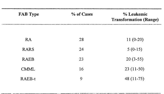
1998).According to the FAB classification, there are five well-defined subtypes:
refractory
anemia
(RA),refractory anemia with ringed sideroblasts
(RARS),refractory anemia
with excess blasts
(RAEB),refractory anemia with excess blasts in transformation
(RAEB-t), andchronic myelomonocytic leukemia
(CMML) (Fauciet al.,
1998). The information on the frequency and the leukemic transformation o f these subtypes are summarized in Table 6. (See also Fig. 2).Table 6 : FAB Classification and Leukemic Transformation in MBS
FAB Type
% of Cases% Leukemic
Transformation (Range)
RA 28 11 (0-20) RARS 24 5 (0-15) RAEB 23 20 (3-55) CMML 16 23 (11-50) RAEB-t 9 48 (11-75)(Adapted from Fauci
et al.,
1998)1.4 Chromosomal Rearrangements in Hematological Malignancies
The first consistent chromosome aberration observed in human neoplasia was the Philadelphia chromosome in CML in 1960. The proof that this is a consequence o f a translocation came in 1973 by the improved chromosome banding techniques. Translocations have one o f two effects. They may lead to the
deregulation
(overexpression) o f oncogenes
by their juxtaposition to enhancer or promoter sequences that are active in the cell type from which the tum or arises, particularly the Ig and TCR enhancers. The alternative molecular consequence o f translocations isgene fusion,
which results in a chimeric oncoprotein, the contribution to whose transforming ability is from both partners (Solomonet a l,
1991).The frequently seen chromosomal rearrangements, which have also been studied in the scope o f this thesis, involve five translocations and one inversion. Besides their frequent involvement in leukemias, the availability o f samples for analysis was also taken into account. The rearrangements analyzed, their fusion gene products, disease association, affected age groups, and frequency in different leukemias is given in Table 7.
Table 7: Chromosomal Rearrangements Analyzed in the Scope of This Study
Rearrangement / Age
Translocation Fusion Gene Disease Group Frequency
CML; ... 90% (CML);5% (P),
t (9;22) (q34; q ll) B C R / A B L AML (Ml) A /P 20% (A); 3% (Ml)
t (8;21) (q22; q22) E T O / A M L I AML (M2); APL A /P 40% (P), 6% (A); 95%
t (15;17) (q22; q21) M Y L / R A R - a AML (M3) A /P 70-100%
t (4;11) (q21; q23) ALL-1 / A F - 4 ALL (lymhoblastic) A /P 100%
t (1;19) (q23; p23) E2A / P B X - 1 ALL (pre-B cell) P 20-25%
inv 16 (pl3; q22) C B F B / M Y H l l AML (M4Eo) A ~ 25%
The Ph chromosome is the result o f a reciprocal translocation between the long arms o f chromosomes 9 and 22. In 1984, Grofifen
et al.
located the breakpoints on chromosome 22 band q l 1 to a region o f 5.8 kb,the breakpoint cluster region
or bcr. Subsequent studies revealed that the bcr is part o f a large gene called theBCR
gene. The bcr contains four exons (bl-b4), and chromosomal breaks occur in two introns between exons b2, b3, and b4. Breakpoints on chromosome 9 are scattered over a distance o f at least 100 kb, but are all located at the 5’ end o f the tyrosine kinase domain o f the c-abl oncogene (Hermanset al.
1987).The normal
BCR
gene occupies a region o f about 135 kb on chromosome 22. It is expressed as mJRNAs o f 4.5 and 6.7 kb, which encodes for the same cytoplasmic 160- kD protein, and contains 23 exons. The Bcr protein contains a unique serine / threonine kinase activity and at least two SH2 binding sites encoded in its first exon and a C-terminal domain that functions as a GTPase activating protein (http://www.ncbi.nlm.gov/htbin-post/Omim/dispmim7151410).BCR
first exon sequences specifically activate the tyrosine kinase and transforming potential o fBCR-
ABL
through interaction o f SH2 domain o f ABL (Pendergastet al.
1991). The wild type Bcr has structural features that suggest a role in signal transduction. In addition to the novel serine / threonine kinase domain, it has an oligomerization domain, a Rho guanine-nucleotide exchange factor (Rho-GEF) homology domain, and acalcium-1.4.11 (9;22) (q34; q l l)
dependent lipid binding site.
Bcr
knockout mice show defects in neutrophil superoxide bursts, suggesting a role forBcr
in the anti-microbial function o f myeloid cells (Raitanoe ta l,
1997).The
ABL
(Abelson strain o f murine leukemia virus) gene is about 225 kb in size and is expressed as either a 6- or 7-kb mRNA transcript, with alternatively spliced first exons, exons la and lb respectively. The 145 kD protein is classified as a nonreceptor tyrosine kinase (http://www.ncbi.nlm.gov/htbin-post/Omim/dispmim7189980). It contains src homology domains SH I, SH2, and SH3. Its carboxy-terminal has a number o f important domains such as nuclear localization signals, a DNA binding domain, a p53 binding site, and an actin binding domain. Abl is expressed in all tissues and is localized in both nucleus and cytoplasm. Mice with targeted disruptions in the c-Abl gene have high neonatal mortality rates and increased susceptibility to infections suggesting a role in B-cell development, but its precise role is unclear (Raitanoet al
., 1997).As a result o f the translocation, the
ABL
gene is transferred from its normal position on chromosome 9 band q34 to the Ph chromosome. This creates a head-to-tailBCR-
ABL
juxtaposition on the Ph chromosome. The fusion gene is transcribed into an 8.5 kb chimeric mRNA, lacking the first exon o fABL.
The hybrid RNA is translated into a hybrid protein product o f 210 IcD (p210®^^‘'^^) and exhibits an in vitro tyrosine kinase activity . The Ph chromosome is also found in ALL (Ph+ ALL). AlthoughABL
is translocated and a new fusion protein p i i s formed, no breakpoints are V
observed in the bcr (Ph+ bcr- ALL). Breakpoints in chromosome 22 occur within the same gene but more 5 ’ o f the bcr. This fusion gene is transcribed into a 7 kb mRNA, encoding a novel fusion protein (Figure 3) (Hermans
et al.
1987).I .AHI.
y
/'v
A
ClBCK
— | -«- tlï cH^ ei]n u l l · - Ü;HI
h---
-«fH»-25 tb ftt-lKT: Ai.i. i i li* M-Uf* CML, ALL AilL ·■ :« V .. CVI„AI..L n^CR-A BL Proteins 22 Ù. Ê
B.iskiN . % i A h ■.'kb Ï F ig u re 3:BCR-ABL
Fusion in t (9,*22) (q34; q l l )(Kurzrock R., and Talpaz M., 1995)
pUK'·*”'"
BCR-ABL transforms hematopoietic cells in vitro and its expression in growth-factor dependent cell lines allows cytokine independent growth. Lethally irradiated mice reconstituted with BCR-ABL expressing BM cells develop different hematological malignancies including a CML-like syndrome (Raitano
et a l,
1997).1.4.2 t (8;21) (q22;q22)
The 8;21 translocation is one o f the most common chromosomal translocations in AML. The chromosomal breakpoints involve
Xh^AMLl
gene on chromosome 21 andETO
gene on chromosome 8. Translocation results in the consistent fusion o f these genes on the der (8) chromosome, resulting in the production o f a novel chimeric gene and message (Figure 4) (Downinget al.,
1993).AM Ll
gene , on chromosome 21 q22, consists o f nine exons and spans more than 150 kb o f genomic DNA. It is highly homologous to the Drosophila segmentation genenm t
and the mouse transcription factor pebp2 (polyoma enhancer binding protein) a subunit gene. The region o f homology, called the Runt domain, is responsible for DNA binding and protein-protein interaction (Miyoshiet al.,
1995). To investigate the normal biologic function o fAMLl in vivo,
Okudaet al.
generated mice carrying a disruptedAM Ll
allele using gene targeting in embryonic stem (ES) cells. Mice lackingam ll
died during midembryonic development, secondary to the complete absence o f fetal-liver derived hematopoiesis. Besides, homozygous ¿Z7w//-deficientcells failed to contribute to hematopoiesis in chimeric animals indicating that,
amll
regulated target genes are essential for definitive hematopoiesis o f all lineages (Okudae ta l ,
1996).ETO
,also calledMTG8
(myeloid translocation gene on 8), contains two DNA- binding zinc finger motifs and several regions that are proline- and serine-rich. It is thought to be a transcription factor due to the presence o f such motifs in other transcription factors (Nucifora G., and Rowley J. D., 1994). The relatively high levels o fETO
in developing brain suggests that it could be involved in the regulation o f some aspect o f neuronal proliferation and differentiation (Ericksonet al.,
1994). In a recent study by Wanget al., ETO
has been shown to have transforming properties. EctopicETO
expression in NIH / 3T3 cells led to foci o f transformation and colony growth in soft agar. £TO-expressing cells induced tumors following injection into irradiated nude mice suggesting an important role in the leukemogenic transforming potential o fxheAMLl-ETO
fusion protein (Wanget a i,
1997).The fusion gene that results from the t (8;21) translocation contains the 5’ region o f
AML I
including the Runt domain fused to almost all o fETO
(Nucifora G., and Rowley J. D., 1994). Both A m ll-Eto and the Amll proteins recognize the same consensus DNA-binding motif, which is found in promoters o f several genes involved in hematopoiesis.BCL-2
is another gene in which this sequence specific binding o fI - V I, IjiX V
3
mb i /
r4 L
:‘ ' C I } J \8
\
/■ X
\
/
m - / f n K r m \M
2 1 ssmJN o r m ai
T r a n s l o c a t i o n
t ( 8 : 2 1 ) ( q 2 2 ; q 2 2)
F ig u re 4: t (8;21) (q22;q22) R earran g em en tboth A M L l and the fusion protein takes place. The elevated levels o f
BCL-2
in cells expressingAMLl-ETO
may prolong their life span and contribute to the development o f t(8;21) leukemia (Klampferet al.,
1996). In a recent study, mice that mimic human t(8;21) were generated. Mice heterozygousfor AMLl-ETO
allele die in midgestation from hemorrhaging in CNS and exhibit severe block in fetal liver hematopoiesis which is very similar to the phenotype obtained by homozygous disruption o f theAM Ll
gene. This indicates thatAMLl-ETO
blocks normalAM Ll
function (Yergeauet al.,
1997).
1.4.3 t(15;17) (q22;q21)
A consistent translocation t(15;17) is present in the blasts o f the majority o f the APL patients. The tw o genes involved are (also called
PML)
on chromosome 15, and R A R -a on chromosome 17. ChimericRAR-a
/MYL
(on the 17q- derivative) andh'lYL
/RAR-a
(on the 15q+ derivative chromosome) are generated as a consequence o f the reciprocal translocation (Figure 5) (Biondietal.,
1992).Retinoic acid receptors (RARs) are nuclear hormone receptors that act as retinoic acid (RA)-inducible transcriptional activators in their heterodimeric form. RA controls fundamental developmental processes, induces terminal differentiation o f myeloid hemotopoietic progenitors, and has tum or and cell growth suppressive activities. The chimeric PM L-RA R -a protein contains most o f the
PML
sequences fused to a largepart
oiRAR-a,
including its DNA and hormone binding domains (Wanget al.,
1998).PML
is an interferon (IFN)-inducibIe gene that encodes a ring-finger protein which is typically concentrated within discrete speckled nuclear structures called PML nuclear bodies or PM L oncogenic domains. Ablation o f murine Pml protein by homologous recombination revealed that PML regulates hemopoietic differentiation and controls cell grow th and tumorigenesis.PML
function is essential for the tumor growth suppressive activity o f RA and for its ability to induce terminal myeloid differentiation o f progenitor cells. ThusPML
is a critical component o f the RA pathway and disruption o f its activity by the translocation may be important in APL pathogenesis (Wang e /a /., 1998).C T D P M L R A R A
T r a n s I O c a t i O n
t ( 15; i 7) ( q 22;q2 1)
iwwÍtó?S8ti¿iewir<Figure 5; t (15; 17) (q22;q21) Rearrangement
(Kurzrock R., and Talpaz M., 1995)
1.4.4 inv 16 (pl3;q22)
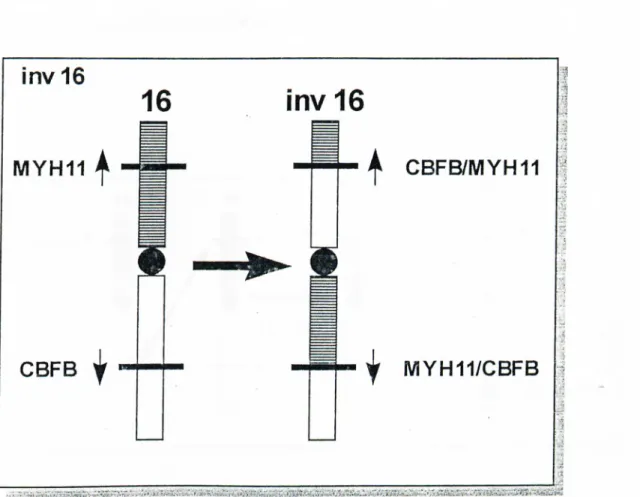
The involvement o f chromosome 16 in leukemia was first reported in 1982 by Arthur and Bloomfield. In 1983, Le Beau
et al
demonstrated chromosome 16 inversion with AML-M4. In the following years, t(16;16) was identified, the breakpoints o f which seemed to be the same as in the inversion (Figures 6 and 7). Molecular studies showed that a fusion gene is generated by inv 16 between theCBFB
gene on q arm and theM Y H ll
gene on p arm (Liuet al.,
1995).Figure 6: inv 16 (pl3;q22) Rearrangement
CBFB
gene encodes a subunit o f the transcription factor complex CBF, also known as PEBP2. PEBP2 was originally identified as a protein in N M 3T3 cells that binds to mouse polyoma virus enhancer. CBF was identified as a protein predominantly expressed in T cells that bind to a conserved “core” site in murine leukemia virus enhancers. B oth PEBP2 and CBF recognize the same core site. The cloning and purification identified them as the same protein. CBF consists o f two subunits: the a subunit, which binds the DNA target, and the |5 subunit, which does not bind DNA directly. At least three a subunits have been identified one o f which is encoded byAM Ll
gene that is disrupted by t(8;21) and t(3;21) (Liuet al.,
1995).Figure 7: t (16; 16) (pl3;q22) Rearrangement
(Adapted fi-om Claxton
e ta l,
1994)The smooth muscle myosin heavy chain (SMMHC) gene
M Y H ll
is a member o f the myosin II family. A typical myosin II protein has an ATPase head, responsible for actin binding and mechanical movement, a hinge region, and a long tail with tandem copies o f a coiled-coil domain that facilitates the filament assembly. The breakpoints identified are within the tail region, fusing the C-terminal portion o f the tail toCBFB
(Liuet al.,
1995).1.4.5 t(4;ll)(q 21;q 23)
The t (4;11) (q21;q23) cytogenetic abnormality is associated with a subset o f ALL. The two genes involved are
AF-4 {FEL)
andALL-1 {MLL,
mixed lineage leukemia or myeloid-lymphoid leukemia,HRX,
homolog o f trithorax,Hrtx
7, human trithorax-like gene 1) on chromosomes 4 and 11 respectively. As a consequence o f the translocation, these genes are fused and transcribed as a chimericALL-1
/AF-4
mRNA (Figure 8) (Ciminoet al.,
1996).A-T hooks i C 4 ; n ) T r a n s lo c a t i o n jM I Sncfingsss T R X h c ^ f e g y C0fl·
L M
L ______
M I L # 1 1 i SP -rich .. , f , "V' ' 1 .. i ■ ' I - L i A f -4 # 4 A T hcxDksi
■ M tS P -f ic h Q Q P - . -L I M LLtf-f-4 d e r ( M ) 1 j ... ... ■. __ 1Figure 8: t (4; 11) (q21;q23) Rearrangement
(Kurzrock R., and Talpaz M., 1995)
The
ALL-1
gene is found rearranged in approximately 10 % o f ALL and in over 5 % o f AML. The gene undergoes fusion with different partners on a variety o f chromosomes such as chromosomes 1, 4, 6, 9 ,10, or 19. The gene, which spans a region on chromosome band llq 2 3 approximately 90 kb in length, consists o f 36 exons (Rasioet al.,
1996).ALL-1
gene belongs to the trithorax gene family o f which the Drosophila trithorax (trx) gene is known to regulate homeotic genes through alternative splicing. Namet al.
demonstrated that, different cell t3q)es transcribeALL-
1
mRNA species lacking exons that generally encode putative regulatory domains such as AT hooks (exon3), repression domain (exon 6), zinc finger motifs (exon 8),and activation domain (exon 18). This suggests that posttranscriptional regulation by alternative splicing may play an important role in
ALL-1
gene expression (Namet al.,
1996). Immunoc5h;ochemical analysis showed that the protein localizes to nuclearstructures in cells with and without llq 2 3 translocations. It is widely expressed in most cell types including hematopoietic cells (Butler
e ta l,
1997).AF-4
(ALL-1 fused gene from chromosome 4), is a serine and proline rich putative transcription factor with a glutamine rich C-terminus. It is expressed normally in both B- and T-lymphoid cell lines . TheALL-1
/AF-4
fusion protein contains a large C- terminal portion o fAF-4
fused to the N-terminal portion o fALL-1
containing its AT hook DNA binding motifs (Morrisseyet a l,
1993).1.4.6 t ( l ; 19) (q23;p23)
The prognostically important 1;19 chromosomal translocation can alter the
E2A
gene on chromosome 19pl3 in childhood B-cell precursor ALL, The breakpoint on chromosome lq23 interrupts a homeobox gene known asPBXl.
As a result, chimeric transcripts are formed that retain the activator domain o f theE2A
gene but substitute a homeobox domain o f the Pbxl protein for the helix-loop-helix DNA binding and dimerization domainofE2A
(Figure 9) (Priviterae ta l,
1992).£ 2A
i rronsacTTr til
T T
HI.H mPBX
B
t(1 ; 19) translocation
HOMEO E 2 A A ^ B X Transact H 0 M £ 0 27ï>p 3 'A 5 'AE
2A ^ B X
I
Tronsoct. I HOrvîEo"Figure 9: t (1;19) (q23;p23) Rearrangement
(Kurzrock R., and Talpaz M., 1995)
C h 19
C h i
T y p e I Ty p e II
T y p e HI
E2A
gene encodes transcription factors o f the helix-loop-helix (HLH) family that are implicated in cell-specific gene expression as part o f dimeric complexes that interact with E box enhancer elements. Its expression is detected in a wide range o f celt lines.E2A
gene gives rise to two proteins, E12 and E47, by differential splicing o f E l 2- and E47- specific basic HLH encoding exons. Both have been implicated in the regulation o f Ig gene expression (Aronheimet al.,
1993).I. 2. MOLECULAR DIAGNOSIS OF HEMATOLOGICAL MALIGNANCIES
2.1 Cytogenetics
Cytogenetics is the study o f the genetic material, genome or chromosomes, o f a cell by cytological means. In molecular cytogenetics, the advantage o f the specificity and base pairing qualities o f DNA is used to look at specific regions o f the genome to which it hybridizes. In hematological malignancies, cytogenetic analysis may be used for: (a) detection o f malignant proliferations, (b) classification o f hematological neoplasms, (c) characterization o f the degree o f neoplastic progression, (d) testing for remission (disappearance o f signs o f cancer), and (e) establishment o f the time o f relapse (Kurzrock
et al.,
1995).The application o f banding techniques; especially Q (quinacrine fluorescence) and G (Giemsa) bandings are used for detailed characterization o f structural rearrangements within chromosomes (Lee
etal.,
1993).Molecular cytogenetics provides a powerful link between molecular genetic analysis and chromosome morphology by allowing to pinpoint structurally aberrant chromosome regions on the molecular level (Lichter
et al.,
1996).Fluorescence In Situ Hybridization (FISH)
In situ hybridization
is the term used to describe a procedure in which labelled DNA is hybridized to cell or tissue preparations on slides. In molecular cytogenetics, this procedure is used to identify chromosomes or chromosome regions containing the probe sequences (Kurzrocket al.,
1995).The advantages o f this technique may be summarized as follows;
a. it allows the study o f chromosome changes by scoring large numbers o f non dividing cells in a relatively short time,
b. it enables better definition o f the nature o f complex structural changes in metaphase cells,
c. it permits estimation o f the size o f abnormal clones in interphase cells, and
d. it may help in determination o f the lineage o f cells carrying a chromosome abnormality (Cuneo
et al.,
1997).Besides FISH, there are other molecular cytogenetics methods such as comparative genomic hybridazation (CGH), which may prove to be very useful for diagnostic purposes in the coming years.
2.2 Im m unological M ethods
In addition to C5dogenetic analysis, a variety o f immunologic techniques have been
developed for more specific and accurate diagnosis. Immunofluorescence and immunoenzymatic techniques allow localization o f cell or tissue antigens on the surface, in the cytoplasm, or in the nucleus. In the direct immunofluorescence techniques, the antigen is identified by a fluorochrome-labelled specific antibody. The more sensitive indirect method utilizes an unlabelled specific primary antibody and allows it to react with the tissue antigen. A fluorochrome conjugated second antibody (anti-immunoglobulin) that binds to the primary antibody is then applied. Immunofluorescence techniques are used mostly to study cells in suspension by means o f fluorescence microscopy or automated flow cj^ometers. There are also immunoenzymatic tests which utilize immunoperoxidase technique, the avidin-biotin complex system, and the immunoalkaline phosphatase technique (Lee
etal.,
1993).2.3 Fusion Transcript Detection
Recurring chromosomal abnormalities are closely associated with particular phenotypes. This is especially the case for structural chromosomal changes seen in leukemias and lymphomas, which have diagnostic and prognostic implications.
Molecular analysis o f these changes not only yields important insights into disease pathogenesis but also provides more precise definition o f disease subsets which is