Preparation and Characterization of Poly(hydroxyethyl
methacrylate-co-poly(ethyl-eneglycol-methacrylate)/Hydroxypropyl-chitosan) Hydrogel Films:
Adhesion of Rat Mesenchymal Stem Cells
Gulay Bayramoglu*,1,K. Can Akcall*,2, Sinan Gultekin2, Erman Bengu3, and M. Yakup Arica1
1Biochemical Processing and Biomaterial Research Laboratory, Faculty of Arts and Sciences, Gazi University,
06500-Teknikokullar-Ankara, Turkey
2Department of Molecular Biology and Genetics, Faculty Science, Bilkent University, 06800 Bilkent-Ankara, Turkey 3Department of Chemistry, Faculty of Science, Bilkent University, 06800 Bilkent-Ankara, Turkey
Received September 5, 2010; Revised December 1, 2010; Accepted December 2, 2010
Abstract: This study examined the effects of the surface properties of the materials, such as the hydroxyl, methyl
and amino groups, on rat bone marrow derived Mesenchymal Stem Cell (MSC) seeding. A series of hydrogels were prepared in film form using 2-hydroxyethyl methacrylate (pHEMA), poly(ethyleneglycol) methacrylate (PEG-MA), and/or hydroxypropyl-chitosan (HPC). The physicochemical properties of these hydrogel films, such as water con-tent, functional groups, contact angle, surface energy and thermal properties were affected by the composition of the materials. The ability of the MSCs to form colonies, as well as their viability on these materials were also analyzed. The water content of the hydrogel films increased with increasing PEG-MA and HPC ratio in the hydrogel. Contact angle measurements of the surface of the hydrogel films demonstrated that all the materials gave rise to a signifi-cantly hydrophilic surface compared to pure pHEMA. The blood protein interactions and platelet adhesion were reduced significantly on the surface of the materials upon the incorporation of PEG-MA compared to the control pure pHEMA and vice versa for HPC. The ability of the MSCs to adhere and form colonies on these materials was also analyzed. The results showed that these materials are suitable candidates to isolate and expand MSCs.
Keywords: hydrogels, contact angle, protein adsorption, mesenchymal stem cells, tissue engineering.
Introduction
The importance of biomaterials has steadily increased in recent years, and the number of polymer applications in tis-sue engineering continues to grow. In addition to the thera-peutic applications, use of biomaterials is also being explored in stem cell maintenance and differentiation. The perfor-mance of a polymeric material relies highly upon the prop-erties of the boundaries in many applications because the interactions between the materials and environments occur chiefly on its surfaces.1-4 The hydroxyethylmethacrylate (pHEMA) hydrogel possesses good biocompatibility as evi-denced by its large scale use in biomedical and biotechno-logical applications such as contact lenses, drug delivery systems, and soft tissue prosthesis.5-9 Adhesion, prolifera-tion, viability and function of anchorage dependent cells were shown to be highly related of the surface properties of the substrate biomaterials used.6 Several studies have been performed to improve biocompatibility of the pHEMA based
hydrogels.3,8 Among them, the grafting of phosphorylcho-line containing moieties10 or incorporation of poly(ethylene oxide) chains to the hydrophilic pHEMA have been reported to reduce protein fouling.11,12 Recently, an alternative approach to modify surface properties of the pHEMA based hydrogel has been proposed.13 According to this method, PEG cou-pled monomer could be introduced with a desired density by adjusting the concentration of PEG carrying co-mono-mer in the polyco-mono-mer structures. Surfaces containing PEG are interesting biomaterials because they exhibit low degrees of protein adsorption and cell adhesion.14-16 PEG is also well known for its extraordinary ability to resist protein adsorp-tion because of its hydrophilicity and unique coordinaadsorp-tion with surrounding water molecules in an aqueous medium.3 Chitosan is a linear polysaccharide derived from partial deacetylation of chitin.15,16 It is considered as an appropriate functional material for biomedical applications because of its high biocompatibility, biodegradability, non-antigenicity, and adsorptive properties.17-23 Chitosan has also many pecu-liar biological activities such as antimicrobial activity and wound-healing acceleration and thus has been receiving *Corresponding Author. E-mail: [email protected]
great interest in tissue engineering as well as in medicine and biotechnology.15 Chitosan has many peculiar biological activities such as antimicrobial activity and wound-healing acceleration and thus prospective applications in the fields of medicine and biotechnology. Chemical modification via etherification with propylene epoxide can improve the water solubility and bioactivities of chitosan. The effects of face chemistry on cell functions depend not only on the sur-face composition but also on the culture conditions and cell type. However, information on the effect of chemical com-position, especially surface charge, on stem cells is very limited.
Stem cells provide alternative treatment options to combat with debilating diseases. Stem cells have the ability of self-renewal and differentiation into mature cells of various lin-eages and are important cell sources for therapeutic applica-tions.24 Among the stem cells, MSCs are multipotent cells, capable of self-renewal and differentiating into multiple lin-eages such as osteocytes, adipocytes, chondrocytes, myo-blasts, and cardiomyocytes.25-27 MSCs are very small fraction of the nucleated cells in the bone marrow, only %0.01-%0.1 of the total population.28 They can easily be isolated from the heterogeneous cell population of bone marrow with their ability to adhere to the culture plate, leading to the for-mation of colonies unlike cells of hematopoietic lineages. MSCs are promising agents for cell-based and regenerative therapies because of their ability not to induce immune reaction in the recipient in addition to the lack of ethical concerns.29 Their use has already been approved in humans to prevent graft rejection and to prevent/control Graft versus Host Diesease (GvHD), as well as in the context of regener-ative medicine with the aim of facilitating tissue repair.30 One drawback of using MSCs, however, is the shortage of these cells. Therefore, it is essential to develop a technology for the efficient preparation and proliferation of cells which are achieved by providing a cell culture substrate as the artifi-cial ECM to prepare large number of stem cells of high quality.
The aim of this work was to evaluate the biocompatibility of the hydrogel films with different compositions as potential tissue engineering scaffolds for MSCs. The hydrogels were prepared from HEMA, PEGMA and HPC. In order to uni-formly combine into hydrogels formulations with improved water-solubility, a water-soluble derivative of chitosan (i.e., hydroxypropyl chitosan, HPC) was prepared and used as a charged component in the formulations. A five different compositions {i.e., p(HEMA-PEG-MA)-1-2, p(HEMA-HPC)-1-2 and p(HEMA-PEG-MA-HPC) were prepared in the presence of an initiator (α,α'-azobisisobutyronitrile; AIBN).
The presented method was chosen so that the PEG and/or HPC can be easily introduced to the polymer structure at a desired density by adjusting the concentration of PEG and/ or HPC polymers in the polymerization mixture. The sur-face properties of the hydrogel films were determined by
measuring the contact angle values to different test liquids, and the surface free energies of the hydrogel films were cal-culated from contact angle data using acid-base method of van Oss.31 System parameters such as properties of hydro-gels (i.e., effect of HEMA:PEG-MA and/or HEMA:HPC ratios on the water content, surface morphology, thermal properties), the interaction with two important blood pro-teins (i.e., human serum albumin and fibrinogen) and plate-let adhesion behavior of the hydrogels were also studied. Finally, the effects of surface properties on the MSCs expan-sion were also evaluated.
Experimental
Materials. 2-Hydroxyethyl methacrylate (HEMA), poly
(ethylene glycol) methacrylate (PEG-MA), chitosan, propy-lene oxide, and α,α'-azobisisobutyronitrile (AIBN) were
obtained from Sigma-Aldrich Chemie GmbH (Germany). HEMA was distilled under reduced pressure before use. Human serum albumin and fibrinogen were supplied from Sigma-Aldrich and used as received. All other chemicals were of analytical grade and were purchased from Merck AG (Germany). The water used in the present work was purified using a Barnstead system (Dubuque, IA, USA).
Modification of Chitosan with Hydroxypropyl Group.
Hydroxypropyl chitosan was prepared as described by Xie
et al.. Chitosan (4.0 g) was mixed with NaOH solution (50%
w/w, 40 mL) and allowed to swell at room temperature for 18 h. It was then incubated in a freezer 24 h at -18oC for alkalization.32 Alkali chitosan and isopropyl alcohol (40.0 mL) were transferred into a 250 mL round-bottomed flask and stirred for 1.0 h at 40 oC. After this period, 80 mL pro-pylene oxide was added in the mixture, and refluxed 2 h at 60oC with continuous stirring. The reaction mixture was adjusted to pH 7.0 with HCl solution. The reaction product was filtered and washed several times by acetone, absolute alcohol and 75% (v/v) alcohol, then dried under vacuum at 60 oC for 48 h.
Preparation of Composite Hydrogel Films. The hydrogel
films (i.e., pHEMA, PEG-MA)-1-2, p(HEMA-HPC)-1-2, and p(HEMA-PEG-MA-HPC)) were prepared by UV-initiated photopolymerization, and were used for culti-vation of MSC. The polymerization was carried out in a flat glass mould (9.0 cm diameter). To check the effect of PEG and HPC ratio on the hydrogels properties and MSC culti-vation efficiency in the initial polymerization mixture dif-ferent HEMA:PEG-MA and HEMA:HPC ratios were used, respectively (Table I). Firstly, AIBN (10 mg) were dissolved in HEMA and then it was mixed with PEG-MA and/or HPC. The resulting mixture was equilibrated at 25oC for 15 min in a waterbath. It was then purged with nitrogen for 2 min and distributed into moulds. It was sealed and exposed to long-wave ultraviolet radiation for 1 h. After polymeriza-tion, all the samples were then washed extensively to remove
residual toxic monomer residues and polymerization impu-rities. Washing was performed in isopropanol and then with purified water in a Soxhlet system. The composite polymer samples were dried in a vacuum oven and then sterilized in an autoclave before use.
Characterizations of Hydrogel Films.
FTIR and SEM Studies: FTIR spectra of the pHEMA,
PEG-MA)-2, HPC)-2, and p(HEMA-PEG-MA-HPC) were obtained by using a FTIR spectro-photometer (Mattson 1000 FTIR, England). The dry poly-mer (about 0.01 g) mixed with KBr (0.1 g) and pressed into a tablet form. The FTIR spectrum was then recorded.
The hydrogel films were coated with a thin layer of gold under reduced pressure and their micrographs were obtained using a scanning electron microscope (SEM) (Model: JEOL, JSM 5600, Tokyo, Japan). MSC cultivated hydrogel films were washed with physiological buffer solution, and then were fixed with 2.0% glutaraldehyde solution for 1.0 h at room temperature. Then, the samples were rinsed with dis-tilled water and freeze dried at 18 ºC for 3 days. Samples were coated under vacuum with a thin film of gold, and then examined in a scanning electron microscope (Carl-Zeiss EVO 40 XVP, Oberkochen, Germany).
Determination of Water Content of the Hydrogels: The
water content of the hydrogel films were determined at 25oC using a gravimetric method. The pre-weighed dry samples were immersed in phosphate buffer solution at pH 7.4, and the swollen hydrogels were removed and weighed on a sensitive balance. The following equation was used to determine the swelling ratio of the hydrogels:
Swelling content (%) = [( Ws-Wd) / Wd] × 100 (1) Where, Ws and Wd are the swollen and dry weights of hydro-gels, respectively.
Contact Angles Studies: The contact angles to different
test liquids (i.e., water, glycerol, and diiodomethane) of the hydrogel materials were measured by sessile drop method at 25oC by using digital optical contact angle meter Phoneix 150 (Surface Electro Optics, Korea). Both the left and the right contact angles and drop dimension parameters were automatically calculated from the digitalized image. The
measurements were the average of five contact angles at least operated on three samples. The free surface energy parameters of the all the hydrogel formulation were calcu-lated using the contact angle data of the probe liquids. The results are analyzed according to acid base method of van Oss et al..31 In this method, the contact angles against at least three liquids with known values of γLW, γ+, and γ- are measured and the superscripts (LW), (+), (-) refers to disper-sive, Lewis acid and base components, respectively. The values for each experiment are put into the following equa-tion:
(1 + cosθ)γ1= 2[(γsLW ×γlLW)½ + (γs+ × γl-)½ + (γs- × γl+)½] (2) total surface energy γTOT is regarded as the sum of Lifshitz-van der Waals and the Lewis acid and base components.
γTOT =γLW + γAB (3)
where γLW designated Lifschitz-van der Waals interaction, reflecting the long-range interactions was calculated from the measured diiodomethane contact angles, and γAB desig-nated such acid-base interactions as hydrogen bonding, γ+ and γ- refer to proton and electron donating character, respectively.
Determination of Anti-fouling Properties of Hydrogel Formulations. The adsorption of human serum proteins
(i.e., HSA and fibrinogen) on the pHEMA, p(HEMA-PEG-MA)-1-2, p(HEMA-HPC)-1-2, and p(HEMA-PEG-MA-HPC) hydrogel films were studied at 37oC for 18 h in a batch sys-tem. The initial concentration of protein was 0.5 mg/mL in the individual adsorption medium. The amount of adsorbed protein on the hydrogels surface was obtained by using the following equation:33
q= [(Co-C) V] / S (4)
Where, q is the amount of protein adsorbed onto hydro-gels surface (ng/cm2); Co and C are the concentrations of the protein in the solution before and after adsorption, respec-tively (mg/mL); V is the volume of the protein solution and
S is the surface area of the hydrogels. The amount of protein
adsorbed on the hydrogel films was determined using a flu-orescence spectrofluorometeric method (fluflu-orescence spec-Table I. Contact Angle Values of Water, Glycerol, and Diiodomethane on the p(HEMA-PEG-MA)-1-2, p(HEMA-HPC)-1-2, and p(HEMA-PEG-MA-HPC) Hydrogel Films
Hydrogel Film Compositions Monomer or Polymer Ratio HEMA:PEG-MA:HPC (v/v/v)
Test Liquids and Their Surface Tensions (γl)
Water (γl=71.3) (θ°) Glycerol (γl=64.0) (θ°) DIM (γl=50.8) (θ°)
pHEMA; (1:0:0) 55.6±1.7 53.6±1.3 39.3±0.8 p(HEMA-HPC)-1; (3:0:1) 47.3±1.1 38.6±0.9 35.1±0.7 p(HEMA-HPC)-2; (1:0:1) 44.7±0.7 34.0±1.2 33.9±0.4 p(HEMA-PEG-MA)-1; (3:1:0) 48.7±1.1 61.6±1.6 34.2±1.3 p(HEMA-PEG-MA)-2; (1:1:0) 46.5±1.6 65.8±0.9 39.6±0.9 p(HEMA-PEG-MA-HPC); (2:1:1) 50.4±1.3 68.9±1.1 37.2±1.0
trofluorometer; Jasco, FP-750, Tokyo, Japan). The fluorescence intensity was measured with excitation and emission at 280 and 340 nm, respectively. Calibration curves were prepared from human serum albumin and fibrinogen and were used in the calculation of protein content of the solutions. For each set of data, the arithmetic mean values and standard deviations were calculated and the margin of error for each data set was determined according to a confidence interval of 95% using the Excel for Windows.
Determination the Amount of Adhered Blood Platelet on the Hydrogel Films. For platelet adhesion studies,
hydrogel films were incubated in PBS (pH 7.4) for 2 h. Fresh human blood was obtained from healthy donors. Platelet rich plasma (PRP) was prepared by centrifugation of the fresh blood (anticoagulated with citrate) at 2,500 rpm at 4 ºC for 5 min. The hydrogel film samples were laid flat in a sterile glass dishes, and were incubated in PRP (3.0 mL) at 37 ºC for 1 h. After this period, the hydrogels were gently washed with PBS solution to remove non-adhering plate-lets. The fixing of adhering platelets was performed with 5.0% glutaraldhyde in PBS buffer at room temperature for 6 h. The composite hydrogels were then washed with PBS buffer, stained with eosin-methylene blue (Wrigth’s stain) and examined in an optical microscope (Nikon, Japan). The platelet counts in PRB samples were also carried out before and after incubation period of the hydrogels and, were determined using an optical microscope. The same proce-dure was used for platelet adhesion studies with pHEMA, PEG-MA)-1-2, HPC)-1-2, and p(HEMA-PEG-MA-HPC) composite hydrogels after contacting with fibrinogen. Hydrogels were incubated for 18 h in fibrinogen solution (0.5 mg fibrinogen in PBS) and contacted with platelet rich plasma as described above.
Determination of Hemolytic Activity of the Hydrogel Films. To determine the effect of polymer compositions on
red blood cells (RBC) hemolysis, oxalated whole blood (0.2 mL) was diluted 15 fold with saline solution (2.8 mL, 0.85% w/v). The hydrogel films were added and incubated for 1.0 h at 37 ºC. After incubation, the RBC was separated by centrifugation at 3,000 rpm for 10 min. The amount of hemoglobin released was determined with UV/Vis spectro-photometer at 545 nm. Hemolytic activity was calculated by subtracting the absorbance of the negative control (saline solution) and dividing by the absorbance caused by distilled water (positive control 100%).
Isolation and Culture of MSCs on Hydrogel Films.
MSCs were obtained from female, nine-week-old, 280-300 g Sprague-Dawley rats. The animals were kept in the animal holding facility of the Department of Molecular Biology and Genetics at Bilkent University under controlled condi-tions at 22oC with 12 h light and 12 h dark cycles. They were provided with unlimited access of food and water. Our experimental protocol was approved by the Animal Ethics Committee of Bilkent University (BILHADYEK).
After the rats were sacrificed by cervical dislocation, a heterogeneous bone marrow cell population was collected from the femurs and tibias by flushing with a 5 mL syringe containing 10% FBS (HyClone, Logan, USA) in DMEM (HyClone). The hydrogel films were placed in 12 well plates and sterilized by exposing short wave UV light for 15 min. Cells from bone marrow were seeded directly on the hydro-gel samples and cultured in MesenCult medium (StemCell Technologies, Vancouver, Canada) with a supplement (20%, w/v; StemCell Technologies) and a penicillin-streptomycin solution (1%, w/v; HyClone). The plates were incubated under CO2 atmosphere (5%) at 37 ºC. After 24 h incubation period, the cells culture medium was changed with fresh one, and the nonadherent cells were removed. The media was changed every 3 days thereafter after washing with sterile 1x PBS. On 14th day, the plates were taken and experiments were performed.
Colony Forming Assay. At 14th day of culturing, MSCs were washed with 1X PBS, then air dried. After fixing with cold methanol for 5 min, MSCs were washed with 1x PBS and giemsa staining reagent (Carlo Erba) was added and left for 5 min. The reaction of giemsa staining was stopped by the addition of tap water.
Results and Discussion
Characterization Studies of Hydrogel Films. Our
objec-tive in the presented work is to determine the effecobjec-tive parameters including microstructure, functional group den-sity, and biological response to develop hydrogel films for potential use as tissue engineering material with improved biocompatibility. In order to reach of these scopes, hydrogel films were synthesized by variation amount of HEMA, PEG-MA, and HPC in the polymerization medium. HPC was pre-pared by the reaction of chitosan with propylene oxide under alkaline conditions (Scheme I).
FTIR spectrum of modified chitosan is recorded and it present new adsorption bands (Figure 1). The absorption band at 1375 cm-1 was assigned as symmetrical deformation vibration of the incorporated CH3 groups on the chitosan
backbone (data not shown). An additional peak at 1100 cm-1 was assigned to C−O stretching vibration in the ether bond of HPC. The stretching vibration bands at 2885 and 1469 cm-1 contributed to the C−H bond became stronger com-pared to that of chitosan. On the other hand, the absorption peaks at 1592 cm-1 belong to the N−H bond was greatly weakened when compared to the chitosan spectra. These results showed that the hydroxypropyl unit was introduced into the HPC structure.
The FTIR spectra of hydrogel films were obtained to illustrate the composition and nature of bond formation within hydrogel formulations (Figure 1). As presented in Figure 1, the FTIR spectra of the pHEMA, p(HEMA-PEG-MA)-2, p(HEMA-HPC)-2, and p(HEMA-PEG-MA-HPC) materials. The FTIR spectra of both pHEMA and p(HEMA-PEG-MA)-2 have the characteristic stretching vibration band of hydrogen-bonded alcohol, O-H, and around 3550 cm-1, and also C=O stretching vibration of ester group appears at 1720 cm-1. On the other hand, several bands appear in the fingerprint region for PEG between 1600 and 1200 cm-1 on the p(HEMA-PEG-MA)-2 structures. These peaks are assigned to -CH2 scissoring band of the PEG at 1450 cm-1, the anti-symmetric and anti-symmetric stretching band -O-CH2-CH2 of PEG at 1360 and 1275 cm-1, respectively. At these regions, pHEMA has also adsorption bands, therefore these bands are overlaps. It should be noted that hydrogen bonded alco-hol O-H stretching band intensity of p(HEMA-PEG-MA)-2 material is higher than that of pure pHEMA due to the incorporation of PEG-MA into the copolymer structure. Incorporation of hydroxypropyl chitosan in the hydrogel structure new adsorption bands are observed at 1375 and
1592 cm-1 which were assigned as stretching vibration and symmetrical deformation vibration of N-H and CH3 groups of HPC, respectively.
The SEM micrographs showed significant difference on the surface morphology of the hydrogel networks. The SEM micrograph of the pHEMA hydrogel surface has a porous structure (Figure 2(A)). The surface of p(HEMA-PEG-MA)-2 hydrogel networks, PEG chains was incorporated with the
co-monomer PEG-MA, exhibited a property of less porous
and smoothness with respect to the surface of pHEMA (Fig-ure 2(B)). When this fig(Fig-ure was examined, its pores had a view of either filled or closed with PEG chains and had a much smoother surface structure compared to pHEMA hydrogel surfaces. The SEM micrograph of p(HEMA-HPC)-2 has more smooth surface with micro-pores compared to pHEMA (Figure 2(C)).
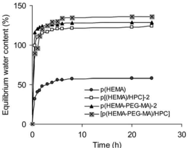
The equilibrium water content of the hydrogels series are presented in Figure 3. The swelling behaviors of the hydro-gel films are significantly affected in the presence of PEG-MA macro-monomer and/or hydroxypropyl chitosan polymer in the hydrogel structures. The hydrogel synthesized from pure HEMA units (i.e., p(HEMA) shows the lowest swell-ing due to the presence of lowest hydroxyl groups in the structure compared to the counterpart hydrogel formula-tions. The composite hydrogels (i.e., p(HEMA-PEG-MA)-2, p(HEMA-HPC)-2) with several pendant hydroxyl groups have higher swelling degrees than those of the pure (pHEMA). The highest value of swelling is obtained for the p(HEMA-Figure 1. The FTIR spectra: (a) p(HEMA), (b)
p(HEMA-PEG-MA)-2, (c) p[(HEMA-HPC)]-2, and (d) p[(HEMA-PEG-MA)/HPC] hydrogels.
Figure 2. Representative SEM micrographs: (A) pHEMA, (B)
PEG-MA-HPC) hydrogels, which contains several pendant hydroxyl groups. The water uptake capacities of the pHEMA, MA)-2, p(HEMA-HPC)-2, and p(HEMA-PEG-MA-HPC) hydrogels were determined between 58% and 136%. Thus, the equilibrium water content increased as the ratio of comonomer (PEG-MA) and/or hydroxypropyl chi-tosan in the polymer structure increases.
Evaluation of Contact Angle Data and Surface Energy Parameters of Hydrogel Films. Biomaterials have to be
optimized, especially regarding to their surface characteris-tics such as surface chemistry, hydrophilicity and surface energy.33 These parameters are critically important for the development of biomaterials in the field of tissue engineering and regenerative medicine. Therefore, contact angle mea-surements are used in the characterization of materials sur-faces to describe the hydrophilicity or to estimate the surface free energy. The wettability of a biomaterial surfaces can be examined by comparing the contact angles for water and diiodomethane since these two solvents are often used as reference liquids in analyses of interaction of polar and apo-lar solvents with solid surfaces. The results of the contact angle measurements of water, glycerol, and diiodomethane on the pHEMA, p(HEMA-PEG-MA)-1-2, p(HEMA-HPC)-1-2, and p(HEMA-PEG-MA-HPC) hydrogels surfaces are presented in Table I. It can be seen that all the composite
hydrogels (i.e., PEG-MA and/or HPC) carrying materials showed lower water contact angles than those of the pure pHEMA. All the investigated samples yielded a different contact angle value. As expected, all the tested hydrogel compositions have polar surfaces. As seen in Table I, for the PEG-MA)-1-2, HPC)-1-2, and p(HEMA-PEG-MA-HPC) hydrogels contact angles of three liquids depend on the PEG-MA and/or HPC ratios of the materials. As the PEG-MA and HPC ratios increase in the hydrogels structure, the water contact angles decrease (Table I).
As presented in Table I, the PEG carrying comonomer incorporated hydrogel materials showed higher glycerol contact angle value than those of the pHEMA. This can be due to increase the density of polar PEG macromolecules on the copolymer surfaces as increase the ratio of PEG-MA co-monomer. On the other hand, HPC incorporated materials showed lover glycerol contact angle values compared to pure pHEMA.
The determined overall surface free energy (γTOT), calcu-lated using the acid base method of van Oss et al.,31 consist-ing of the sum of the Lifschitz-van der Waals (γLW) and the acid-base components (γAB) applies for all investigated sam-ples at different values (Table II). As seen in the table, pHEMA, p(HEMA-PEG-MA)-1-2, p(HEMA-HPC)-1-2, and p(HEMA-PEG-MA-HPC) hydrogels formulations seem to exhibit “amphoteric” character. The basic parameters (γ-) of the p(HEMA-PEG-MA)-1-2 and p(HEMA-PEG-MA-HPC) are highly larger compared to the acidic parameter (γ+). The relatively high basic (γ-) component of the surface energy is caused by the electron ion pairs of oxygen atoms contained in the copolymer (hydroxyl, carbonyl, and carboxyl func-tionalities), which are effective in Lewis base sites.34-38 It is interesting to observe that the base parameter of the p(HEMA-PEG-MA)-1-2 materials significantly increased from 41.92 to 51.35 mN/m2 whereas acid parameter slightly decreased from 0.28 to 0.69 mN/m2 when the ratio of PEG-MA increased in the hydrogel formulation. On the other hand, opposite trend was observed for HPC incorporated counterparts. The acid parameters of the p(HEMA-HPC)-1-2 are increased from 1.39 to 3.31 mN/m2 materials, whereas base parame-ters for p(HEMA-HPC)-1-2 are decreased from 24.59 to 20.16 mN/m2, respectively, as the ratio of HPC increased in
Figure 3. The equilibrium water content of the hydrogels films.
Table II. Surface Free Energy Parameters (mN/m) of pHEMA, PEG-MA)-1-2, HPC)-1-2, and p(HEMA-PEG-MA-HPC) Hydrogel Films according to the van Oss et al. Method
Hydrogel Film Compositions γTOT [mN/m] γd [mN/m] γp [mN/m] γ+ [mN/m] γ- [mN/m] Polarity (%)
pHEMA 45.04 39.96 5.08 0.27 24.70 11.27 p(HEMA-HPC)-1 53.61 41.99 11.62 1.39 24.59 21.68 p(HEMA-HPC)-2 58.51 42.51 16.29 3.31 20.16 27.84 p(HEMA-PEG-MA)-1 49.25 42.40 6.85 0.28 41.92 13.91 p(HEMA-PEG-MA)-2 51.71 39.81 11.89 0.69 51.35 22.30 p[(HEMA-PEG-MA-HPC) 55.53 40.99 14.54 1.01 48.05 26.18
the hydrogel formulations. Thus, all these parameters should be effective in determining the surface energy properties of the pHEMA, p(HEMA-PEG-MA)-1-2, p(HEMA-HPC)-1-2 hydrogel films when contacted with serum proteins and blood cells. It should be noted that water absorbance behav-ior of the pHEMA, p(HEMA-PEG-MA)-1-2, p(HEMA-HPC)-1-2 hydrogel films, presented in Figure 3, also has a similar trend as contact angles studies since more hydrophilic sur-face was to absorb water more easily. This study demon-strated that by incorporation PEG carrying comonomer to the hydrogel structure, the hydrophilicity and biocompati-bility could be improved. As presented in Figure 3, an increase in the amount of more hydrophilic PEG chains prevents the copolymer from collapsing as much as the pure pHEMA hydrogel and this behavior should be related to the balance between Lifschitz-van der Waals (γLW) and the Lewis acid-base (γAB) forces of the biomaterials. As expected, all the investigated materials exhibit different acid base compo-nents (γAB) of the surface free energy due to the various amounts of pendant PEG groups of the copolymers back-bone. It should be noted that the contact angle degree decrease for water on the p(HEMA-PEG-MA)-1-2, hydro-gel formulations accompanied by an increase in (γAB) value and polarity of the materials. On the other hand, the Lifs-chitz-van der Waals (γLW) component of the surface free energy of the p(HEMA-PEG-MA)-1-2 hydrogels formula-tions also slightly increased as the PEG density increased on the copolymer backbone. As reported earlier, surface wetta-bility is recognized as a critical factor for cell behavior, and cells tend to attach better to hydrophilic surfaces than to hydrophobic surfaces.39 In order to investigate this effect in our system, we investigated the adhesion behavior of MSCs on the highly hydrophilic composite hydrogels with the dif-ferent surface energy parameters and polarities.
Evaluation of Antifouling Properties of Hydrogel Films.
The chemical composition can determine the biomaterial properties and subsequently influence protein adsorption, cell attachment and proliferation. Among these factors, the chemical composition is important for the determination of biocompatibility of the materials. In order to determine the effect of chemical composition on the protein adsorption of the pHEMA based hydrogels, a series of composite poly-mers have been prepared from PEG containing monomer and hydroxypropyl chitosan. Those polymers could easily be combined with the hydrophilic networks structure of the pHEMA biomaterials.40 The adsorption properties of major blood proteins (i.e., albumin and fibrinogen) on the p(HEMA-PEG-MA)-1-2, p(HEMA-HPC)-1-2, and p(HEMA-PEG-MA-HPC) hydrogel films were tested in a batch system using pure pHEMA as a control materials. Hydrogel samples were incubated with PBS (pH 7.4) containing 0.5 mg/mL of indi-vidual plasma protein for a period of 18 h at 37 ºC. The tested two proteins reached to their maximum adsorption values on the pHEMA and composite hydrogel materials
after 10 h and remained constant after this period. The amount of adsorbed serum albumin and fibrinogen on the samples surfaces were presented in Table III. Some of the composite polymers include a hydroxypropyl-chitosan chains and/or PEG side-chains. As observed in the table, the HPC chains facilitate adsorption of blood proteins the PEG side-chains provide a steric barrier, avoiding the adsorption of proteins onto the material surfaces. The results show that addition of the HPC to the pHEMA network structure decreased the blood compatibility of pHEMA. On the other hand, PEG side-chains containing polymer (with hydrogen acceptor sides) exhibits the greatest improvement in bio-compatibility of the materials.41 It should be noted that the amount of adsorbed protein on the HPC containing hydrogels increased as the HPC ratio increased in the hydrogel networks. This may be due to increased affinity of blood protein to positively charged HPC chains in the network structure of the films. Thus, these results show that PEG side-chains contain-ing composite materials to have the best anti-foulcontain-ing prop-erties for the tested blood proteins compared to pHEMA and p(HEMA-HPC)-1-2 materials. The isoelectric points of the blood proteins are between 4.0 and 6.9, therefore, these proteins are carrying negative charge at blood pH 7.4. The relatively high basic (γ-) component of the PEGylated hydrogels could create repulsive force against to the nega-tively charged blood proteins. Thus, an increase in the PEG-MA monomer ratio could improve the antifouling properties of the materials. As seen in Table III, the amount of adsorbed major serum proteins on the p(HEMA-PEG-MA)-1-2 materials were reduced compared to pure pHEMA. The reduction in the adsorbed amount of protein was mainly due to the repulsion of Lewis base component of PEG.16 In the present work, the hydrophilic PEG chains should be moved from interior to the surface of the materials because of material bulk should be less hydrophilic than that of the aqueous surrounding, thus all PEG introduced on the hydro-gels showed a good antifouling surface properties. There were not significant differences in the levels of protein adsorption as a function of the PEG content of the hydrogels as observed for water content and contact angles studies, Table III. Amount of HSA and Fibrinogen Adsorbed on the pHEMA, p(HEMA-PEG-MA)-1-2, p(HEMA-HPC)-1-2, and p(HEMA-PEG-MA-HPC) after 18 h Incubation Period at 37 ºC
Hydrogel Film Compositions HSA (ng/cm2) Fibrinogen (ng/cm2)
pHEMA 356±09 341±3 p(HEMA-HPC)-1 465±12 503±11 p(HEMA-HPC)-2 734±14 857±23 p(HEMA-PEG-MA)-1 278±11 221±09 p(HEMA-PEG-MA)-2 174±07 153±13 p(HEMA-PEG-MA-HPC) 398±17 362±06
and these results were in good agreement with the related literatures.42-45 It should be noted that the amounts of absorbed albumin and fibrinogen on surfaces of hydrogel films with different chemical compositions may be helpful for the rational design of cell culture surfaces to elicit specific cell responses.
Hemolytic Activity of Hydrogel Films. The
biocompati-bility of the biomaterials formulations p(HEMA-PEG-MA)-1-2, p(HEMA-HPC)-1-p(HEMA-PEG-MA)-1-2, and p(HEMA-PEG-MA-HPC) were assessed in vitro by using hemolysis tests. The hemolytic activities of the biomaterials with isotonic saline solution are as the blank. As expected complete hemolysis was observed in distilled water which was the positive control but was negligible in physiological saline solution (negative control). In the hemolysis test, both HPC incorporated p(HEMA-HPC)-1-2 formulations were incubated with blood 7.8% and 12.1% hemolysis was observed. On the other hand, PEG containing formulation p(HEMA-PEG-MA)-1-2 showed low degree of hemolytic activity about 1.3% and 0.9%, respectively. However, the effect of incorporation of PEG was not pronounced for p(HEMA-PEG-MA-HPC) biomaterial formulations, combination of HPC with PEG in the polymer networks still proved to have a significant posi-tive effect and the degree of hemolysis was about 3.7%. The hemolytic activity tests suggest that presence of HPC increased the hemolytic activity compared to other tested formula-tions, and so the optimal HPC content of the hydrogels for tissue engineering needs to be adjusted. The p(HEMA-PEG-MA-HPC) hydrogels can be feasible for use as scaffolds for tissue engineering.
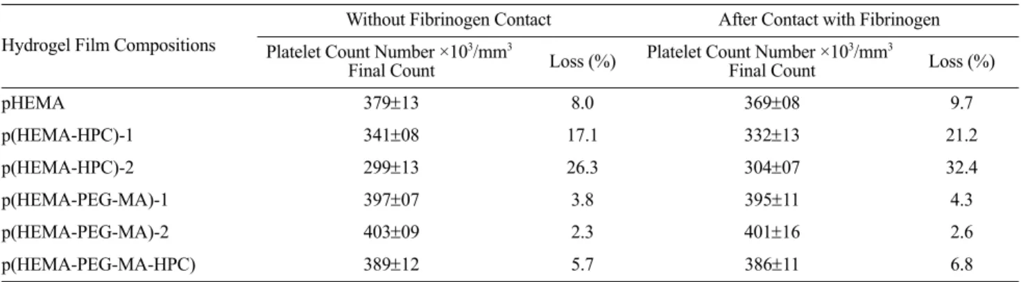
Interaction of Hydrogel Films with Blood Platelets.
Table IV summarizes hematological data obtained from in
vitro platelet adhesion studies with pHEMA,
p(HEMA-PEG-MA)-1-2, p(HEMA-HPC)-1-2, and p(HEMA-PEG-MA-HPC) hydrogels materials before and after the contact with fibrin-ogen. Both HPC containing materials have more influence with the adhesion of the platelets. The results show that materials with HEMA:HPC ratio of 1:1 (p(HEMA-HPC)-2) exhibits much higher platelets adhesion (about 26.3% plate-lets loss) compared to all other tested materials (Table IV)
under the same experimental conditions. Also, the adhesion of platelets decreased with increasing PEG side-chains con-taining ratio in the hydrogel formulations but increased with increasing HPC ratio. In the previous studies, chitosan con-taining interpenetration network has been used as an adsor-bent for separation of protein from aqueous medium due to its high contents of amino and hydroxyl functional groups.45,46 The PEG side-chains onto copolymer membranes can be expected to reduce the interaction between blood (plasma protein and cells) and the membrane surface. As expected, PEG side-chains containing membranes showed reduced plate-let adhesion compared to pHEMA, and p(HEMA-HPC)-1-2, and p(HEMA-PEG-MA-HPC) formulations. It should be noted that the platelet adhesion of a PEG containing copoly-mer membrane depends on amounts of the PEG ratio of the membrane. As seen in Table IV the incorporation PEG to the pHEMA networks improves its biocompatibility as evaluated in terms of platelet adhesion and protein adsorp-tion. As seen from Table IV, the surfaces of the PEG intro-duced biomaterials series surface are resistant to the adhesion of platelets. It is well accepted that PEG is resis-tant to adhesion of blood components (i.e., plasma proteins and cells), and the specific character of PEG can be explained by creating a surface similar to biological macro-molecules and cells.39,42,44 On the other hand, the biomaterial samples which were incubated with fibrinogen for 18 h, showed slightly higher platelet adsorption than those of the non-fibrinogen containing counterpart. In this case, the presence of platelet-adhesive protein (i.e., fibrinogen) results in a slight increase of adhesion of platelets. As seen in Table IV, the increase in the percent platelet adhesion on the pure pHEMA is more pronounced compared to PEGylated mate-rials.
Here, the solubility of chitosan is also improved with hy-droxylpropyl group attachment on the C-2 and/or C-6 car-bon atom of the glucose amine units of the chitosan chains. Whereas some of the pendant amino group of the glucose amine unit of the chitosan chains were modified into sec-ondary amino groups. Chitosan chains have net positive charges with presence of the primary and secondary amino Table IV. Platelet Adhesion on the pHEMA, p(HEMA-PEG-MA)-1-2, p(HEMA-HPC)-1-2, and p(HEMA-PEG-MA-HPC)
Hydrogel Film Compositions
Without Fibrinogen Contact After Contact with Fibrinogen Platelet Count Number ×103/mm3
Final Count Loss (%) Platelet Count Number ×10
3/mm3
Final Count Loss (%)
pHEMA 379±13 8.0 369±08 9.7 p(HEMA-HPC)-1 341±08 17.1 332±13 21.2 p(HEMA-HPC)-2 299±13 26.3 304±07 32.4 p(HEMA-PEG-MA)-1 397±07 3.8 395±11 4.3 p(HEMA-PEG-MA)-2 403±09 2.3 401±16 2.6 p(HEMA-PEG-MA-HPC) 389±12 5.7 386±11 6.8
groups. On the other hand, sialic acid is a molecule which is responsible for the net negative surface charge of platelets. Sialic acid is the most prevalent sugar of the glycolipids and glycoproteins on the mammalian cell surface and is the key epitope recognized as essential for a number of pathogenic infections. Thus, the ionic nature of the modified chitosan causes it to bind with sialic acid in phospholipids of plate-lets cell membranes, resulting adhesion of plateplate-lets on the HPC containing materials.
Bone Marrow Isolated MSCs Grown on Hydrogel Films.
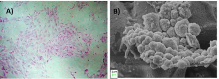
The growth of MSCs were investigated on the pHEMA, PEG-MA)-1-2, HPC)-1-2, and p(HEMA-PEG-MA-HPC) hydrogel films, which were produced com-binations of the three different monomers/polymer (Table I). Our purpose was to evaluate the suitability of these bio-materials in MSCs’ attachment and maintenance since MSCs have been shown to have very important roles in regenera-tive medicine and tissue engineering.47 However MSCs are rare cells28 and novel seeding materials are required to improve the yield of MSC maintenance. Thus, we investigated whether MSCs were able to attach the biomaterials after 14 days of culture by checking the attachment of the cells and by the colony forming assay. It has been shown previously by us and other researchers that 14 days culture of MSCs with commercially available medium (Stem Cell Technolo-gies) are required to isolate MSCs from the heterogeneous bone marrow stromal cells.48 Our results showed the attach-ment of MSCs after 14 days of culture on p(HEMA-HPC)-1, p(HEMA-PEG-MA)-1-2, and p(HEMA-PEG-MA-HPC) (Figure 4(A-D), respectively). In addition, when the MSCs were stained with Giemsa to reveal their CFU-F activity, we found that they were capable of forming colonies suggest-ing that these biomaterials are compatible for MSC cultur-ing (Figure 5(A)). Furthermore, SEM analysis of MSCs on
p(HEMA-PEG-MA)-2 hydrogel film formulation clearly demonstrate the colony forming capacity which is unique for MSCs (Figure 5(B)), whereas no colony formation was observed when MSCs were grown on pHEMA and p(HEMA-HPC)-2 hydrogel films (data not shown). Using stem cells as a therapeutic agent has been receiving tremendous inter-est as it provides opportunities to combat with debilitating diseases. Our data showed or the first time that p(HEMA-HPC)-1, p(HEMA-PEG-MA)-1, p(HEMA-PEG-MA)-2, and p(HEMA-PEG-MA-HPC) are suitable formulations to use in isolating and expanding MSCs. In this study, we aimed to understand whether MSCs would be attaching to these newly synthesized hydrogel films. Our data on MSCs are qualitative rather than quantitative and therefore clearly warrant new studies regarding to the effect of these hydro-gel films on the differentiation and proliferation rate of MSCs. It may induce differentiation and/or block this event all together. Even though MSCs are multipotent cells, their interaction with these polymers may cause them to become pluripotent and increase their differentiation capacity. Their role on differentiation and proliferation are currently being conducted in our laboratory. It should be also noted that some biomaterials might toxic to the cells or they not allow to diffusion of nutrients and waste product of cells to the medium49 because of the combination of different polymer, which might be the reason of problem in observation of cells grown in some of the biomaterials.
The interaction mechanism of chitosan with biological macromolecules is generally considered due to the amino group at the C-2 position of the glucose amine residue, this is, the ionic nature of chitosan. The hydroxypropyl chitosan is a derivative of chitosan and is mainly anionic nature at neutral conditions so the adsorption and binding of cationic groups are effective to explain its interaction mechanism with the biological molecules. At physiological pH 7.4, the degree of protonation of NH2 is very low that is the repulsion of NH3+ is weak so the strong intermolecular and intramo-lecular hydrogen bond results in formation of hydrophobic micro area in polymer chain. Whereas, the hydroxyl groups in the modified chitosan polymer chain is strongly hydro-philic. Therefore, the polymer chains have hydrophobic and Figure 4. Growth of bone marrow derived mesenchymal stem cells
on biomaterials with different compositions: (A) p(HEMA/HPC)-1, (B) p(HEMA-PEG-MA-1), (C) p(HEMA-PEG-MA-2), and (D) p(HEMA-PEG-MA/HPC).
Figure 5. (A) Representative CFU-activity of mesenchymal stem
cells on p(HEMA-PEG-MA-2; (B) Exemplified SEM image of mesenchymal stem cells grown on the p(HEMA-PEG-MA)-2 hydrogel films.
hydrophilic parts. The amphiphilic structure provides struc-ture affinity between the cell membrane protein of the MSC and chitosan derivative. The HPC-contained surface might provide attractive electrostatic interaction between the posi-tively charged surface and the negaposi-tively charged cells.
We examined the ability of the p(HEMA-PEG-MA)-1-2, p(HEMA-HPC)-1-2, and p(HEMA-PEG-MA-HPC) hydro-gel to support cell adhesion and cell growth. The images of all the PEG containing gels showed that following seeding, the cells spread on the polymer surface, and gradually adhere to hydrogels within 14 days. In contrast to the hydrogel pre-pared by combining pHEMA with HPC via interpenetrating polymerization of HEMA monomers, no colony formation was observed with the p(HEMA-HPC)-2 formulation. When the content of HPC increased, cell attachment was inhibited, and this represents strong experimental evidence that the amount of incorporated HPC is the key to significantly inhibit cell growth and colony formation. Moreover, different com-position of HEMA and PEG-MA might be used in order to produce more suitable biomaterials because it was shown that PEG-MA containing HEMA provided a good condition for MSCs isolation from the rat bone marrow. As previously reported, incorporation of PEG polymer on the biomaterials surface may also improve the attachment of MSCs on bio-materials.50
Conclusions
Use of biomaterials and technologies has become one of the leading areas in science and creates a local environment which enables cells to promote their proliferation and differ-entiation.41 Several established methods are presently avail-able for in vitro isolation and differentiation of MSCs from bone marrow. In this study, novel composite hydrogel for-mulations based on HEMA (monomer), PEG-MA (macro-monomer) and hydroxypropyl chitosan were prepared by UV-initiated photopolymerization; different amount of PEG-MA, HPC and/or combinations were used to prepare and characterize scaffolds for mesenchymal stem cells which represent one of the most accessible sources of stem cells for therapeutic use today. The idea of growing and then in
vivo delivering stem cells by using different biomaterials
which play a key role in designing and creating substitutes for ECM is very tempting. The success would inevitably result in the realization of several processes such artificial bone, chondrocyte or muscle tissues that are considered to be science fiction not long ago.
The polarities and the surface free energies of the biocom-patible materials were determined by contact angle mea-surements. Both water contact angle and water content measurements demonstrated that the hydrophilicity of the materials was increased by introduction of comonomer PEG-MA into copolymer structure. The protein adsorption and platelet adhesion studies demonstrated that the introduction
of PEG-MA into pHEMA structure significantly improved the biocompatibility of the material by improving anti-foul-ing properties to proteins and cell adhesion. All copolymer series showed good blood compatibility than those of the pHEMA. The reported PEG-MA containing material was effective to improve the biocompatibility and anti-fouling properties of the pHEMA polymer.
References
(1) V. Ruggeri, I. Francolini, G. Doneli, and A. Piozzi, J. Biomed.
Mater. Res. Part A, 81, 287 (2007).
(2) L. De Bartolo, S. Morelli, A. Bader, and E. Drioli,
Biomateri-als, 23, 2485 (2002).
(3) A. Y. Kwok, G. G. Qiao, and D. H. Solomon, Chem. Mater.,
16, 5650 (2004).
(4) E. Sykova, P. Jendelova, L. Urdzikova, P. Lensy, and A. Hejcl,
Cell Mol. Neurobiol., 26, 1111 (2006).
(5) M. Y. Arica, D. Tuglu, M. M. Basar, D. Kilic, G. Bayramoglu, and E. Batislam, J. Biomed. Mater. Res. B: Appl. Biomater., 86, 18 (2008).
(6) C. R. Jenney and J. M. Anderson, J. Biomed. Mater. Res., 44, 206 (1999).
(7) A. Larsson, T. Ekblad, O. Andersson, and B. Liedberg,
Bio-macromolecules, 8, 287 (2007).
(8) S. Abraham, S. Brahim, K. Ishihara, and A. Guiseppi-Elie,
Biomaterials, 26, 4767 (2005).
(9) Y.-M. Lim, H.-J. Gwon, J.-H. Choi, J. Shin, Y.-C. Nho, S. I. Jeong, M. S. Chong, Y.-M. Lee, I. K. Kwon, and S. E. Kim,
Macromol. Res., 18, 29 (2010).
(10) G.-H. Hsiue, S.-D. Lee, P. C.-T. Chang, and C.-Y. Kao, J.
Biomed. Mater. Res., 42, 134 (1998).
(11) G. Bayramo lu, E. Batislam, and M. Y. Arica, J. Appl. Polym.
Sci., 112, 1012 (2009).
(12) X. Gong, L. Dai, H. J. Griesser, and A. W. H. Mau, J. Polym.
Sci. Part B: Polym. Phys., 38, 2323 (2000).
(13) J. P. Montheard, M. Chatzopoulos, and D. Chappard, J.
Mac-romol. Sci. MacMac-romol. Rev., 32, 1 (1992).
(14) A. K. Bajpai, Polym. Int., 56, 231 (2007).
(15) J.-S. Park and Y.-W. Cho, Macromol. Res., 15, 513 (2007). (16) M. Y. Arica, G. Bayramoglu, B. Arica, K. Ito, and Y. Yagci,
Macromol. Biosci., 5, 983 (2005).
(17) Y. I. Woo, M. H. Lee, H.-L. Kim, J.-C. Park, D.-W. Han, J. K. Kim, K. Tsubaki, K.-H. Chung, S. O. Hyun, and Y.-I. Yang,
Macromol. Res., 18, 90 (2010).
(18) G. Bayramoglu and M. Y. Arica, Macromol. Symp., 203, 213 (2003).
(19) J. Ma, H. Wang, B. He, and J. Chen, Biomaterials, 22, 331 (2001).
(20) F. A. Sheikh, N. A. M. Barakat, M. A. Kanjwal, S. J. Park, D. K. Park, and H. Y. Kim, Macromol. Res., 18, 59 (2010). (21) J. L. Drury and D. J. Mooney, Biomaterials, 24, 4337 (2003). (22) T.-W. Son, B.-G. Kim, Y.-M. Park, H.-S. Lim, and O. K. Kwon,
Macromol. Res., 14, 267 (2006).
(23) R. A. A. Muzzarelli, Carbohydr. Polym., 76, 167 (2009). (24) A. J. Friedenstein, R. K. Chailakhyan, N. V. Latsinik, A. F.
Panasyuk, and I. V. Keiliss-Borok, Transplantation, 47, 331 gˇ
(1974).
(25) A. J. Friedenstein, R. K. Chailakhjan, and K. S. Lalykina, Cell
Tissue Kinet., 3, 393 (1970).
(26) L. da Silva Meirelles, A. I. Caplan, and N. B. Nardi, Stem
Cells, 26, 2287 (2008).
(27) Z. Tokcaer-Keskin, A. R. Akar, F. Ayaloglu-Butun, E. Terzio-glu-Kara, S. Durdu, U. Ozyurda, M. Ugur, and K. C. Akcali,
Can. J. Physiol. Pharmacol., 87, 143 (2009).
(28) M. F. Pittenger, A. M. Mackay, S. C. Beck, R. K. Jaiswal, R. Douglas, J. D. Mosca, M. A. Moorman, D. W. Simonetti, S. Craig, and D. R. Marshak, Science, 284, 143 (1999). (29) J. M. Karp and G. S. Leng-Teo, Cell Stem Cell, 4, 206 (2009). (30) M. E. Bernardo, F. Locatelli, and W. E. Fibbe, Ann. N Y Acad.
Sci., 1176, 101 (2009).
(31) C. J. van Oss, Colloids Surf. A, 78, 1 (1993).
(32) W. Xie, P. Xu, Q. Liu, and J. Xue, Polym. Bull., 49, 47 (2002). (33) G. Bayramoglu and M. Y. Arlca, Int. J. Biol. Macromol., 37,
249 (2005).
(34) A. Bismarck, M. E. Kumru, and J. Springer, J. Colloid Surf.
Sci., 217, 377 (1999).
(35) R. S. Faibish, W. Yoshida, and Y. Cohen, J. Colloid Surf. Sci.,
256, 341 (2002).
(36) C. J. van Oss, J. Mol. Recognit., 16, 177 (2003).
(37) G. Bayramoglu, M. Yilmaz, and M. Y. Arica, Colloids Surf.
A, 243, 11 (2004).
(38) W.-F. Lee and W.-J. Lin, J. Polym. Res., 9, 23 (2002). (39) Y. I. Woo, M. H. Lee, H.-L. Kim, and J.-C. Park, Macromol.
Res., 18, 90 (2010).
(40) B. K. Mann and J. L. West, J. Biomed. Mater. Res., 60, 86 (2002).
(41) R. Ward, J. Anderson, R. McVenes, and K. Stokes, J. Biomed.
Mater. Res., 80, 34 (2007).
(42) G. Bayramoglu, B. Kaya, and M. Y. Arlca, Chem. Eng. Sci.,
57, 2323 (2002).
(43) M. Ebert, R. Ward, J. M. Anderson, R. McVenes, and K. Stokes,
J. Biomed. Mater. Res. A, 75, 175 (2005).
(44) M. Y. Arlca, M. Yllmaz, E. Yalln, and G. Bayramo lu, J.
Membr. Sci., 240, 167 (2004).
(45) G. Bayramoglu, J. Appl. Polym. Sci., 88, 1843 (2003). (46) Z. Tokcaer-Keskin, Z. G. Dikmen, F. Ayaloglu-Butun, S. Gultekin,
S. M. Gryaznov, and K. C. Akcali, Stem Cell Rev., 6, 224 (2010).
(47) N. B. Ivanova, J. T. Dimos, C. Schaniel, J. A. Hackney, K. A. Moore, and I. R. Lemischka, Science, 298, 601 (2002). (48) M. Pevsner-Fischer, V. Morad, M. Cohen-Sfady, L.
Rousso-Noori, A. Zanin-Zhorov, S. Cohen, I. R. Cohen, and D. Zipori,
Blood, 109, 1422 (2007).
(49) S. R. Adamson and B. Schmidli, Canadian J. Chem. Eng.,
64, 531 (2009).
(50) C. Lin and K. Anseth, J. Am. Pharm. Assoc., 26, 631 (2008). gˇ
![Figure 2. Representative SEM micrographs: (A) pHEMA, (B) p[(HEMA-PEG-MA)]-2, and (C) p[(HEMA)/HPC]-2 hydrogels.](https://thumb-eu.123doks.com/thumbv2/9libnet/5959243.124484/5.892.476.797.150.545/figure-representative-sem-micrographs-phema-hema-hema-hydrogels.webp)