Ankara Üniv Vet Fak Derg, 65, 331-334, 2018
Short Communication / Kısa Bilimsel Çalışma
First report of avian infectious laryngotracheitis infection in broiler
breeders in Turkey
İnci Başak KAYA, Mehmet AKAN
Ankara University, Faculty of Veterinary Medicine, Department of Microbiology, Ankara, Turkey.
Summary: Infectious laryngotracheitis (ILT) is a very serious respiratory disease of poultry all over the world. For the last
decade, many countries have reported the ILT disease. However, there is no report about ILT disease in broiler breeders in Turkey. In this study, we investigated ILT in broiler breeders on a poultry farm in Turkey. For this purpose, five samples were investigated from twenty-eight week old broiler breeders on a poultry farm, which consists of 5 flocks with 34.500 broiler breeders. The presence of ILT virus was determined by PCR targeting ICP4 gene (infected-cell polypeptide 4) and characterized by sequencing. This is the first report of the ILT in broiler breeders in Turkey.
Keywords: Avian infectious laryngotracheitis, broiler breeder, ICP4 gene, poultry, sequencing.
Türkiye’de broyler damızlık tavuklarda infeksiyöz laringotracheitis infeksiyonunun ilk bildirimi
Özet: İnfeksiyöz laringotracheitis (ILT) tüm dünyada kanatlı hayvanların ciddi solunum sistemi hastalığıdır. Son on yılda birçok
ülkede ILT infeksiyonu bildirilmiştir. Ancak, Türkiye’de broyler damızlıklarda ILT infeksiyonu bildirimi bulunmamaktadır. Bu çalışmada, 28 haftalık broyler damızlık bulundurulan 34.500 kapasiteli 5 kümesten alınan 5 örnek incelendi. ILT virusunun varlığı, ICP4 geninin (infected-cell polypeptide 4) PZR ile çoğaltılması ve sekans analizi ile belirlendi. Bu çalışma, Türkiye’de broyler damızlık tavuklarda ILT varlığının ilk bildirimidir.
Anahtar sözcükler: Avian infeksiyöz laringotracheitis, broyler damızlık, ICP4 geni, kanatlı, sekans.
Infectious laryngotracheitis (ILT) is an acute and contagious upper respiratory disease that only affects chickens, which leads to economic losses in poultry industries worldwide. The causative agent of the disease is Gallid herpesvirus-1 (GaHV- 1), also commonly referred as infectious laryngotracheitis virus (ILTV). The virus has a similar genome structure with the herpes simplex virus (HSV) and classified in Herpesviridae family. The ILTV has a genome of linear and approximately 150 kb double-stranded DNA (8). The symptoms of the disease are nasal discharge, mucoid tracheitis, sinusitis, conjunctivitis, reduced egg production, gasping, expectoration of bloody mucus, coughing and dyspnea. Additionally, asymptomatic latent infections can also be observed (12).
Virus isolation is one of the detection methods for ILTV but it is time-consuming. Serological tests, such as conventional Enzyme-linked immunosorbent assay (ELISA), Fluorescent antibody technique (FAT) which do not have enough sensitivity have also been used for detection. PCR-based molecular methods have also been used and preferred for the detection of viral DNA (12).
Restriction fragment length polymorphism (RFLP) analyses of the viral genes thymidine kinase (TK), ICP4, glycoprotein E (gE), glycoprotein G (gG) and UL47 have been used to identify GaHV-1 isolates and for epidemiological investigations. In addition, the sequence analysis of these genes has revealed outbreak-related isolates in some countries (3). Another protocol which amplifies two regions of the ICP4 gene and sequence analysis of these regions has also been used in veterinary laboratories (3).
According to the World Organization for Animal Health (OIE) and World Animal Health Information Database disease timelines (WAHID, 2013), reports from the Secretariat of the Pacific Community of the Food and Agricultural Organization (SPC-FAO, 2013), the Caribbean Animal Health Network (CaribVet, 2012) and PubMed searches ILT disease has spread all over the world (6). However, there is no data about the positive ILTV in broiler breeders in Turkey. This is the first case that has reported ILTV infection in the broiler breeders in Turkey.
İnci Başak Kaya - Mehmet Akan 332
Figure 1. General symptoms observed in a broiler breeder chicken infected with ILT. Şekil 1. ILTV ile infekte bir broyler damızlık tavukta gözlenen genel semptomlar.
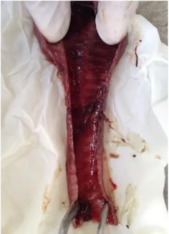
Figure 2. Hemorrhage and exudates in trachea. Şekil 2. Trakeada hemoraji ve eksudat.
ILTV was investigated in 5 trachea samples taken from twenty-eight week old broiler breeders which were not vaccinated against ILT on a poultry farm located in the eastern Mediterranean region of Turkey in 2016, which
consists of 5 flocks with 34.500 broiler breeders. Suspected chickens from which the samples were taken showed several signs of the disease before death such as dyspnea, coughing, gasping and breathing through the mouth (Figure 1). The mortality rate was 6.7% and the egg production decreased 13.4%. In necropsy examination, hemorrhagic tracheas with exudate mucus were observed in some of the samples (Figure 2).
Total DNA extracted trachea samples obtained from five broiler breeders. Briefly, 20 mg of each tissue samples added an appropriate volume of solution homogenized with Qiagen TissueLyser II. Then the extraction procedure was applied by a commercial kit (Genomic DNA Purification, Thermo Fisher Scientific, USA) following the manufacturer’s recommendations. The PCR was performed using specific primers (ICP4-1F ACT-GAT-AGC-TTT-TCG-TAC-AGC-ACG-3’ and ICP4-1R 5’-CAT-CGG-GAC-ATT-CTC-CAG-GTA-GCA-3’) to amplify a 688 bp fragment; (ICP4-2F 5’-CTT-CAG-ACT-CCA-GCT-CAT-CTG-3’ and ICP4-2R 5’-AGT-CAT-GCG-TCT-ATG-GCG-TTG-AC-3’) to amplify a 635 bp fragment according to Chacon & Ferreira (3). The PCR reaction was performed containing 0.2 µM of each primer, 0.2 mM dNTPs (10 mM dNTP mix; (Thermo Fisher Scientific, USA), 3 mM of MgCl2 (Thermo Fisher Scientific, USA), 2.5 µl PCR reaction buffer, 2U of Taq DNA polymerase (Thermo Fisher Scientific; EP0402), and nuclease-free water to a final volume of 25 µl. In the reaction, 2 µl of DNA was used as a template. Thermal cycling conditions were as follows: strand separation at 95°C for 5 min, followed by 35 cycles of 95°C for 45 seconds, 65°C for 45 seconds, and 72°C for 45 seconds.
Ankara Üniv Vet Fak Derg, 65, 2018 333
Finally, there was 7 minutes at 72°C for further strand extension. Then, 10 µl of the amplified PCR product was separated by electrophoresis on 1.5% agarose gel (Promega Corporation, USA) with 4 µl of SafeView Classic (Applied Biological Materials, Canada) in Gel Electrophoresis Apparatus with 90v for 45 minutes (3).
Sequencing of the two regions of ICP4 gene from three positive ILTV samples was also performed. The PCR products were purified using ExoSAP-IT (USB Corporation) which was added 2 µl per 5 µl sample and incubated 37°C for 30 minutes, 80°C for 15 minutes. Followed by the cycle sequencing process with BigDye Direct Cycle Sequencing Kit (Applied Biosysytems) according to the manufacturer’s instructions. Then, amplicons were purified with Sephadex G-50 (Sigma-Aldrich) and sequenced on Applied Biosystems 3500 Genetic Analyzer (Applied Biosystems). For all sequence analysis CLC Main Workbench version 6.7.1 was used.
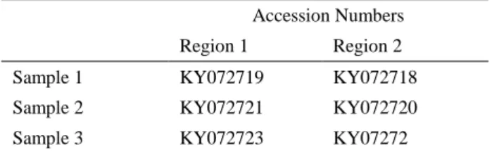
Three positive samples from five dead broiler breeders were detected by PCR and characterized by sequencing. Sequences of all samples were compared with the reference sequences in GenBank with BLASTn and accession numbers were also deposited in GenBank (Table 1).
Table 1. GenBank accession numbers of samples taken for 2 regions of ICP4 gene.
Tablo 1. ICP4 geninin 2 bölgesi için alınan GenBank kabul numaraları.
Accession Numbers Region 1 Region 2
Sample 1 KY072719 KY072718
Sample 2 KY072721 KY072720
Sample 3 KY072723 KY07272
The sequence analysis of the virus is provided for faster, more sensitive and more reliable diagnose of the disease and also to discriminate the infection from other respiratory infections such as avian influenza infection, Newcastle disease. On the other hand, DNA sequencing of ICP4 gene can be used to differentiate isolates of virulence and vaccine (3) and sequence analysis also gives an advantage to improve vaccines in the area. According to the BLASTn sequence results, all samples investigated in this study had 99% sequence identity with wild-type strains sequenced from different countries and also with the tissue culture origin (TCO) and chicken embryo origin (CEO) vaccine sequences which are used as live vaccines against ILT disease in other countries. Since ILT live vaccines are not used in Turkey we assessed the sequence results of all samples as wild-type strains.
ILT disease still causes economical losses in the world poultry industry. Over the past decade, ILT disease
has been reported in several countries such as Australia (1) USA, China, Italy (9), Finland, Peru, Ecuador, Costa Rica (7), Europe, Southeast Asia (5). Although there is only one study which was carried out in 2007 and reported the ILT disease in hens (white Leghorns) in Elazığ, Turkey (4), there is no data about the situation in broiler breeders. This is the first report which shows ILT disease in broiler breeders in Turkey. This study recommends the amplification of two regions of the ICP4 gene and sequence analysis of these regions to diagnose ILTV. Beside this, since TCO/CEO vaccine sequences were found to be similar to wild-type strain sequences, it can be concluded that these vaccines could be used to control the ILT in Turkey.
References
1. Agnew-Crumpton R, Vaz P, Devlin J, et al. (2016): Spread of the newly emerging infectious laryngotracheitis viruses in Australia. Infect Genet Evol, 43, 67-73. 2. Caribbean Animal Health Network (CaribVet) (2012):
Infectious laryngotracheitis monograph. Retrieved july
20,2012, from
http://www.caribvet.net/en/diseases/infectious-laryngotracheitis/monograph.
3. Chacón J, Ferreira A (2009): Differentiation of field isolates and vaccine strains of infectious laryngotracheitis virus by DNA sequencing. Vaccine, 27, 6731-6738. 4. Gulacti I, Eroksuz Y, Bulut H, et al. (2007): Outbreak of
clinical infectious laryngotracheitis in Turkey. Vet Rec, 160, 554-555.
5. Hidalgo H (2003): Infectious laryngotracheitis: A review. Braz J Poultry Sci, 5, 157-168.
6. Menendez K, García M, Spatz S, et al. (2014): Molecular epidemiology of infectious laryngotracheitis: a review. Avian Pathol, 43, 108-117.
7. OIE (2016): Disease outbreak maps. http://www.oie.int/wahis_2/public/wahid.php/Diseaseinfor mation/Diseaseoutbreakmaps?disease_type_hidden=0&dis ease_id_hidden=-999&selected_disease_name_hidden=--- Terrestrial+---&disease_type=0&disease_id_terrestrial=-999&disease_id_aquatic=-999&selected_start_day=1& selected_start_month=1&selected_start_year=2016&select ed_end_day=1&selected_end_month=12&selected_end_y ear=2016 (Erişim tarihi: 10.04.2017)
8. Ou S, Giambrone JJ (2012): Infectious laryngotracheitis virus in chickens. WJV, 1, 142.
9. Piccirillo A, Lavezzo E, Niero G, et al. (2016): Full genome sequence-based comparative study of wild-type and vaccine strains of infectious laryngotracheitis virus from Italy. PLoS ONE, 11, p.e 0149529.
10. Secretariat of the Pacific Community (SPC) Food and Agricaltıra Organization (FAO) (2013): B302- avian infectious laryngotracheitis. AHP Disease Manual.
Retrieved January 29, 2013, from
http://www.spc.int/lrd/ext/Disease_Manual_Final/b302__a vian_infectious_laryngotracheitis.html#auto_top.
11. World Animal Health Information Database (WAHID) (2013): Avian Infectious laryngotracheitis disease
İnci Başak Kaya - Mehmet Akan 334
timelines. Retrieved August 21, 2013, from http://www.oie.int/wahis_2/public/wahid.php/Diseaseinfor mation/Diseasetimelines.
12. Zhao Y, Kong C, Cui X, et al. (2013): Detection of infectious laryngotracheitis virus by real-time PCR in naturally and experimentally infected chickens. PLoS ONE, 8, p.e 67598.
Geliş tarihi. 14.04.2017 / Kabul tarihi: 12.06.2017 Address for correspondence:
Dr. İnci Başak KAYA
Ankara University, Faculty of Veterinary Medicine, Department of Microbiology,
06110, Dışkapı, Ankara, Turkey. e-mail:[email protected]