Hepatogenous photosensitization in Akkaraman lambs: special
emphasis to oxidative stress and thrombocytopenia
Naci OCAL1, Ali Evren HAYDARDEDEOĞLU2, Miyase CINAR3, Oguz KUL4, Mustafa TÜRK5 1 University of Kirikkale, Faculty of Veterinary, Department of Internal Medicine, Kirikkale; 2 University of Ankara, Faculty of
Veterinary, Department of Internal Medicine, Ankara; 3 University of Kirikkale, Faculty of Veterinary, Department of Biochemistry,
Kirikkale; 4 University of Kirikkale, Faculty of Veterinary, Department of Pathology, Kirikkale; 5 University of Kirikkale, Faculty of
Arts and Sciences, Section of Biology, Department of Molecular Biology, Kirikkale, Turkey.
Summary: In this study, a total of 24 Akkaraman lambs with clinical signs that are reminiscent of photosensitization composed the experimental group (Group I). Additional 10 healthy lambs were included as controls (Group II). We were aimed to make definitive diagnosis of photosensitization, which can be confused with blue tongue, big head and sunburns and to establish etiology as primary or hepatogenous. In total blood analyses, the leukocyte count was higher, but thrombocyte count was lower (thrombocytopenia) in Group I compared to Group II. The lower MCV indicates presence of a case of microcytosis. Furthermore, affected lambs had significantly higher levels of phylloerythrin, γ-glutamyl transpeptidase, aspartate aminotransferase, alkaline phosphatase, cholesterol and serum urea nitrogen. Histopathological lesions included periaciner necrosis, periportal fibrosis and biliary duct hyperplasy. The grazing field, where the lambs had been grazing, was very rich in Tribulus terrestris, a hepatotoxic plant. Based on the increased levels of phylloerythrin, γ-glutamyl transpeptidase, aspartate aminotransferase and alkaline phosphatase as well as pathological findings in liver, the cases were diagnosed as hepatogenous photosensitization. In addition, presence of
Tribulus terrestris in the grazing pasture strongly supported the diagnosis. Increased levels of serum malondialdehyde in affected
lambs showed an ongoing oxidative stress. In addition, thrombocytopenia in such cases should be considered as a risk factor for disseminated intravascular coagulation (DIC). Thus in addition to a semptomatical treatment protocol, use of antioxidants, anti-coagulants, and liver protective agents shoud be taken into consideration in hepatogenous photosensitization in lambs.
Key words: Lamb, malondialdehyde, microcytosis, oxidative stress, photosensitization, Tribulus terrestris, thrombocytopenia,
Akkaraman kuzularda hepatojen fotosensitizasyon: oksidatif stres ve trombostopeniye özel vurgu
Özet: Bu çalışmada, klinik olarak fotosensitizasyona benzeyen semptomlar gösteren farklı cinsiyetten 24 (Grup I) ve 10 tane de sağlıklı (Grup II) Akkaraman kuzu çalışmanın hayvan materyalini oluşturdu. Kuzularda mavi dil, big head (koca kafa) ve güneş yanığı ile karışabilen fotosensitizasyonun kesin tanısının yapılması, etiyolojik olarak primer veya sekonder (hepatojen) tip olup olmadığının ortaya konması amaçlandı. Sağlıklı kuzulara göre hastaların lökosit sayısının yüksek, trombosit sayısının ise düşük (trombositopeni) olduğu saptandı (P<0.001). Ayrıca düşük MCV değeri mikrositozis olarak değerlendirildi. Biyokimyasal analizlerde hasta kuzuların filloeritrin, γ-glutamil transpeptidaz, aspartat aminotransferaz, alkalin fosfataz, kolesterol değerlerinin P<0.001 ve serum üre nitrogen değerinin ise P<0.01 düzeyinde yüksek olduğu belirlendi. Histopatolojik incelemede periasiner nekrozis, periportal fibrozis ve bilier kanal hiperplazisi ortaya kondu. Yapılan mera taramasında, kuzuların otlatıldığı tarlaların bitki örtüsünün demir dikeni (Tribulus terrestris) bitkisi yönünden zengin olduğu belirlendi. Karciğerdeki patolojik bulgulara ve filloeritrin, γ-glutamil transpeptidaz, aspartat aminotransferaz ve alkalin fosfataz düzeylerindeki artışlara dayanarak tablonun hepatojen photosensitizasyon olduğu, karaciğer hasarının etiyolojisinde demir dikeni bitkisinin sorumlu olduğu kanısına varıldı. Ayrıca fotosensitizasyonlu kuzularda serum malondialdehid değerinin önemli düzeyde yüksek olduğu ve dolayısıyla patogenezde oksidatif stresin önemli bir yer tuttuğu, trombositopeninin ise koagulasyon bozukluğu (DIC) gelişimi için önemli bir risk faktörü olarak dikkate alınmasının gerektiği düşünüldü. Bu nedenle semptomatik sağaltıma ek olarak, antioksidantların, pıhtılaşma düzenleyicilerin ve karaciğer koruyucuların kullanılmasının tedaviye yeni bir boyut kazandırabileceği söylenebilir.
Anahtar sözcükler: Demir dikeni, fotosensitizasyon, kuzu, malondialdehid, mikrositozis, oksidatif stres, trombositopeni
Introduction
Photosensitization is defined as hypersensitivity reaction of non-pigmented and short-haired regions of the skin containing photodynamic agents which are
exposed to the sunlight (3). Photosensitization is a worldwide problem and can affect any species. However, it is most commonly seen in sheep, goats, cattle, and horses. Although the incidence of death due to
photosensitization is very rare; it causes significant economical losses as a result of weight loss, udder lesions, myiasis and secondary infections (7, 21).
Photosensitization is frequently classified according to the source of the photodynamic agent: primary or type I photosensitivity, type II or secondary (hepatogenous) photosensitivity, and abberant endogenous pigment synthesis or type III photosensitivity (23). Hepatogenous photosensitization occurs when the liver is damaged by toxins, infectious agents or neoplasms, so that the liver cannot sufficiently excrete phylloerythrin. Subsequently, the photodynamic agent phylloerythrin levels increase in the blood and then accumulate in the skin (13, 23). Brachiaria decumbens (5), Tribulus terrestris (22), and Narthecium ossifragum (29) are well known hepatotoxic plants due to their steroidal saponin content, causes an outbreak of sporadic hepatogenous photosensitization in small ruminants. Hepatogenous photosensitization is the most frequently seen photosensitivity type in livestock animals (7). The clinical signs associated with photosensitivity are similar regardless of the cause. However, a photosensitization case should properly diagnosed whether or not the case is hepatogenous by measuring the levels of bilirubin, liver enzymes, bile acids, (23) and the circulating phylloerythrin (19, 20). Some of the clinical signs of photosensitization in sheep can also be seen in blue tongue and clostridial infections (big head), and sun burns (3, 23). While different therapies have been applied according to photosensitization type, there are no fully effective treatment regime (7, 9). Firstly, establisment of photosensitization type is essential on the definition of treatment protocol. Complete recovery from primary photosensitization usually occurs provided the animal does not return to eating the photosensitizing plant. However, hepatogenous photosensitization has a poor prognosis because of the underlying liver disease.
In this study, we aimed to make a definitive diagnosis of an endemic case in lambs during a summer seasion with clinical signs that are reminiscent of photosensitization.
Materials and Methods
This study was conducted in the Mid-Anatolia region. A total of 24 Akkaraman lambs of three different herds grazing in cultivated and non-cultivated grazing-lands and with clinical signs that are reminiscent of photosensitization composed the experimental group (Group I). In addition, 10 healthy lambs were included as a control group (Grup II). The lambs were of either sex and at the age of 4-5 months. Their body weight varied from 25 kg to 40 kg. Lambs included in the study were examined and body temperature, heart rate and respiratory rates were recorded. Urine and blood samples were collected from all lambs in either group. Affected lambs were transferred to sheep pens with no sun
exposure. For symptomatical treatment, the following treatment protocol was applied: intramuscular single dose of penicillin (Deposilin, İ.E ULAGAY, Turkey) at a dose of 60.000 IU/kg, intravenous flunixin meglumin (Fluvil, VİLSAN) at a dose of 1.1 mg/kg for two days, and intravenous polyionic fluid with glucose (Laktaring, %5 glucose, İ.E. ULAGAY, Turkey) 80 ml/kg/day for three days. The blood samples were also collected from Group I after the three-day symptomatic treatment. Total and differential blood counts were made using a MS9 cell counter (MS9-3-Melet Schloesing Laboratories, France). Serum malondialdehyde (MDA), aspartate aminotransferase (AST), alkaline phosphatase (ALP), gamma glutamyl transpeptidase (GGT), total protein (TP), albumin, creatinine, serum urea nitrogen (BUN), cholesterol, triglyceride, creatine kinase, glucose, and calcium were measured spectrophotometrically (Shimadzu UV-1700, Japan). Plasma phylloerythrin was measured using fluorescence spectrofluorometry using a Shimadzu RF-1507 device according to the protocol established by Scheie et al., (19). Urine samples were qualitatively analyzed by a deepstix (Multistix 10 SG, 2300A, Bayer). The edema fluid obtained from the head regions and sera were evaluated for big head and blue tongue diseases, respectively. During the one-week period of the disease, clinically effected two lambs were necropsied. The grazing field, where the lambs had been grazing, was very rich in Tribulus terrestris which contains steroidal saponin.
Clinical data and total blood counts were analyzed statistically by Man Withney U test. The biochemical data were analyzed One way Anova test. Data were expressed as mean ± std. A p value less than 0.05 was considered significant.
Results
In lambs of Group I, the clinical findings of photosensitivity varied from per acute (Figure 1) to progressed (Figure 2) lesions. The per acute symptoms included photophobia, local temperature increase in the head, redness and swelling in the ear pinna, eye lids, lips, and extremities. The progressed lesions included sloughing off the skin over the ear pinna, face, muzzle, extremities (photodermatitis), nasal serous discharge and black coloured diarrea in lambs.
The lambs with per acute signs were placed indoor houses and a symptomatical treatment was applied. Subsequently, the per acute signs resolved. However, the same clinical signs of photosensitivity relapsed when the animals were exposed to the sunlight. The body temperature of the lambs in Group I (40.78±0.27 °C) were significantly higher compared to that of Grup II (39.54±0.48 °C) (p<0.01). In qualitative examination of the urine samples, varying degrees of bilirubinuria, 1(+) and 3(+), were detected in 9 out of 24 (37,5%) lambs of Group I.
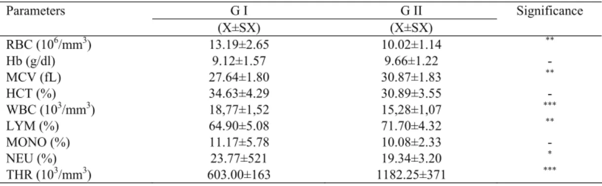
The total blood counts were presented in Table 1. Erythrocyte (RBC), mean corpuscular volum (MCV), lymphocyte (LYM) values were significantly different between Group I and Group II (p<0.01). Importantly, a dramatical increase in leukocyte count (WBC) (p<0.001) and a dramatical decrease in thrombocyte count (THR) (p<0.001) were determined (Table 1).
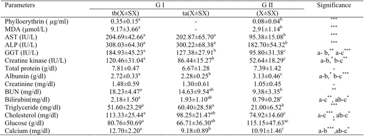
The biochemical data were presented in Table 2. In afftected lambs, phylloerythrin, MDA, AST, ALP, GGT and cholesterol (p<0.001) and BUN (p<0.01) were significantly higher compared to those of Group I. On the other hand, the albumin value was significantly lower (p<0.01) (Table 2).
Histopathological lesions in the liver of the both lambs included diffuse vacuolar-hydropic degeneration in the hepatocytes, severe periacinar necroses, and Kupffer’s cell hyperplasy. There were numerous lymphocytes and plasma cell accumulations in the portal area. Portal fibrosis was especially exaggregated around the hyperplasic bile ducts (Figure 3). In the skin of affected regions, vacuolar degenerations and necrosis in epidermal layer was observed. In the dermis, edema, macrophage infiltrations and fibrinoid degeneration in the middle arteries Figure 1. Peracute clinical case of photosensitization, sweeling of the face, edema in the ear pinna and palpebrae. Lamb, 4 months old. (The picture taken by Naci Ocal)
Şekil 1. Dört aylık bir kuzuda yüzde, göz kapaklarında ve kulaklarda kızarıklık ve ödem ile karekterize fotosensitizasyonun perakut bulguları.
Figure 2. Severe and progressive lesions characterized by eroded skin end covered by crusts. Lamb, 5 months old. (The picture taken by Naci Ocal)
Şekil 2. Beş aylık bir kuzuda erezyon ve kabuklanmayla sonuçlanmış şiddetli ve progressive deri lezyonları.
Table 1. The total blood counts in affected (GI) and healty (GII) lambs. Tablo 1. Hasta (GI) ve sağlıklı (GII) kuzuların total kan sayımı sonuçları.
G I G II Parameters (X±SX) (X±SX) Significance RBC (106/mm3) 13.19±2.65 10.02±1.14 ** Hb (g/dl) 9.12±1.57 9.66±1.22 - MCV (fL) 27.64±1.80 30.87±1.83 ** HCT (%) 34.63±4.29 30.89±3.55 - WBC (103/mm3) 18,77±1,52 15,28±1,07 *** LYM (%) 64.90±5.08 71.70±4.32 ** MONO (%) 11.17±5.78 10.08±2.33 - NEU (%) 23.77±521 19.34±3.20 * THR (103/mm3) 603.00±163 1182.25±371 *** *= p<0.05, **= p<0.01, ***= p<0.001
Figure 3. Degeneration and necrosis of the hepatocytes, periportal fibrosis and bile duct epithelia hyperplasia (arrows) are prominent. Megalocytes having broad and eosinophilic cytoplasm and twin nuclei (arrow heads). Hematoxylin eosin, X 180. (The picture taken by Oguz Kul)
Şekil 3. Hepatositlerde dejeneratif ve nekrotik değişiklikler, periportal fibrozis ile safra kanalı hiperplazisi (oklar). Çift çekirdekli, geniş ve eozinofilik sitoplazmaya sahip megalositler (okbaşı). Hematoksilen eosin, X180
were prominent. In one out of two necropsied lambs, hyperemia and alveolar edema in the lungs and diffuse hemosiderosis in the spleen were also noticed.
In microbiological examination, edema fluid obtained from the head regions were negative for Clostridium sordellii and C. novyi. Likewise, the serum samples were negative for blue tongue.
The grazing field, where the lambs had been grazing, was very rich in Tribulus terrestris which is a toxic agent, causing hepatogenous photosensitization in lambs. (Figure 4).
Discussion and Conclusion
The differential diagnosis in photosensitization is difficult as it can be clinically confused with actual sunburn, blue tongue and big head (Clostridial infection, Clostridium. sordellii, C. novyi) (3, 23). We also had difficulty in diagnosing especially the first cases. In differential diagnosis, we eliminated sun burn, big head
and blue tongue based on sudden occurence of the symptoms in the present cases (23), presence of the symptoms in both sexes and based on serological data, respectively. Furthermore, the increase in plasma phylloerythrin levels and histopathological findings of the liver damage indicated that the present cases were hepatogenous photosensitization related to liver dysfunctions (4, 6, 14). Inaddition, diagnosis was supported by observing the grazing field, which was very rich in Tribulus terrestris.
Histopathological findings included severe hepatocyte necrosis in the liver and hyperplasia of the bile duct along with periportal fibrosis (Figure 3). Such lesions were characteristics of hepatotoxicity casued by ingestion of Tribulus terrestris. These findings are charecteristic to hepatogenous photosensitization and comparable to those reported by Sean et al., (22), Wisløff et al., (29), and Witte et al., (30)
Leukocytosis and increase in body temperature, common laboratory findings, are frequently occur in infections or during the inflammatory processes. Opasina (14) claimed that the body temperature in sheep showing the symptoms of photosentisation was in normal range (38.7±0.7 0C). In contrary, the affected lambs in our
study had increased body temperature (40.78±0.27 °C) and WBC compared to healthy lambs (Table 1). We thought the incerase in body temperature in leukocyte counts was most likely associated with the inflammatory process in photosentization (9, 18, 23).
Bilirubinuria along with icterus was reported in hepatogenous photosentization in sheep (22) and in cattle (30). In our study, we detected varying degrees of bilirubinuria in qualitative examination of urine samples, but hemoglobinuria and a conspicious case of icterus were not present in our cases. This condition can be explained by the occurrence of bilirubinuria without icterus or icterus occurs long after bilirubinuria. This finding is also supportive of the reports by Opasina (14). Table 2. The biochemical data in affected (GI) and healty (GII) lambs.
Tablo 2. Hasta (GI) ve sağlıklı (GII) kuzuların biyokimyasal analiz sonuçları.
G I G II Parameters tb(X±SX) ta(X±SX) (X±SX) Significance Phylloerythrin ( µg/ml) 0.35±0.15a - 0.08±0.04b *** MDA (µmol/L) 9.17±3.66a - 2.91±1.14b *** AST (IU/L) 204.69±42.66a 202.87±65.70a 95.38±15.08b *** ALP (IU/L) 308.03±64.30a 300.22±68.38a 182.70±54.32b *** GGT (IU/L) 184.93±45.23a 127.38±27.91b 95.80±31.38c a- b,** a-c***
Creatine kinase (IU/L) 120.46±31.04a 86.44±15.27b 52.64±18.29c a-b,* b-c**
Total protein (g/dl) 7.81±0.47 6.67±1.28 7.39±1.42 -
Albumin (g/dl) 2.72±0.33a 2.28±0.25b 3.13±0.46c a-b,* b-c***
Creatinine (mg/dl) 1.48±0.59 1.30±0.61 1.05±0.45 -
BUN (mg/dl) 18.23±4.47a 14.63±9.54ab 9.38±3.35b **
Bilirubin(mg/dl) 2.18±1.50a 1.93±1.10ab 0.79±0.28c a-c**, ab-c*
Triglyceride (mg/dl) 51.60±23.29a 60.40±28.58a 21.00±6.52b ***
Cholesterol (mg/dl) 113.33±25.44a 98.25±21.47ab 74.92±14.60c a-c***, ab-c*
Glucose (g/dl) 80.76±50.69a 66.71±36.30ab 115.15±47.63ac *
Calcium (mg/dl) 12.70±2.20a 9.18±0.89b 10.91±1.46c a-b***,ab-c*
Explanation: means with different superscripts within the same row differ significantly (p<0.05). * = p<0.05, ** = p<0.01, *** = p<0.001, tb = before treatment, ta = after treatment
Figure 4. Tribulus terrestris founded in grazing pastures (The picture taken by Naci Ocal)
Şekil 4. Kuzuların otlatıdığı tarlalarda bulunan demir
Bilirubinuria in hepatogenous photosentization is an indicative of liver damage and bile duct disorders (25).
Although RBC counts and PCV were within the physiological limits, but they were significantly higher in affected lambs compared to healthy ones. This elevations might have occurred due to a relative increase in hemoconcentration as a consequence of clinical dehydratation related to inappetence (8). Although normal levels of RBC and Hb values and the lower levels of MCV in affected lambs does not indicate a case of microcytic anemia, but lower MCV may indicate a case of microcytosis. Such a condition can be explained by the opinion of Tvedtend et al., (26) that a case of hypochromic microcytosis can develop without changing of erytrocyte counts at the beggining stages of anemia. In addition, disorder in iron metabolism due liver damage might have also contributed to this condition.
Lecha et al., (10) has been documented that in human patients of photosensitivity with erythropoietic protoporphyria, trombocytopenia along with a mild case of microcytic anaemia occurs at a rate of 20%-60% as a consequence of impaired erythropoiesis. Likewise, trombocytopenia associated with microcytosis was detected in affected lambs of our study (Table 1). However, the mechanism of trombocytopenia accompanied with microcytosis in lambs with hepatogenous photosensitization is difficult to explain in lambs with a similar mechanism mentioned above due to absence of a sufficient literature generated in ruminants. However, oxidative stress that plays an important role in pathogenesis of photosensitization might have contributed to this condition (9, 11, 25).
Thrombocytopenia and leukocytosis are considered as an important parameter for diagnostic purposes. Phylloerythrin, a potent photosensitizer and an efficient source of singlet oxygen, is considered as one of the important photodynamic agents in hepatogenous photosensitivity diseases in sheep (25). Singlet oxygen causes an intense inflammatory reaction in the regional blood vessels and surroundings, leading to tissue necrosis by the effects of severe oxidative stress (5, 9, 10). This situation is assumed to be source of thrombocytopenia due to migration and aggregation of platelets at the region with inflammed vasculature (16, 24). The liver damage might be an additional factor contributing to thrombocytopenia in photosensitization (15).
The results of biochemical analyses indicated that the level of phylloerthrin, the diagnostic parameter for hepatogenous photosensitization, was significantly higher (P<0.001) in Group I compared to Group II. An experimental study by Scheie et al., (19) indicated that the clinical signs of hepatogenous photosensitization became evident when the plasma phylloerythrin reached the level of 0.3µg/ml. In support to this condition, the plasma phylloerythrin level in lambs with photosensitization in our study was 0.35±0.015µg/ml. In
normal conditions, phylloerythrin is conjugated by the liver and excreted into bile. However, liver damages or dysfunctions interfere with the hepatic elimination of phylloerythrin (7, 20). Increase levels of phylloerythrin in circulation and eventually in skin due to hepatic damages and subsequent excitation of phylloerythrin by the sun light resulted in clinical signs of hepatogenous photosensitization (7, 9). Wondrak et al., (28) claimed that singlet oxygen and other reactive oxygen species (ROS) play important roles in phototoxicity reactions. In a support to this claim, Tønnesen et al., (25) detected a high level of serum singlet oxygen in lambs with hepatogenous photosensitization. In paralel to these views, the lambs with hepatogenous photosensitization in our study had increased levels of serum MDA, a marker of oxidative stress and an indicative of lipid peroxidation of ROS in the cell membrane and low density lipoproteins (1, 27). Thus, increased levels of MDA should be considered as an important criterum for pathogenesis and treatment strategies in hepatogenous photosensitization in lambs.
Compared to Group II, Group I had higher levels of hepatocellular damage indicators (Table 2), including hypoalbuminemia (2.28±0.25 g/dl), hypercholesterolemia (113.33±0.25 mg/dl) and aspartate aminotransferase, and indicators of hepatobiliar disorders including gamma glutamyl transpeptidase, alkaline phosphatase and total bilirubin. These findings along with high level of phylloerythrin confirmed the case of hepatogenous photosensitization. Likewise, such laboratory findings are comparable to those of Witte et al., (30) and Sean et al., (22). Compared to healthy lambs, the affected lambs high triglyceride levels in addition to hypercholesterolemia. Increased fat mobilisation due to inappetence as well as hepatobiliar dysfunction, which is frequent in hepatogenous photosensitization, could be listed among the factors contributing to high triglyceride levels in lambs with photosensitization in our study (8). The serum urea nitrogen value was slightly higher in Group I compared to Group II. This slight increase was considered as a relative increase indicating prerenal azotemia related to dehydration. However, the creatine level (1.48±0.59 mg/dl) was between the physiological limits, indicating absence of an important kidney disorder (8).
Following a treatment protocol for three day, serum biochemical parameters were positively improved as the severity of dehydration was reduced, but they did not return to normal levels (Table 2). Thus, as Knight and Walter (9) suggest that hepatobiliar disorders in hepatogenous photosensitization do not heal in a short period of time, and even can be irreversible. Similarly, this view is also supported by high serum aspartate aminotransferase, alkaline phosphatase and gamma glutamyl transpeptidase levels in affected lambs in our study. Apart from the serum biochemical parameters discussed above, the other serum parameters tested
including serum glucose, calcium, total protein values were found between the physiological limits in both groups although some fluctuations were observed (Table 2).
Pathogenesis of the disease should be taken into account in treatment. Increased levels of serum malondialdehyde in affected lambs showed an ongoing oxidative stress. In addition, trombositopenia in such cases should be considered as a risk factor for disseminated intravascular coagulation (DIC) (2, 12, 17). Thus in addition to a semptomatical treatment protocol, use of antioxidants, anti-coagulators, and liver protective agents shoud be taken into consideration in hepatogenous photosensitization in lambs.
References
1. Aksoy H, Koruk M, Akçay F (2003): The relationship
between serum malondialdehyde and ceruloplasmin in chronic liver disease. Turk J Biochem,28, 32-34.
2. Bick RL (1994): Disseminated intravascular coagulation:
Objective criteria for diagnosis and management. Med
Clin N Am, 78, 511-543.
3. Blowey RW, Weaver AD (2003): Photosensitization
(photosensitive dermatitis). 24-25. In: Color Atlas of Diseases
and Disorders of Cattle, 2nd ed. Mosby, RDC Group, China. 4. Bonnefoi M, Braun JP, Bézille P, LeBars J, Sawadogo
G, Thouvenot JP (1989): Clinical biochemistry of
sporidesmin natural intoxication (Facial Eczema) of sheep. J Clin Chem Clin Bio, 27, 13-18.
5. Brun A, Sandberg S (1991): Mechanisms of photosensitivity
in porphyric patients with special emphasis on erythropoietic protoporphyria. J Photoch Photobi B, 10, 285-302.
6. Flåǿyen A, Borrebek B, Nordstoga K (1991): Glycogen
accumulation and histological changes in the liver of lambs with alveld and experimental sporidesmin intoxication. Vet Res Commun, 15, 443-453.
7. Flåǿyen A (2007): Photosensitization. 338-342. In: ID Aitken (Ed), Diseases of sheep, 4th Ed. Blackwell, UK. 8. Kerr MG (2002): Veterinary Laboratory Medicine, Clinical
Biochemistry and Haematology, 2nd Ed. Blackwell, UK.
9. Knight AP, Walter RG (2003): Plants affecting the skin
and liver. In: A Guide to Plant Poisoning of Animals in
North America, Teton New Media, Jackson WY.
10. Lecha M, Puy H, Deybach JC (2009): Erythropoietic
protoporphyria (Review). Orphanet Journal of Rare
Disease doi: 10.1186/1750-1172-4-19.
11. Lund JE (2000): Toxicologic effects on blood and bone
marrow. 44-49. In: FF Bernard, GZ Joseph, CJ Nemi
(Eds), Schalm’s Veterinary Hematology. Lippincott Williams and Wilkins, Philadelphia.
12. Mammen EF (1994): Coagulation defects in liver disease. Med. Clin. N. Am. 78, 545-554.
13. Mayland HF, Cheeke PR (1995): Forages-induced
animal disorders. 121-135. In: The Science of Grassland
Agriculture, 5th Ed. Ames, Iowa.
14. Opasina BA (1985): Photosensitization juandice
syndrome in West African dwarf sheep and goats grazed on Brachiaria decumbens. Trop Grasslands, 19,120-123.
15. Prater MR (2000): Acquired Coagulopathy II: Liver
Disease. 560-563. In: FF Bernard, GZ Joseph, CJ Nemi
(Eds). Schalm’s Veterinary Hematology, Lippincott Williams and Wilkins, Philadelphia.
16. Russel KE, Grindem CB (2000): Secondary
Thrombocytopenia. 487-493. In: FF Bernard, GZ Joseph,
CJ Nemi (Eds). Schalm’s Veterinary Hematology, Lippincott Williams and Wilkins, Philadelphia.
17. Rutherford CJ, Frenkel EP (1994): Thrombocytopenia:
Issues in diagnosis and therapy. Med Clin N Am, 78, 555-575.
18. Scheie E, Flåǿyen A, Moan J, Berg K (2002):
Phylloerythrin: Mechanisms for cellular uptake and location, photosensitisation and spectroscopic evaluation.
New Zeal Vet J, 50, 104-110.
19. Scheie E, Ryste EV, Flåǿyen A (2003a): Measurement of
phylloerythrin (phytoporphyrin) in plasma or serum and skin from sheep photosensitised after ingestion of Narthecium ossifragum. New Zeal Vet J, 51, 99-103.
20. Scheie E, Smith BL, Cox N, Flåǿyen A (2003b):
Spectroflourometric analysis of phylloerythrin (phytoporphyrin) in plasma and tissues from sheep suffering from facial eczema. New Zeal Vet J, 51, 104-110.
21. Scott DW (1993): Enviromental Skin Diseases,
Photodermatitis. 904-906. In: JL Howard (Ed). Current
Veterinary Therapy 3, Food Animal Practice. WB Saunders Company, Philadelphia.
22. Sean PMcD, Woodbury AH, Galey FD, Wilson DW, East N, Bracken E (1994): Hepatogenous photosensitization
of sheep in California associated with ingestion of Tribulus tevrestris (puncturae vine). J Vet Diagn Invest, 6, 392-3965.
23. Tennant, BC (1997): Hepatic Function, Hepatic
Photosensitivity. 334-335. In: JJ Kaneko, JW Harvey, ML
Bruss (Eds). Clinical Biochemistry of Domestic Animals, 5th Ed. Academic Pres, USA.
24. Thompson, AR, Harker LA (1983): Manual of hemostasis
and thrombosis, FA Davis (Ed). 3rd. Ed. Philadelphia.
25. Tønnesen, HH, Mysterud I, Karlsen J, Skulberg OM, Laane CM, Schumacher T (2010): Detection of singlet
oxygen in blood serum samples of clinically healthy lambs and lambs suffering from alveld disease. Vet Res
Commun, 34, 347-357.
26. Tvedten H, Weiss DJ (2000): Classification and laboratory
evaluation of anemia. 143-150. In: FF Bernard, GZ Joseph,
CJ Nemi (Eds), Schalm’s Veterinary Hematology, Lippincott Williams and Wilkins, Philadelphia.
27. Webb C, Twedt D (2008): Oxidative stress and liver
disease. Vet Clin N Am Small Anim Pract, 38, 125135.
28. Wondrak GT, Jacobson MK, Jacobson EL (2006):
Endogenous UVA-photosensitizers: mediators of skin photodamage and novel targets for skin photoprotection.
Photoch Photobi Sci, 5, 215-237.
29. Wisløff H, Wilkins AL, Scheie E, Flaøyen A (2002):
Accumulation of sapogenin conjugates and histological changes in the liver and kidneys of lambs suffering from alveld, a hepatogenous photosensitization disease of sheep grazing Narthecium ossifragum. Vet Res Commun. 26,
381-396.
30. Witte ST, Curry SL (1993): Hepatogenous photosensitization
in cattle fed a grass hay. J Vet Diagn Invest, 5, 133-136. Geliş tarihi: 16.07.2012 / Kabul tarihi: 06.11.2012
Address for correspondence: Naci Ocal
University of Kırıkkale, Faculty of Veterinary, Department of Internal Medicine, Campus-71451, Yahsihan/Kırıkkale, Turkey