Address for correspondence: Dr. Ela Kadıoğlu, Gazi Üniversitesi Tıp Fakültesi Eczacılık Fakültesi Toksikoloji Bölümü, 06330 Ankara-Türkiye
Phone: +90 312 202 30 83 Fax: +90 312 222 23 26 E-mail: [email protected]; [email protected] Accepted Date: 12.02.2016 Available Online Date: 26.04.2016
©Copyright 2016 by Turkish Society of Cardiology - Available online at www.anatoljcardiol.com DOI:10.14744/AnatolJCardiol.2016.6697
Ela Kadıoğlu, Gülten Taçoy*, Eren Özçağlı
1, Kaan Okyay
2, Mehmet K. Akboğa
3, Atiye Çengel*, Semra Şardaş
4Departments of Toxicology Faculty of Pharmacy and *Cardiology Faculty of Medicine, Gazi University; Ankara-Turkey
1Department of Toxicology, Faculty of Pharmacy, İstanbul University; İstanbul-Turkey 2Department of Cardiology, Medical School, Başkent University; Ankara-Turkey
3Department of Cardiology, Türkiye Yüksek İhtisas Training and Research Hospital; Ankara-Turkey 4Department of Toxicology, Faculty of Pharmacy, Marmara University; İstanbul-Turkey
The role of oxidative DNA damage and GSTM1, GSTT1, and hOGG1
gene polymorphisms in coronary artery disease risk
Introduction
Coronary artery disease (CAD), which is one of the most frequent causes of death, is associated with chronic inflamma-tion of the arterial intima and is characterized by the formainflamma-tion of atherosclerotic plaques (1). Epidemiological studies have demonstrated that CAD is a multifactorial disease caused by one or more genes in combination with environmental factors, including gene–environment interactions (2). Oxidative DNA damage has been investigated in CAD cases, and some evi-dence of increased reactive oxygen species (ROS) formation and impaired antioxidant protection during disease progres-sion has been provided (3–6). Moreover, some genetic variants have been associated with the risk of developing CAD (7, 8).
Therefore, a study of the interactions between the polymor-phisms of several genes, especially the ones coding phase I and phase II detoxification enzymes such as CYPs or glutathi-one S-transferase (GSTs), and oxidative DNA damage may be helpful in understanding the mechanism of this atherosclerotic disease. The GST gene family, which encodes phase II metabo-lizing enzymes, plays an important role in the biotransforma-tion and detoxificabiotransforma-tion of different xenobiotics and endogenous compounds. Since many GST genes are polymorphic, there has been considerable interest in determining whether particular allelic variants are associated with an altered risk (or outcome) of a variety of diseases, including CAD (9, 10).
Two polymorphic genes of this family, GSTM1 and GSTT1, present null alleles that consequently do not produce the
res-Objective: Atherosclerotic coronary artery disease (CAD) appears to be a multifactorial process caused by the interaction of environmental risk factors with multiple predisposing genes. Therefore, in this study we aimed to determine the role of oxidative DNA damage and some variations in glutathione S-transferase (GSTM1 and GSTT1) and DNA repair (hOGG1) genes in CAD risk.
Methods: A case-control study was conducted on 59 individuals who had undergone coronary angiographic evaluation. Of these, 29 were patients diagnosed with CAD (mean age =61.5±10.3) and 30 were controls examined for reasons other than suspected CAD and who had an-giographically documented normal coronary arteries (mean age =60.4±11.6). Basal DNA damage as well as pyrimidine and purine base damage were evaluated in peripheral blood lymphocytes using the modified comet assay. Polymerase chain reaction-restriction length polymorphism (PCR-RFLP)-based assay was used for genotyping.
Results: Basal DNA damage levels in patients [9.16 (3.26)] were significantly higher than those in controls [7.59 (3.23); p=0.017], and basal DNA and pyrimidine base damage levels were significantly correlated with disease severity based on Gensini scoring (r=0.352, p=0.006; r=0.318, p=0.014, respectively). However, no significant differences were observed in terms of oxidized DNA bases between patients and controls. The frequencies of studied genotypes (GSTM1, GSTT1, and hOGG1) were similar between groups.
Conclusion: The results of this study pointed out the role of DNA damage in CAD and its severity. However, GSTM1, GSTT1, and hOGG1 gene polymorphisms seemed to have no effect on individual susceptibility for disease progression. (Anatol J Cardiol 2016; 16: 931-8)
Keywords: coronary artery disease, DNA damage, GSTM1, GSTT1, hOGG1
pective enzyme when the genotype is homozygous. The poly-morphisms of these two enzymes are often studied together for many diseases. These enzymes convert many electrophilic toxic chemicals, which damage macromolecules including DNA, to less harmful compounds through glutathione conjugation. Many carcinogenic and toxic molecules that produce DNA damage can contribute to the development of CAD. The polymorphisms of GSTM1 and GSTT1 genes may affect enzymatic activity and susceptibility to CAD. On the other hand, the polymorphisms of DNA repair genes may also be associated with insufficient DNA repair and may influence individual susceptibility to CAD. Base excision repair (BER) is a DNA repair pathway that operates on small lesions such as oxidized or reduced bases, fragmented or non-bulky adducts, or those produced by methylating agents (11). hOGG1 is a DNA glycosylase that recognizes and removes the altered base (8-OHdG) in the BER pathway. It is encoded by the hOGG1 gene localized on chromosome 3p25. Among several polymorphisms of the hOGG1 gene, Ser326Cys polymorphism is one of the most important, in which C/G polymorphism at posi-tion 1245 in exon 7 results in an amino acid substituposi-tion of serine with cysteine in codon 326 (12).
The alkaline comet assay, which detects single-strand breaks (SSBs) as well as alkaline-labile sites, is one of the most promis-ing biomarkers. Recently, it has been modified to analyze oxida-tive DNA damage using lesion-specific enzymes [(endonuclease III (Endo III) and formamidopyrimidine glycosylase (Fpg)] and has been accepted as a reliable biomarker for oxidative stress (13).
In the present study, we aimed to investigate the roles of oxi-dative DNA damage and some polymorphisms in oxioxi-dative stress (GSTM1 and GSTT1) and DNA repair (hOGG1 Ser326Cys) genes in terms of individual susceptibility to CAD. We also investigated the interaction between oxidative DNA damage and the poly-morphisms of these genes in CAD patients.
Methods
Subjects
The study population consisted of 59 patients who had un-dergone coronary angiography. Of these, 29 patients had an-giographically documented CAD (24 men, 5 women; mean age, 61.5±10.3 years); 30 patients with angiographically documented normal coronary arteries were considered as the control group (24 men, 6 women; mean age, 60.4±11.6 years). Significant CAD was defined as a stenosis of ≥50% in any of the main coronary arteries, and control subjects were enrolled if they had neither a history nor clinical evidence of CAD. The severity of CAD was de-termined by the number of affected vessels (one-, two-, or three-vessel disease). The severity of the disease was also evaluated by calculating the Gensini score (14, 15).
Details of patients and controls that were recorded at the time of sampling included age, gender, smoking status, a family history of CAD, a history of hypertension, diabetes, and hyperlipid-emia, and serum lipid profiles [low-density lipoprotein cholesterol
(LDL), high-density lipoprotein cholesterol (HDL), total cholester-ol, and triglyceride levels]. Exclusion criteria were the presence of neoplastic disease, acute or chronic inflammatory disease, and immunological disease and a history of recent major surgi-cal procedures. Medications used by patients generally included calcium channel blockers, angiotensin-converting enzyme (ACE) inhibitors, nitrates, statins, and oral aspirin. None of the patients or control subjects were receiving any antioxidant drugs such as vitamins. Written informed consent was obtained from all partici-pants, and the study was approved by the local ethics committee.
Blood sampling
Lymphocytes were obtained from venous blood samples supplemented with heparin for the comet assay. Blood for ge-notyping was collected in tubes coated with EDTA. After col-lection and during transport, the samples were kept at 4°C and protected from light. The comet assay was started no later than 24 h after sampling. Blood samples for genotyping were stored at –80°C until use. Samples from patients and controls were con-currently handled.
Modified comet assay
From each individual, 2 mL of whole blood was added to 1% Mg2+-free phosphate-buffered saline (PBS) and underlayed
with Histopaque 1077 (Sigma, Steinheim, Germany). Cells were immediately centrifuged at 200 xg for 3 min at 4°C. Lympho-cytes were retrieved from just above the boundary between PBS and Histopaque, added to PBS, and centrifuged at 200 xg for 3 min at 4°C.
The basic alkaline technique was followed, with modifica-tions for the detection of oxidized bases using bacterial repair endonucleases, as previously described (16). Before elec-trophoresis, the lymphocytes were treated with either buffer to examine basal DNA damage or Endo III or Fpg to examine pyrimidine or purine base damage, respectively. Freeze-dried Endo III (specific for oxidized pyrimidines) and Fpg (recognizes altered purines, including 8-oxoGua) were kindly provided by Dr. Andrew Collins, University of Oslo, Oslo, Norway. The percent-age of DNA in the tail (TD%, percentpercent-age of tail DNA) was taken as a measure of DNA break frequency. TD% was assessed in 50 cells using the Comet Assay III image analysis system (Percep-tive Instruments, UK). Analysis was blindly performed by a slide reader. Subtracting the TD% without enzyme incubation (i.e., strand breaks) from the TD% with enzyme incubation gives the net amount of damage, which is represented by pyrimidine (Endo III sites) or purine base damage (Fpg sites) (13).
Genotype analysis
Genomic DNA, which was isolated from frozen peripheral blood samples using the sodium perchlorate/chloroform extrac-tion method (17), was used as a template in PCR-based geno-typing analysis. Genotypes for the selected genes were deter-mined by the PCR/RFLP method.
GSTM1 gene
The most important polymorphism encodes a partial gene deletion at the GSTM1 locus on chromosome 1p13.3 (GSTM1 null genotype), resulting in the complete absence of GSTM1 enzyme activity. For detecting this deletion, the PCR primers used were Primer 1 (AA1-forward): 5’-CGC CAT CTT GTG CTA CAT TGC CCG-3’; Primer 2 (AA2-reverse): 5’-ATC TTC TCC TCT TCT GTC TC-CCG-3’; and Primer 3 (AA3-reverse): 5’-TTC TGG ATT GTA GCA GAT CA-3’ (Thermo, Ulm, Germany). PCR was performed in a total volume of 10 µL consisting of 100 ng of genomic DNA, 0.25 mM dNTP (Fermentas Hanover, MD, USA), 1.5 mM MgCl2, 0.4 µM of each
primer, and 1 unit of Taq DNA polymerase (Fermentas) in 1x PCR buffer. The PCR program consisted of a 2-min initial denaturation step at 94°C, followed by 35 cycles of 1-min denaturation at 94°C, 1 min 30 s of annealing at 53°C, and 2-min elongation at 72°C. The final elongation was for 10 min at 72°C. The specific size of the GSTM1 gene PCR product (231 bp) and a control band (158 bp) was assessed after electrophoresis on a 2% agarose gel.
GSTT1 gene
The GSTT1 gene, located on chromosome 22q11.23, exhibits a deletion polymorphism similar to that of the GSTM1 gene. GSTT1 genotyping was determined by PCR using the forward primer: 5’-AGG CAG CAG TGG GGG 5’-AGG ACC-3’ and reverse primer: 5’-CTC ACC GGA TCA TGG CCA GCA-3’. GSTP1 primers (GSTP1-forward: 5’-ACC CCA GGG CTC TAT GGG AA-3’ and GSTP1-reverse: 5’-TGA GGG CAC AAG AAG CCC CT-3’) were also included as controls in the PCR mixture to see the independent amplification of each sample. PCR was performed in a total volume of 10 µL consisting of 100 ng of genomic DNA, 0.2 mM dNTP (Fermentas Hanover, MD, USA), 2 mM MgCl2, 0.25 µM of each primer, and 1 unit of Taq
DNA polymerase (Fermentas) in 1x PCR buffer. The PCR program consisted of a 5-min initial denaturation step at 94°C, followed by 30 cycles of 1-min denaturation at 94°C, 1-min annealing at 60°C, and 2-min elongation at 72°C. The final elongation was for 10 min at 72°C. The specific size of the GSTT1 gene PCR product (138 bp) and a control band (158 bp) was assessed after electro-phoresis on a 2% agarose gel. Null genotypes were scored after GSTP1 amplifications were confirmed.
hOGG1 gene
For the genotyping of codon 326 of hOGG1, the following prim-ers were designed to encompass the Ser326Cys polymorphic site: forward 5’: 5’-ACT GTC ACT AGT CTC ACC AG-3’; hOGG1-reverse 3’: 5’-TGA ATT CGG AAG GTG CTT GGG GAA T-3’ (Thermo, Ulm, Germany). PCR was performed in a total volume of 20 µL consisting of 100 ng of genomic DNA, 0.25 mM dNTP (Fermen-tas Hanover, MD, USA), 1.5 mM MgCl2, 0.3 pmol of each primer,
and 1 unit of Taq DNA polymerase (Fermentas) in 1x PCR buffer. The PCR program consisted of a 2-min initial denaturation step at 94°C, followed by 33 cycles of 15-s denaturation at 94°C, 30-s an-nealing at 60°C, and 35-s elongation at 72°C. The final elongation was for 10 min at 72°C. PCR yielded a 207-bp product. After PCR,
a 10-µL aliquot of the amplification product was digested (over-night at 37°C) by 2 units Fnu4HI (NEB, England, UK). The digestion products were separated after electrophoresis on a 3% NuSieve agarose gel, and the DNA fragments were visualized with 10 mg/ mL ethidium bromide. Consequently, the Ser/Ser, Ser/Cys, and Cys/Cys genotypes resulted in 207-bp; 207-, 107-, and 100-bp; and 107- and 100-bp digestion products, respectively.
Statistical analysis
Data analysis was performed using SPSS for Windows, ver-sion 11.5. Whether the continuous variables were distributed nor-mally or not, they were determined using the Shapiro–Wilk test. Data were shown as mean±standard deviation or median (IQR) for continuous variables, where appropriate. The differences in medians between the control and CAD groups were compared by the Mann–Whitney U test. Nominal data were evaluated by the continuity correction chi-square or Fisher’s exact test, where applicable. Univariate logistic regression analyses were applied for determining the associations between each continuous vari-able and the presence of CAD. Odds ratios and 95% confidence intervals were calculated. Degrees of association between the continuous variables were evaluated by Spearman’s rank correla-tion test. Whether each genotype (i.e. GSTM1, GSTT1 and hOGG1) has a statistically significant effect on the presence of CAD was evaluated by multiple logistic regression analyses after adjustment for age, gender, the presence of hypertension, and smoking habits. Adjusted odds ratios and 95% confidence intervals were also cal-culated. A p value of <0.05 was considered statistically significant.
Results
The demographic and clinical characteristics of patients and controls are presented in Table 1. A statistically significant association was found between age and the presence of CAD (p=0.02, OR=1.06 95% CI=1.01–1.12). The number of hyperten-sive subjects in the patient group was significantly higher than that in the control group (p=0.015, OR=4.64, 95% CI=1.48–14.54). Seven patients had a family history of CAD, whereas none of the controls had a family history of CAD (p=0.005). No significant differences were observed between the groups with respect to gender, smoking habits, and other risk factors.
The distribution and frequency of GSTM1, GSTT1, and hOGG1 genotypes for patients and controls are given in Table 2. Among the control population, the frequency of selected polymorphic vari-ants were GSTM1 (null), 0.59; GSTT1 (null) 0.21; hOGG1 (Cys/Cys), 0.07, and these frequencies were not different from those (0.47, 0.17, and 0.07, respectively) in the studied patient population. The results of multiple logistic regression analyses after adjustment for age, gender, the presence of hypertension, and smoking habits are also given in Table 2. No deviation from the Hardy–Weinberg equilibrium was seen for any of the genotypes studied.
Significant differences were observed in basal DNA damage in terms of TD% between patients and controls (Table 3). Basal
DNA damage levels in patients [9.16 (3.26)] were significantly higher than those in controls [7.59 (3.23); p=0.017]. The addition of Endo III or Fpg enzymes caused an increase in SSBs in both patients and controls, demonstrating the presence of pyrimi-dine and purine base damage in DNA. However, these increases were not statistically different between the groups (p>0.05).
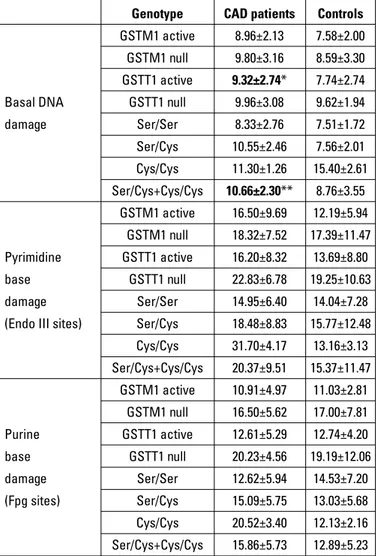
The influences of GSTM1, GSTT1, and hOGG1 genotypes on oxidative DNA damage parameters are presented in Table 4. No significant differences were observed between individuals car-rying the GSTM1, GSTT1, and hOGG1 variant or the wild genotype in terms of oxidative DNA damage parameters in both controls and patients. However, basal DNA damage levels were statisti-cally different in patients as compared with in controls carrying the same genotypes. Patients carrying the GSTT1 active genotype or Ser/Cys + Cys/Cys variants of the hOGG1 showed higher levels of basal DNA damage compared with controls carrying the same genotypes (p=0.014 and 0.022 respectively).
The correlations between the Gensini score as a marker of CAD severity and oxidative DNA damage parameters are presented in Table 5. There were significant correlations between the Gensini score and basal DNA damage (r=0.352, p=0.006) and pyrimidine base damage r=0.318, p=0.014), whereas no significant correlation was observed for purine base damage (p>0.05).
Table 1. Demographic and clinical characteristics of patients and controls
CAD patients (n=29) Controls (n=30) OR (95%CI) P
Age, years±SD 61.5±10.3 54.4±11.6 1.06 (1.01–1.12) 0.020† Gender, Men/Women, n (%) 24 (82.8)/5 (17.2) 24 (80)/6 (20) 0.83 (0.22–3.10) 1.000‡ Hypertension, n (%) 17 (60.7) 7 (25) 4.64 (1.48–4.54) 0.015‡ Diabetes mellitus, n (%) 11 (39.3) 11 (40.7) 0.94 (0.32–2.77) 1.000‡ Hyperlipidemia, n (%) 9 (32.1) 7 (25.9) 1.35 (0.42–4.36) 0.833‡ Smoking, n (%) 16 (55.2) 11 (40.7) 1.79 (0.62–5.17) 0.417‡ Family history, n (%) 7 (24.1) 0 (0) NC 0.005¶ Total cholesterol, mg/dL 187.7±36.1 188.3±62.1 1.00 (0.99–1.01) 0.965† LDL-cholesterol, mg/dL 101.7±21.3 102.8±44.5 0.99 (0.98–1.01) 0.918† HDL- cholesterol, mg/dL 45.3±10.4 47.3±19.3 0.99 (0.95–1.03) 0.649† Triglyceride, mg/dL 265.2±344.4 164.4±84.5 1.00 (0.99–1.01) 0.230† Gensini score 54.1±36.8 7.4±29.9 1.06 (1.03–1.10) <0.001†
‡Continuity correction chi-square test; ¶Fisher's exact test; †Univariate logistic regression analyses; CAD - coronary artery disease; HDL - high–density lipoprotein cholesterol; LDL - low-density lipoprotein cholesterol; NC - not calculated
Table 2. Distribution n (%) of GSTM1, GSTT1 and hOGG1 genotypes in patients and controls
CAD patients Controls OR (95% CI) P Adjusted* OR (95% CI) P
GSTM1 Active 12 (41.4) 16 (53.3) Null 17 (58.6) 14 (46.7) 1.619 (0.578–4.534) 0.510† 0.325 (0.082–1.293) 0.111 GSTT1 Active 23 (79.3) 25 (83.3) Null 6 (20.7) 5 (16.7) 1.304 (0.350–4.858) 0.950† 1.005 (0.200–5.041) 0.995 hOGG1 Ser/Ser 15 (51.7) 17(56.7) Ser/Cys 12 (41.4) 11 (36.7) 1.236 (0.423–3.615) 0.909† 1.230 (0.327–4.631) 0.759 Cys/Cys 2 (6.9) 2 (6.7) 1.133 (0.142–9.065) 1.000‡ 0.510 (0.038–6.861) 0.612
*Adjusted for age, gender, presence of hypertension and smoking habits; †Continuity correction chi-square test; ‡Fisher's exact test; CAD - coronary artery disease
Table 3. Oxidative DNA damage in patients and controls. Values are median (IQR)
CAD patients Controls P
Basal DNA damage 9.16 (3.26) 7.59 (3.23) 0.017 Pyrimidine base damage
(Endo III sites) 15.04 (12.07) 11.02 (6.98) 0.08 PBD (Fpg sites) 12.38 (6.98) 12.24 (4.80) 0.55
Discussion
In the present study, we investigated two different types of biomarkers in CAD patients. We assessed a biomarker of effect (DNA damage) and a biomarker of susceptibility (gene polymorphism) for CAD risk. Our results significantly high-lighted increased basal DNA damage levels in CAD patients with respect to controls. However, neither pyrimidine nor
pu-rine base damage levels, which reflect oxidative DNA dam-age, were significantly different between the groups. On the other hand, basal DNA and pyrimidine base damage levels were significantly correlated with disease severity based on Gensini scoring. Although there were some significant differ-ences in terms of basal DNA damage between the patients and controls carrying the same alleles, genotype analysis did not reveal a clear association between any of the studied SNPs (GSTM1, GSTT1, and hOGG1) and CAD. Although the re-sults of our study pointed out the effects of age, hypertension, and family history as possible risk factors for CAD, none of these factors affect the frequency of the polymorphisms after adjusting the results for age, gender, the presence of hyper-tension, and smoking habits.
The association between DNA damage and CAD was in-vestigated in several studies, and there is increasing evidence that DNA damage plays an important role in atherogenesis. Botto et al. (5) investigated micronucleus (MN) frequency in CAD patients and found that MN levels were higher in these patients than in controls. They also observed a relationship between increased MN frequencies and disease severity. In a follow-up study, major adverse cardiovascular events (MAC-Es) such as heart failure, stroke, or cardiovascular death were reported in CAD patients for a period of 51 months, and MN levels were found to be significantly higher in patients who had MACEs than in those who did not (18). Many factors can cause DNA damage, including mainly oxidative stress. Oxida-tive stress to lipids and DNA caused by free radicals is known to be one of the several fundamental mechanisms that are responsible for developing atherosclerosis and cancer (19). Some possible atherogenic mechanisms, such as increasing ROS production, the potentiation of LDL oxidation, and the diminished release of nitric oxide, were suggested in vari-ous studies (20, 21). Oxidative stress-related studies in CAD patients have usually been performed by measuring the lipid peroxidation or antioxidant capacity in plasma (4, 22). Howev-er, the number of studies on oxidative DNA damage in CAD pa-tients is limited, and the information in the literature regarding the role of DNA damage in the etiology of CAD is inadequate. In the present study, we observed significantly increased lev-els of basal DNA damage but not oxidative DNA damage in the lymphocytes of CAD patients. This finding indicates possible damage to the DNA of these patients; however, this damage, as detected by the modified comet assay, was not a result of oxidized DNA bases. Moreover, basal DNA damage levels in patients were positively correlated with disease severity. Similar studies using the alkaline comet assay have been pre-viously conducted for investigating oxidative DNA damage in CAD patients. Botto et al. (6) demonstrated that increased lev-els of SSBs and oxidized DNA bases correlated with the sever-ity of CAD in patients. Demirbağ et al. (4, 23) investigated DNA damage using the standard alkaline comet assay (not
modi-Table 4. Influences of GSTM1, GSTT1 and hOGG1 genotypes on oxidative DNA damage parameters in patients and controls. Values are mean±SD
Genotype CAD patients Controls
GSTM1 active 8.96±2.13 7.58±2.00
GSTM1 null 9.80±3.16 8.59±3.30
GSTT1 active 9.32±2.74* 7.74±2.74
Basal DNA GSTT1 null 9.96±3.08 9.62±1.94
damage Ser/Ser 8.33±2.76 7.51±1.72 Ser/Cys 10.55±2.46 7.56±2.01 Cys/Cys 11.30±1.26 15.40±2.61 Ser/Cys+Cys/Cys 10.66±2.30** 8.76±3.55 GSTM1 active 16.50±9.69 12.19±5.94 GSTM1 null 18.32±7.52 17.39±11.47 Pyrimidine GSTT1 active 16.20±8.32 13.69±8.80 base GSTT1 null 22.83±6.78 19.25±10.63 damage Ser/Ser 14.95±6.40 14.04±7.28
(Endo III sites) Ser/Cys 18.48±8.83 15.77±12.48
Cys/Cys 31.70±4.17 13.16±3.13 Ser/Cys+Cys/Cys 20.37±9.51 15.37±11.47 GSTM1 active 10.91±4.97 11.03±2.81 GSTM1 null 16.50±5.62 17.00±7.81 Purine GSTT1 active 12.61±5.29 12.74±4.20 base GSTT1 null 20.23±4.56 19.19±12.06 damage Ser/Ser 12.62±5.94 14.53±7.20 (Fpg sites) Ser/Cys 15.09±5.75 13.03±5.68 Cys/Cys 20.52±3.40 12.13±2.16 Ser/Cys+Cys/Cys 15.86±5.73 12.89±5.23
*P=0.014 (vs controls); **P=0.022 (vs. controls); Mann-Whitney U test. CAD - coronary artery disease
Table 5. Correlation between oxidative DNA damage parameters and Gensini score
Correlation coefficient (r) Significance (P)
Basal DNA damage 0.352 0.006
Pyrimidine base damage 0.318 0.014
(Endo III sites)
PBD, (Fpg sites) 0.110 0.408
fied with repair enzymes) in CAD patients and reported higher SSBs and lower plasma total antioxidant capacity levels in patients with acute coronary syndrome than those in patients with stable angina and healthy subjects. In another study, contrary to our findings, significantly increased levels of oxi-dized pyrimidine and purine bases were found; however, no significant difference was reported in terms of SSBs between CAD patients and controls (24). Recently, Rajesh et al. (3) as-sessed DNA damage using the standard comet assay in a rather large population of CAD patients and reported signifi-cantly higher levels of DNA damage as well as malondialde-hyde (MDA) and nitrite/nitrate levels in patients than those in controls.
In the present study, the roles of GSTM1, GSTT1, and hOGG1 gene polymorphisms and their relationship with oxi-dative DNA damage were also investigated in CAD patients. The GSTM1 null genotype is very common among Caucasians and is found in approximately 50% of the population (25). A homozygous deletion of the GSTT1 gene (null genotype) is found in 10%–20% of Caucasians, making them unable to per-form GSTT1-mediated detoxification reactions (26). Genes coding for DNA repair molecules, such as hOGG1, have been proposed as candidate susceptibility genes because of the importance of maintaining genomic integrity. (11). Our results regarding the frequencies of studied gene polymorphisms are consistent with those of previous Turkish studies (27, 28). However, our results did not show any statistical difference between CAD patients and controls in terms of GSTM1, GSTT1, and hOGG1 variant allele frequency. There are some previous studies investigating the effect of these polymorphisms on the risk of developing CAD, and the fi ndings are contradic-tory. In a recent study, similar to our findings, the frequency of the GSTM1 and GSTT1 null alleles did not differ among CAD patients and controls; however, a combined effect of CYP1A1 and GSTT1 variant genotypes was reported for the risk of de-veloping CAD (7). Similarly, Bazo et al. (29) did not detect any association between GSTM1 and GSTT1 gene polymorphisms and coronary atherosclerosis in a Brazilian population. On the other hand, Girisha et al. (30) reported a decreased frequency of the GSTT1 null genotype and no significant association with GSTM1 genotypes in CAD patients compared with in controls. An association between GST polymorphisms and CAD risk is demonstrated mostly in studies investigating the interaction of these polymorphisms with cigarette smoking. In three dif-ferent studies, a strengthened effect of GST-null genotypes on smoking-induced CAD risk was reported, although GST null genotypes did not differ between patients and controls (10, 31, 32). Similar results were obtained in a large cohort study pointing out a positive interaction with the GSTT1 null geno-type (8).
hOGG1 is one of the polymorphic repair genes in the BER pathway, which has a critical role in removing damaged DNA
bases. It catalyzes the cleavage of the glycosylic bond be-tween the modified base and the sugar moiety; as a result, an abasic apurinic or apyrimidinic (AP) site occurs in the DNA. Studies on the role of hOGG1 gene polymorphism in CAD risk are limited. Two recent studies on a Taiwanese popula-tion demonstrated contradictory results. Wang et al. (33) re-ported an association between hOGG1 polymorphism and an increased risk of CAD, whereas Shyu et al. (34) found no as-sociation. In a cohort study, 98 subjects suffering from severe atherosclerotic lesions were followed up for 14 years, and an adverse effect on survival was detected in subjects having the double GSTM1 and GSTT1 deletion, although the frequen-cies of both null alleles were not different from the general population. In the same study, the survival rate was not in-fluenced by hOGG1 polymorphism (35). Gökkuşu et al. (36) investigated the role of different DNA repair enzyme polymor-phisms in progressing CAD, and they reported that XRCC3 and hOGG1 genetic variants may be risk factors that may lead to the development of CAD. Another study on a Chinese popula-tion revealed similar results in coronary ectasia patients, with a significant association of hOGG1 Ser326Cys polymorphism. The study also reported higher 8-OHdG levels in subjects car-rying the hOGG1 variant allele (37).
During the past few years, a large number of studies have focused on individual susceptibility for predicting the risk of developing some common diseases such as cancer and ath-erosclerosis. A variety of factors can contribute to variations in the individual response to environmental mutagenic agents, thus causing some individuals to be susceptible to these dis-ease and others to be resistant. Moreover, combinations of biomarkers are valuable in assessing the individual risk of diseases (38, 39).
Study limitations
The small sample size limited us from analyzing more fac-tors involving the risk of CAD, such as smoking. Therefore, these preliminary findings need to be validated in larger popu-lations.
Conclusion
In this study, we demonstrated the role of DNA damage in CAD; however, no significant differences were observed in terms of oxidized DNA bases between patients and con-trols. Therefore, we can conclude that the DNA damage we observed does not originate from ROS. In the present study, the studied genotypes (GSTM1, GSTT1, and hOGG1) showed similar frequencies between groups. Although there were some significant differences in terms of basal DNA damage between the patients and controls carrying the same alleles, genotype analysis did not reveal a clear association between any of the studied SNPs and CAD.
Conflict of interest: None declared. Peer-review: Externally peer-reviewed.
Authorship contributions: Concept – All authors; Design – All authors; Supervision – E.K., G.T., E.Ö.; Fundings – S.Ş.; Materials – E.K., G.T., E.Ö., K.O., M.K.A.; Data collection &/or processing – E.K., G.T., E.Ö., K.O., M.K.A.; Analysis &/or interpretation – E.K., G.T., E.Ö.; Literature search – All authors; Writing – E.K.; Critical review – All authors.
References
1. Ross R. Atherosclerosis is an inflammatory disease. Am Heart J 1999; 138: 419-20. Crossref
2. Hauser ER, Pericak-Vance MA. Genetic analysis for common complex disease. Am Heart J 2000; 140: 36-44. Crossref
3. Rajesh KG, Surekha RH, Mrudula SK, Prasad Y, Sanjib KS, Prathiba N. Oxidative and nitrosative stress in association with DNA damage in coronary heart disease. Singapore Med J 2011; 52: 283-8. 4. Demirbağ R, Yılmaz R, Koçyiğit A. Relationship between DNA
damage, total antioxidant capacity and coronary artery disease. Mutat Res 2005; 570: 197-203. Crossref
5. Botto N, Rizza A, Colombo MG, Mazzone AM, Manfredi S, Masetti S, et al. Evidence for DNA damage in patients with coronary ar-tery disease. Mutat Res 2001; 493: 23-30. Crossref
6. Botto N, Masetti S, Petrozzi L, Vassalle C, Manfredi S, Biagini A, et al. Elevated levels of oxidative DNA damage in patients with coronary artery disease. Coron Artery Dis 2002; 13: 269-74. 7. Taşpınar M, Aydos S, Sakırağaoğu O, Düzen IV, Yalçınkaya A,
Öz-tuna D, et al. Impact of genetic variations of the CYP1A1, GSTT1, and GSTM1 genes on the risk of coronary artery disease. DNA Cell Biol 2012; 31: 211-8. Crossref
8. Olshan AF, Li R, Pankow JS, Bray M, Tyroler HA, Chambless LE, et al. Risk of atherosclerosis: interaction of smoking and glutathione S-transferase genes. Epidemiology 2003; 14: 321-7. Crossref
9. Strange RC, Spiteri MA, Ramachandran S, Fryer AA. Glutathione-S-transferase family of enzymes. Mutat Res 2001; 482: 21-6. 10. Kim SJ, Kim MG, Kim KS, Song JS, Yim SV, Chung JH. Impact of
glutathione S-transferase M1 and T1 gene polymorphisms on the smoking-related coronary artery disease. J Korean Med Sci 2008; 23: 365-72. Crossref
11. Goode EL, Ulrich CM, Potter JD. Polymorphisms in DNA repair genes and associations with cancer risk. Cancer Epidemiol Bio-markers Prev 2002; 11: 1513-30.
12. Kohno T, Shinmura K, Tosaka M, Tani M, Kim SR, Sugimura H, et al. Genetic polymorphisms and alternative splicing of the hOGG1 gene, that is involved in the repair of 8-hydroxyguanine in dam-aged DNA. Oncogene 1998; 16: 3219-25.
13. Collins AR, Duthie SJ, Dobson VL. Direct enzymic detection of endogenous oxidative base damage in human lymphocyte DNA. Carcinogenesis 1993; 14: 1733-5. Crossref
14. Güven M, Güven GS, Öz E, Özaydın A, Batar B, Ulutin T, et al. DNA repair gene XRCC1 and XPD polymorphisms and their association with coronary artery disease risks and micronucleus frequency. Heart Vessels 2007; 22: 355-60. Crossref
15. Gensini GG. A more meaningful scoring system for determining the severity of coronary heart disease. Am J Cardiol 1983; 51: 606.
16. Kadıoğlu E, Kocabaş NA, Demircigil GC, Çoşkun E, Özcağlı E, Durmaz E, et al. Assessment of individual susceptibility to base-line DNA and cytogenetic damage in a healthy Turkish population: evaluation with lifestyle factors. Genet Test Mol Biomarkers 2012; 16: 1157-64.
17. Johns MB Jr, Paulus-Thomas JE. Purification of human genomic DNA from whole blood using sodium perchlorate in place of phe-nol. Anal Biochem 1989; 180: 276-8.
18. Federici C, Botto N, Manfredi S, Rizza A, Del Fiandra M, Andre-assi MG. Relation of increased chromosomal damage to future adverse cardiac events in patients with known coronary artery disease. Am J Cardiol 2008; 102: 1296-1300. Crossref
19. Park E, Kyoung Park Y, Kim SM, Lee HJ, Kang MH. Susceptibility to oxidative stress is greater in Korean patients with coronary artery disease than healthy subjects. J Clin Biochem Nutr 2009; 45: 341-6. 20. Cavalca V, Cighetti G, Bamonti F, Loaldi A, Bortone L, Novembrino
C, et al. Oxidative stress and homocysteine in coronary artery dis-ease. Clin Chem 2001; 47: 887-92.
21. Mahmoudi M, Mercer J, Bennett M. DNA damage and repair in atherosclerosis. Cardiovasc Res 2006; 71: 259-68.
22. Mutlu-Türkoğlu U, Akalın Z, İlhan E, Yılmaz E, Bilge A, Nişancı Y, et al. Increased plasma malondialdehyde and protein carbonyl lev-els and lymphocyte DNA damage in patients with angiographically defined coronary artery disease. Clin Biochem 2005; 38: 1059-65. 23. Demirbağ R, Yılmaz R, Gür M, Koçyiğit A, Çelik H, Güzel S, et al.
Lymphocyte DNA damage in patients with acute coronary drome and its relationship with severity of acute coronary syn-drome. Mutat Res 2005; 578: 298-307. Crossref
24. Yurdakul S, Özben B, Bilge AK, Türkoğlu UM, Arkaya S, Nişancı Y. Oxidative DNA damage is significantly correlated with flow-medi-ated dilation in patients with coronary artery disease. J Investig Med 2008; 56: 925-30. Crossref
25. Hirvonen A. Polymorphisms of xenobiotic-metabolizing enzymes and susceptibility to cancer. Environ Health Perspect 1999; 107: 37-47. Crossref
26. Norppa H. Genetic polymorphisms and chromosome damage. Int J Hyg Environ Health 2001; 204: 31-8. Crossref
27. Ada AO, Süzen SH, İşçan M. Polymorphisms of cytochrome P450 1A1, glutathione S-transferases M1 and T1 in a Turkish popula-tion. Toxicol Lett 2004; 151: 311-5. Crossref
28. Işbir O, Görmüş U, Ergen HA, Çakmakoğlu B, Kahraman OT, Baykan N. XPD and hOGG1 gene polymorphisms in reperfusion oxidative stress. Genet Mol Res 2011; 10: 3157-62. Crossref
29. Bazo AP, Salvadori D Jr, Salvadori RA, Sodre LP, da Silva GN, de Camargoa EA, et al. DNA repair gene polymorphism is associated with the genetic basis of atherosclerotic coronary artery disease. Cardiovasc Pathol 2011; 20: 9-15.
30. Girisha KM, Gilmour A, Mastana S, Singh VP, Sinha N, Tewari S, et al. T1 and M1 polymorphism in glutathione S-transferase gene and coronary artery disease in North Indian population. Indian J Med Sci 2004; 58: 520-6.
31. Masetti S, Botto N, Manfredi S, Colombo MG, Rizza A, Vassalle C, et al. Interactive effect of the glutathione S-transferase genes and cigarette smoking on occurrence and severity of coronary artery risk. J Mol Med 2003; 81: 488-94. Crossref
32. Tamer L, Ercan B, Çamsarı A, Yıldırım H, Çiçek D, Sucu N, et al. Glutathione S-transferase gene polymorphism as a susceptibil-ity factor in smoking-related coronary artery disease. Basic Res
Cardiol 2004; 99: 223-9. Crossref
33. Wang CL, Lin TH, Lin HY, Sheu SH, Yu ML, Hsiao PJ, et al. The 8-oxoguanine glycosylase I (hOGG1) Ser326Cys variant affects the susceptibility to multi-vessel disease in Taiwan coronary ar-tery disease patients. Thromb Res 2010; 126: 319-23. Crossref
34. Shyu HY, Shieh JC, Ji-Ho L, Wang HW, Cheng CW. Polymorphisms of DNA Repair pathway genes and cigarette smoking in relation to susceptibility to large artery atherosclerotic stroke among ethnic Chinese in Taiwan. J Atheroscler Thromb 2012; 19: 316-25. 35. Izzotti A, Piana A, Minniti G, Vercelli M, Perrone L, De Flora S.
Survival of atherosclerotic patients as related to oxidative stress and gene polymorphisms. Mutat Res 2007; 621: 119-28. Crossref
36. Gökkuşu C, Çakmakoğlu B, Daşdemir S, Tulubas F, Elitok A, Tam-er S, et al. Association between genetic variants of DNA repair genes and coronary artery disease. Genet Test Mol Biomarkers 2013; 17: 307-13. Crossref
37. Hsu PC, Wang CL, Su HM, Juo SH, Lin TH, Voon WC, et al. The hOGG1 Ser326Cys gene polymorphism and the risk of coronary ectasia in the Chinese population. Int J Mol Sci 2014; 15: 1671-82. 38. Au WW. Usefulness of biomarkers in population studies: From exposure to susceptibility and to prediction of cancer. Int J Hyg Environ Health 2007; 210: 239-46.
39. Norppa H. Genetic susceptibility, biomarker respones and cancer. Mutat Res 2003; 544: 339-48. Crossref