Case Report
ABSTRACT
This study aimed presenting a case of a 64-year-old woman with a rare diagnosis of myofibroblastoma (MFB). MFB is one of the rare, benign, spindle-like stromal tumors arising from the connective tissue of the breast. MFBs are often confused with fibroadenomas and hamartomas because of their benign characteristic appearance on breast imaging and are diagnosed after excisional biopsies. Their differential diagnosis with malignant neoplasia of the breast is important because of their wide morphological spectrum. Our case also demonstrated a breast mass with benign imaging characteristics and a needle core biopsy revealing a benign, spindle-like stromal tumor. The pathological examination performed after the excision of the lump demonstrated a collagenous-/ fibrous-type MFB. This case report emphasizes the rare but important place of MFB variants of the breast in the differential diagnosis of breast mass. Keywords: Myofibroblastoma, spindle cell, stromal tumor, breast mass, phyllodes tumor, fibroadenoma, hamartoma
Myofibroblastoma of the Breast
Hüseyin Özgür Aytaç
1, Filiz Aka Bolat
2, Tuba Canpolat
2, Ayşin Pourbagher
31Department of General Surgery, Başkent University Faculty of Medicine, Adana, Turkey 2Department of Pathology, Başkent University Faculty of Medicine, Adana, Turkey 3Division of Radiodiagnosis, Başkent University Faculty of Medicine, Adana, Turkey
Address for Correspondence:
Hüseyin Özgür Aytaç, e-mail: [email protected] Accepted: 20.07.2015Received: 29.05.2015
J Breast Health 2015; 11: 192-4
DOI: 10.5152/tjbh.2015.2655
192
Introduction
Myofibroblastoma (MFB) is a rare spindle cell tumor arising from breast stroma. Toker et al first defined it in 1981 (1). It is seen in both sexes at older age (2). Several subtypes of MFB have been defined such as; classic, cellular, collagenous / fibrous, lipomateous, infiltrative, myxoid, epithelioid, and decidua-like variant (2). Clinically they are often mistaken for fibroadenoma and hamartomas since they have a regular and non-invasive appearance on mammography and breast ultrasound (2). We aimed to report a rare collagenous / fibrous type MFB case with emphasis on differential diagnosis.
Case Presentation
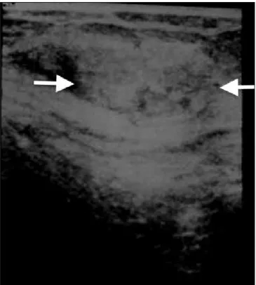
A 64-year-old woman was admitted with complaints of a growing mass in her left breast. The patient had no co-morbidities other than hypertension. She gave birth to three children and nursed them, did not use birth control or hormonal drugs, and had been in menopause for 18 years. There was no family history of breast cancer. On physical examination, a 3 cm in diameter, hard, well-circumscribed, partially mobile mass was detected in the upper outer quadrant of the left breast 3 cm away from the areola. The right breast and both axilla were normal. A 1.2x3 cm non-calcified oval-shaped mass was observed on mediolateraloblique and craniocaudal mammography views in the upper outer quadrant of the left breast. It did not contain fatty tissue and had partially indistinct margin on mammography (Figure 1). On ultrasonography, this mass was oval-shaped with mixed echogenicity (Figure 2). The lesion was BI-RADS category 4. The radiologic differential diagnosis of this lesion included fibroadenoma, fibroadenolipoma, and angiolipoma. Ultrasound guided core needle biopsy was performed. Tissue blocks obtained from biopsy were fixed in neutral buffered 10% formalin for 24 hours and were stained with hematoxylin-eosin (HE). On histological sections, hyalinized-collagenized stroma and small number of inflammatory cells were detected with spindle cells that stain positive for desmin, CD34 and vimentin. The preliminary diagnosis was “benign spindle cell stromal tumor” thus; excision of the lesion was planned. The mass was excised with a safety margin of one cm. The tissue fixation and dying process de-scribed above was repeated in the same manner for tissue blocks prepared from the excision. On histological section, a tumor lesion form-ing nodules that was separated from the surroundform-ing breast tissue with a fine pseudocapsule was observed. The described lesion consisted of spindle cells forming short fascicles mixed with hyalinized bright eosinophilic bands in wide areas. Mitotic activity was not detected in the lesion. Variable amount of fatty tissue and patchy perivascular lymphocytic infiltration was observed within the lesion (Figure 3).
On immunohistochemical examination, spindle cells showed wide-spread positive staining with CD34, vimentin, and desmin, and focal positive staining with smooth muscle actin and h-caldesmon. Spindle cell nuclei were positive for Estrogen receptor (ER) and Progesterone receptor (PR), and negative for S-100 protein and keratin. Ki-67 index was 1%. With these findings, the lesion was accepted as collagenous/ fibrous-type MFB. An informed consent was obtained for this case report.
Discussion and Conclusions
Myofibroblastoma that is a rare, benign mesenchymal neoplasm of the breast arising from myofibroblasts and fibroblasts, presents with soli-tary, hard, painless, mobile and slow-growing mass.
Histopathologically classic, cellular, collagenous / fibrous, lipomate-ous, infiltrative, myxoid, epithelioid, and decidua-like variants have been identified (2). On immunohistochemical evaluation, MFB typically stains positive with CD34 and vimentin, desmin, SMA, and displays variable staining with CD99 and bcl-2. In the presence of smooth muscle differentiation in mammary fibroblasts, it may exhibit focal h-caldesmon expression. In most cases, positive staining with ER, PR and androgen receptors is observed (3).
Since MFB has many variants, differential diagnosis of breast carcino-mas, benign and malignant spindle cell neoplasms may be required. Epithelioid type MFB can be confused with invasive lobular breast carcinoma, and cellular-type MFB can be mistaken for metaplastic breast carcinoma, negative staining for keratin helps in the differen-tial diagnosis. Other spindle cell lesions that should be considered in the differential diagnosis include solitary fibrous tumors of the breast, benign spindle cell lipomas, low-grade myofibroblastic sarcomas and phyllodes tumors, leiomyomas, angiomyolipomas, nodular fasciitis, desmoid-type fibromatosis, benign and malignant myxoid lesions, and low-grade sarcomas (4).
Myofibroblastoma has no specific radiological findings. On mammog-raphy, they usually appear as well-defined, round or oval lesions with-out calcification. On ultrasound, they are viewed as well-defined hy-poechoic or mixed echogenic solid lesions (5). Radiological differential diagnoses include fibroadenoma, the most common solid mass in wom-en, and angiolipoma. Fibroadenoma and angiolipomas may represent with mixed density on mammography, fat density within soft tissue density. Visualization of both tumors on mammography is dependent on the adipose tissue content within the lesion. In our case, radiolucent areas consisting of adipose tissue was not observed on mammography. Myofibroblastoma is a rare, benign soft tissue tumor of the breast. They are usually diagnosed after excision of the suspicious mass due to their indolent growth course and absence of specific radiological findings. Exclusion of malignancies that could be misdiagnosed as variants other than the classic type is especially important. Knowl-edge on MFB variants is essential in differentiating benign and / or malignant spindle cell lesions.
Figure 1. Left mediolateraloblique and craniocaudal mammography views showing a non-calcified, oval-shaped mass that is 1.2x3 cm in size in the upper outer quadrant. It has slightly irregular borders (arrow), BI-RADS 4
Figure 3. Collagenous / fibrous type myofibroblastoma that is separated from the surrounding breast tissue with a pseudocapsule, rich in collagen, with relatively small spindle cells, and containing fat and perivascular lymphoid infiltration (arrows) (hematoxylin and eosin x40)
Figure 2. Mixed echogenicity on ultrasound in the oval-shaped mass
193
Informed Consent: Written informed consent was obtained from patients who participated in this study.
Peer-review: Externally peer-reviewed.
Yazar Katkıları / Author Contributions: Concept - H.Ö.A, F.A.B.; Design - H.O.A, T.C.; Supervision - F.A.B, A.P.; Funding - F.A.B, A.P.; Materials - H.Ö.A., T.C.; Data Collection and/or Processing - H.Ö.A., F.A.B.; Analysis and/or Interpretation - H.Ö.A., A.P.; Literature Review - H.Ö.A., F.A.B.; Wri-ter - H.Ö.A., T.C.; Critical Review - A.P., F.A.B.
Conflict of Interest: No conflict of interest was declared by the authors. Financial Disclosure: The authors declared that this study has received no financial support.
References
1. Toker C, Tang CK, Whitely JF, Berkheiser SW, Rachman R. Benign spindle cell breast tumor. Cancer 1981; 48:1615-1622. [CrossRef]
2. Mele M, Jensen V, Wronecki A, Lelkaitis G. Myofibroblastoma of the breast: Case report and literature review. Int J Surg Case Rep 2011; 2:93-96. (PMID: 22096693) [CrossRef]
3. Magro G, Bisceglia M, Michal M. Expression of steroid hormone receptors, their regulated proteins, and bcl‐2 protein in myofibroblastoma of the breast. Histopathology 2000; 36:515-521. (PMID: 10849093) [CrossRef]
4. Özkayalar H, Kabukçuoğlu F, Tanık C, Eken KG, Eryılmaz ÖT, Yetkin G. Memenin mezenkimal tümörleri. Şişli Etfal Hastanesi Tıp Bülteni 2010; 44:11.
5. Yoo EY, Shin JH, Ko EY, Han B-K, Oh YL. Myofibroblastoma of the Female Breast Mammographic, Sonographic, and Magnetic Resonance Imaging Findings. J Ultrasound Med 2010; 29:1833-1836.