Address for Correspondence: Dr. Sevinç Sarınç Ulaşlı, Afyon Kocatepe Üniversitesi Tıp Fakültesi, Göğüs Hastalıkları Anabilim Dalı, İzmir Karayolu 7. Km 03200 Afyon-Türkiye
Phone: +90 505 307 36 58 Fax: +90 272 246 33 22 E-mail: [email protected] Accepted Date: 13.12.2013 Available Online Date: 16.04.2014
©Copyright 2014 by Turkish Society of Cardiology - Available online at www.anakarder.com DOI:10.5152/akd.2014.4687
Tuğce Şahin Özdemirel, Sevinç Sarınç Ulaşlı
1, Begüm Yetiş
2, Emir Karaçağlar
2, Nilüfer Bayraktar
3, Gaye Ulubay
4 Clinic of Chest Diseases, Atatürk Chest Diseases and Thoracic Surgery Training and Research Hospital; Ankara-Turkey1Department of Pulmonary Diseases, Faculty of Medicine, Afyon Kocatepe University; Afyon-Turkey
Departments of 2Cardiology, 3Biochemistry and 4Pulmonary Diseases, Faculty of Medicine, Başkent University; Ankara-Turkey
Effects of right ventricular dysfunction on exercise capacity and
quality of life and associations with serum NT-proBNP levels in COPD:
an observational study
A
BSTRACTObjective: During the course of chronic obstructive pulmonary disease (COPD), pulmonary hypertension (PH) and right ventricular (RV) failure may develop due to elevated afterload of the RV. In those patients, exercise capacity is reduced due to pulmonary and cardiac limitations. We investigated relationships between serum N-terminal of proB-type natriuretic peptide (NT-proBNP) and RV functions with exercise capacity and quality of life in patients COPD.
Methods: An observational case-control study was conducted. We enrolled 31 moderate and severe COPD patients, and 20 subjects without chronic diseases as control group. Parameters reflecting the right ventricular diastolic and systolic functions by echocardiography along with serum NT-proBNP levels were assessed. Cardiopulmonary exercise testing and Short Form-36 (SF-36) were applied.
Results: Serum NT-proBNP levels were higher in COPD patients than control group (p=0.003). Serum NT-proBNP level was found to be related with pulmonary arterial pressure. Serum NT-proBNP levels were negatively correlated with anaerobic threshold oxygen uptake (AT VO2) and peak oxygen uptake (PVO2) values. Early ventricular filling velocity (Em) was lower in COPD patients. Em wave was significantly correlated with O2 pulse. There was a positive relationship between tricuspid E/A ratio and VO2 value at AT. SF-36 domains of physical functioning, general health and role limitation due to physical disorder were significantly correlated with AT VO2, PVO2 and O2 pulse.
Conclusion: Exercise limitation may be predicted by assessment of right ventricule functions and NT-proBNP levels and exercise limitation impairs quality of life in COPD patients. (Anadolu Kardiyol Derg 2014; 14: 370-7)
Key words: COPD, pulmonary hypertension, NT-proBNP, cardiopulmonary exercise testing, right ventricular dysfunction, quality of life.
Introduction
Chronic obstructive pulmonary disease (COPD) is a major cause of morbidity all over the world and characterized by irre-versible airway obstruction (1). Impaired exercise capacity can result from ventilatory limitation, respiratory muscle weakness and abnormal gas exchange together with cardiac limitation in COPD patients. Pulmonary hypertension and right heart failure may develop in the course of COPD. Pulmonary hypertension (PH) with increased right ventricle load, failure and elevated pulmo-nary vascular resistance is a cause of early death. Development of PH is also associated with poor quality of life and exercise limitation. Diastolic dysfunction is defined as inconvenient increase in right ventricle diastolic pressure with insufficient
fill-ing of right ventricle. Right ventricular dysfunction can be seen before the development of pulmonary hypertension and cor pul-monale in COPD patients (2-4).
N-terminal of proB-type natriuretic peptide (NT-proBNP) is a precursor of brain natriuretic peptide (BNP). Measurement of NT-proBNP level is more reliable than measurement of BNP due to the short half life of BNP. N-terminal proB-type natriuretic peptide (NT-proBNP) is an important and helpful indicator in differentiating congestive heart failure from lung diseases (5). However, elevated NT-proBNP levels have been found in patients with COPD and in patients with right ven-tricular dysfunction (6, 7). Moreover, increased NT-proBNP levels can be seen in many diseases such as left ventricular dysfunction, right ventricular dysfunction secondary to
pul-monary diseases, infectious diseases, endocrinological dis-orders, and high output status without decreased left ven-tricular ejection fraction (8). Therefore measurement of NT-pro BNP levels can also be useful in the diagnosis of impaired right ventricular functions in COPD patients.
Dyspnea is a major symptom limiting exercise capacity in COPD patients (1). Cardiopulmonary exercise testing is a nonin-vasive tool in the diagnosis of pulmonary and cardiovascular diseases and also helps to determine disease severity.
Although significance of serum NT-proBNP in COPD patients has been investigated previously, there has been no study inves-tigating the relationship between serum NT-proBNP and exer-cise capacity in COPD patients according to our knowledge. We hypothesize that serum NT-proBNP level might indicate impaired right ventricular functions, quality of life and exercise limitation in COPD patients.
The goal of this study was to investigate relationships between serum NT-proBNP level and right ventricular functions with exercise capacity and quality of life in patients with COPD.
Methods
A case control observational study was designed. The Ethics Committee of our university certified the study protocol and study was conducted within research project as KA09/274.
Study population
Patients with COPD followed in department of pulmonary diseases outpatient clinic between August 2009 and August 2010 were consecutively included. All participants provided informed consent. COPD was diagnosed based on the criteria defined in Global Initiative for Chronic Obstructive Lung Disease (GOLD) guidelines (1).
Thirty-one patients with moderate, severe and very severe COPD patients during stable period and 20 healthy individuals were recruited to the study. Subjects in the control group were older than 40 years of age without any medical disorders.
COPD patients during acute exacerbation period, patients with asthma, hypoxia, pneumonia, left ventricular systolic dys-function, valvular pathology, arrhythmia, renal failure, diffuse parenchymal lung diseases, lung carcinoma and other malig-nancies, neuromuscular disorders affecting exercise capacity and subjects younger than 40 years of age were all excluded from the study.
Study protocol
Pulmonary function tests, echocardiography, plasma NT-pro BNP measurement, CPET and short form-36 (SF-36) question-naire were performed to all subjects.
Pulmonary function tests
Pulmonary function tests (PFTs) were performed with a clinical spirometer (SensorMedics Vmax spectra 229, Bilthoven, The Netherlands). Maximal expiratory flow maneuver was
per-formed by patients and control subjects. Forced expiratory vol-ume at 1 second (FEV1) and forced vital capacity (FVC) values were obtained and FEV1/FVC was calculated. Standard PFTs including spirometry and lung volumes were evaluated accord-ing to the previously described guidelines (9). Patients with post-bronchodilator FEV1 <80% of predicted value, FEV1/FVC <70%, and irreversible airflow obstruction were recruited to the study (10). Post-bronchodilator FEV1 values were used to define dis-ease stage according to the GOLD severity classification.
Echocardiography
Tissue Doppler and 2-dimensional echocardiographic mea-surements of 31 patients with moderate, severe and very severe COPD patients during stable period and 20 healthy individuals were carried out in the supine position according to the recom-mendations of American Society of Echocardiography with Acuson Sequoia C-256 (Acuson Corporation, California, USA) cardiac ultrasound machine and 3.5 MHz probe (11). Apical 4-chamber, and parasternal long-axis views were obtained. The left ventricular ejection fraction (EF%) was calculated using the modified Simpson’s rule (12).
Mean pulmonary arterial pressure (PAP) was figured out by the formula of 79- (0.45X pulmonary artery acceleration time) (13, 14).
Right ventricular diastolic velocities were obtained by using pulsed tissue Doppler imaging [early ventricular filling -early diastolic wave (Em) and late diastolic wave during atrial con-traction (Am)]. Peak early (E) and late (A) diastolic velocities of tricuspid valve were recorded during apical 4-chamber view. Assessment of right ventricular (RV) diastolic function was car-ried out by pulsed Doppler of the tricuspid inflow and tissue Doppler of the lateral tricuspid annulus. Grading of RV diastolic dysfunction was performed. Patients with tricuspid E/A ratio <0.8 was determined as grade 1 RV diastolic dysfunction and a tricuspid E/A ratio of 0.8 to 2.1 with an E/e’ ratio >6 as grade 2 RV diastolic dysfunction (pseudonormal filling). We had no subject with a tricuspid E/A ratio >2.1 with deceleration time <120 ms. Tricuspid Annular Plane Systolic Excursion (TAPSE) - was mea-sured from the tricuspid lateral annulus (15, 16).
Cardiopulmonary exercise test
Cycle ergometer (Ergo-metrics 900; SensorMedics Bilthoven, The Netherlands) was used for cardiopulmonary exercise test (CPET). All subjects underwent symptom-limited exercise with a facemask (Rudolph Face Mask for Exercise Testing; Hans Rudolph Inc., Kansas City, MO, USA) fixed on face. After three-min baseline resting period records, a three-three-min warm-up peri-od (60 rpm was the maintainance pedaling rate) was started. And then incremental work (10-15 W elevation for each minute) was applied (17). An automated exercise testing system (Desktop Diagnostics/CPX; Medical Graphics Corporation, St. Paul, MI, USA) was used for collecting data. The maximum work rate for half a minute was saved. During CPET, continious monitorization of 12 lead electrocardiography, blood pressure, and pulse oxy-gen saturation was performed. Peak oxyoxy-gen uptake (PVO2 mL/
kg/min), peak CO2 output, and VE/VCO2 values were evaluated. The two-slope method was used to determine anaerobic thresh-old (AT L/min). The equation of Wasserman et al. (17) was used to determine age-predicted values. Symptoms like fatigue, dys-pnea, dizziness for ending the test were also noted.
The SF-36 questionnaire
The SF-36 questionnaire was used to evaluate quality of life (QOL) (18, 19). This questionnaire has been validated for COPD patients previously. The subjects’s daily routine activities, social life and exercise performance were determined with 36 ques-tions of this item. Main eight domains as physical function, social function, physical and emotional role function, mental health, bodily pain, vitality, and general health perception were found. A computer algorithm was used to score the responses to the SF-36 (20).
NT-proBNP measurement
Blood samples were drawn in tubes containing EDTA. Plasma NT-proBNP was measured with electrochemilumines-cence immunoassay on an Elecsys 2010 system (Roche Diagnostics, Mannheim, Germany). NT-proBNP concentrations were measured as pg/mL. Serum NT-proBNP was evalauted as normal when below 84 pg/mL for males and 155 pg/mL for females.
Statistical analysis
Statistical analysis was accomplished using SPSS (SPSS ver-sion 20.0; SPSS Inc., Chicago, IL, USA). Data was expressed as mean values±SD and median (range min-max) for continious vari-ables, percentage for categorical variables in descriptive analysis. Distribution of continuous variables were checked with
Shapiro-Parameters COPD patients Control subjects P
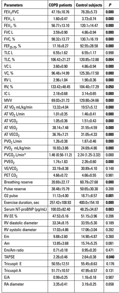
FEV1/FVC 47.19±10.76 78.20±5.73 0.000 FEV1, L 1.60±0.47 3.73±0.74 0.000 FEV1, % 56.77±13.10 120.1±14.47 0.000 FVC L 3.59±0.90 4.86±0.94 0.000 FVC, % 98.32±13.77 130.7±16.19 0.000 FEF25-75, % 17.16±8.27 92.55±28.58 0.000 TLC L 6.55±1.62 6.55±1.17 0.998 TLC, % 106.42±21.27 120.85±12.88 0.004 VC L 3.60±0.90 4.86±0.94 0.000 VC, % 96.48±14.99 125.30±17.50 0.000 RV L 2.96±1.04 1.90±0.36 0.000 RV, % 133.42±48.45 104.40±17.29 0.050 IC L 2.18±0.68 3.14±0.65 0.000 MVV 69.03±31.73 129.80±34.66 0.000 AT VO2 mL/kg/min 13.32±4.04 19.57±5.12 0.000 AT VO2 L/min 1.01±0.35 1.46±0.41 0.000 AT VCO2 1.05±0.36 1.51±0.43 0.000 AT VEO2 38.14±7.48 31.55±4.59 0.000 AT VECO2 36.76±7.21 31.05±4.33 0.000 PVO2 L/min 1.26±0.38 1.87±0.46 0.000 PVO2 mL/kg/min 16.93±3.86 24.69±4.86 0.000 PVCO2 L/min* 1.46 (0.56-11.2) 2.24 (1.25-3.32) 0.000 PVEO2 1.74±1.83 2.26±0.60 0.000 VE/VCO2 33.19±8.38 30.89±4.10 0.148 PET CO2 4.66±0.72 4.66±0.55 0.981 Breathing reserve 20.69±22.17 60.79±27.68 0.000 Pulse reserve 38.48±15.79 50.65±30.30 0.288 O2 pulse 11.13±4.00 18.71±8.57 0.001 Exercise duration, sec 257.42±108.93 400.0±154.10 0.000 Serum NT-proBNP (pg/mL) 100.03±82.40 48.25±34.87 0.003 RV EF, % 47.52±5.16 51.15±3.96 0.206 RV diastolic diameter 33.34±8.15 33.55±5.30 0.189 RV systolic diameter 17.03±4.86 17.06±3.04 0.282 Em 9.88±2.60 14.90±4.87 0.360 Am 13.85±3.68 15.74±5.25 0.081 Em/Am ratio 0.71±0.18 0.95±0.20 0.471 TAPSE 2.26±0.46 2.64±0.38 0.040 Tricuspit E 50.55±12.51 55.45±8.63 0.176 Tricuspit A 51.77±10.57 47.95±8.57 0.131 E/A 0.99±0.25 1.16±0.18 0.907 RA diameter 3.35±0.41 3.19±0.25 0.658 Table 2. Pulmonary function test, cardiopulmonary exercise testing, serum NT-proBNP. echocardiography and SF-36 results of study population
COPD patients Control subjects (n=31) (n=20) P
Age, mean±SD, years* 61.2±7.5 59.3±7.0 0.671 Gender F/M n (%)** 3 (9.7) / 28 (90.3) 8 (40) / 12 (60) 0.015 Weight (mean±SD)*, kg 74.4±17.2 75.4±12.8 0.831 Height (mean±SD)*, cm 166.9±8.4 165.9±7.1 0.645 BMI (mean±SD)*, kg/m2 26.5±4.6 27.3±3.9 0.508
Pack year (mean±SD)* 48.5±16.0 26.4±18.0 0.001 Smoking history n (%)**
Active smoker 12 (38.7) 6 (30.0)
Ex-smoker 17 (54.8) 5 (25.0) 0.004 Never smoked 2 (6.5) 9 (45.0)
Data was expressed as mean values±SD for age, weight, height, BMI and pack year. Data was expressed as percentage for gender and smoking history.
BMI - body mass index; COPD - chronic obstructive pulmonary disease; F/M - female/male *Student’s t-test.
**Pearson chi-square test and Fisher Exact test.
Table 1. Demographic data of study population
Wilk test. Homogenity of variances was analysed with Levene test. Mean of two groups with normally distributed variances were compared with Student’s t test. Comparisons of parameters with-out normal distribution between two groups were made by Mann-Whitney U test. Pearson chi-square test and Fisher exact test were used for the analysis of categorical variables. Spearman rho cor-relation coefficient was used to determine corcor-relations between parameters without normal distribution and Pearson correlation coefficient was used to determine correlations between parame-ters with normal distribution. P<0.05 was accepted as significant.
Results
Baseline characteristics
Demographic data of study population is demonstrated at Table 1. There was significant difference between patient and control groups in terms of smoking status (p=0.004). No signifi-cant statistical difference was found in terms of body mass index of patient and control group (p=0.508).
Pulmonary function tests and CPET results
FEV1 (liter and %), FVC (liter and %), FEV1/FVC ratio, TLC (%), VC (liter and %), FEF25-75 (%) and MVV values were decreased in patient group significantly (p=0.000) (Table 2).
We found significant differences between patient and control groups in terms of AT VO2, AT VCO2, AT VEO2, AT VECO2 values, PVO2,VE, breathing reserve, O2 pulse and exercise duration (p=0.000) (Table 2). Serum NT-proBNP values were significantly
higher in COPD patients than control group (mean serum NT-proBNP: 100.03±82.40; 48.25±34.87 respectively, p=0.000) (Table 2).
Echocardiographic assessment results
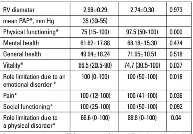
Echocardiographic assessment results are demonstrated at Table 2. TAPSE values were significantly different between RV diameter 2.98±0.29 2.74±0.30 0.973 mean PAP*, mm Hg 35 (30-55) Physical functioning* 75 (15-100) 97.5 (50-100) 0.000 Mental health 61.62±17.88 68.18±15.30 0.474 General health 49.94±18.24 71.95±10.51 0.518 Vitality* 66.5 (20.5-90) 74.7 (30.5-100) 0.037 Role limitation due to an 100 (0-100) 100 (50-100) 0.018 emotional disorder *
Pain* 100 (12-100) 100 (41-100) 0.036 Social functioning* 100 (25-100) 100 (50-100) 0.092 Role limitation due to 66.6 (0-100) 88.8 (0-100) 0.04 a physical disorder*
Data was expressed as mean±SD for parameters with normal distribution and *median for parameters with skewed distribution (range min-max).
*Comparisons of these parameters between two groups were made by Mann-Whitney U test. Other parameters of two groups were compared with Student’s t-test.
Am - late diastolic wave during atrial contraction; AT - anaerobic threshold; CPET - cardiopulmonary exercise test; COPD - chronic obstructive pulmonary disease; Em - early diastolic wave; FEF25-75% - forced expiratory flow at 25-75%; FEV1 - forced expiratory volume
at 1 second; FVC - forced vital capacity; IC - inspiratory capacity; MVV - maximum voluntary ventilation; NT-pro BNP - N-terminal of pro B-type natriuretic peptide; PAP - pulmonary arterial pressure; PET CO2 - partial pressure of end-tidal carbon dioxide; PVCO2 - peak CO2
output, PVO2-peak oxygen uptake; RA diameter - right atrium diameter; RV diameter - right
ventricle diameter; RV EF - right ventricle ejection fraction; RV - residuel volume; SF-36 - short form-36; TAPSE - tricuspid annular plane systolic excursion; TLC - total lung capacity; VC - vital capacity; VECO2 - ventilatory equivalent for CO2; VEO2 - ventilatory equivalent for O2.
NT-Pro BNP Parameters rho P mean PAP* 0.651 0.016 Tricuspit E -0.126 0.377 Tricuspit A 0.090 0.530 E/A -0.186 0.190 TAPSE 0.005 0.974 Em -0.154 0.281 Am 0.068 0.635 Em/Am rate -0.182 0.202 RV EF% -0.093 0.514 RA diameter 0.079 0.584 RV diameter 0.185 0.194 RV diastolic diameter 0.140 0.329 RV systolic diameter 0.111 0.438 Cigarette pack year 0.354 0.025 Age 0.422 0.002 FEV1 L -0.439 0.001 FEV1, % -0.269 0.049 FVC, % -0.295 0.035 IC L -0.407 0.003 VC L -0.544 0.000 AT VO2 mL/kg/min -0.462 0.001 AT VO2 L/min -0.372 0.008 AT VCO2 -0.372 0.008 PVO2 L/min -0.441 0.001 PVO2 mL/kg/min -0.527 0.000 O2 pulse -0.388 0.005 VE/VCO2 -0.041 0.776 PET CO2 0.071 0.621
*Spearman Correlation analysis was performed.
Other correlations were performed with Pearson Correlation Analysis
Am - late diastolic wave during atrial contraction; AT - anaerobic threshold; CPET - cardiopulmonary exercise test; COPD - chronic obstructive pulmonary disease; Em - early diastolic wave; FEV1 - forced expiratory volume at 1 second; FVC - forced vital capacity; IC -
inspiratory capacity; MVV - maximum voluntary ventilation; NT-pro BNP - N-terminal of pro B-type natriuretic peptide; PAP - pulmonary arterial pressure; PET CO2 - partial pressure of
end-tidal carbon dioxide; PVCO2 - peak CO2 output, PVO2 - peak oxygen uptake; RA
diameter - right atrium diameter; RV diameter - right ventricle diameter; RV EF - right ventricle ejection fraction; TAPSE - tricuspid annular plane systolic excursion; VC - vital capacity; VECO2 - ventilatory equivalent for CO2, VEO2 - ventilatory equivalent for O2.
Table 3. Relationships between NT-proBNP level with smoking, age, pulmonary function test, echocardiography, and cardiopulmonary exercise testing results in COPD patients
patient and control groups (2.26±0.46 cm; 2.64±0.38 cm respec-tively, p=0.040). Moreover COPD patients had reduced right ventricular Em wave, Em/Am ratio and tricuspit E wave (Table 2). When we graded right ventricle diastolic dysfunction in study population, 6 (19.3%) COPD patients had grade 1 right ventricle diastolic dysfunction, 8 (54.8%) COPD patients had grade 2 dia-stolic dysfunction and the rest of COPD patients and control subjects had normal right ventricle diastolic functions.
SF-36 results
Domains of role limitation due to an emotional disorder, pain and role limitation due to a physical disorder were significantly different between patient and control groups (Table 2).
Correlation analyses
When we correlated serum NT-proBNP level of COPD patients with cigarette consumption, pulmonary arterial pres-sure, pulmonary function test, cardiopulmonary exercise testing, echocardiographic assessments, we found negative correla-tions between serum NT-proBNP level with pulmonary function test results, AT VO2, AT VCO2, PVO2, and O2 pulse; positive cor-relations between serum NT-proBNP level with pulmonary arte-rial pressure and cigarette consumption (Table 3).
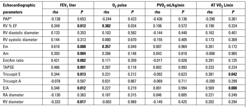
There were significant correlations between FEV1 (liter) with Em, Am, Em/Am ratio, TAPSE, tricuspit E, E/A ratio and right ven-tricle diameter; IC and VC (liter) with Em, Am waves, Em/Am ratio and TAPSE values in COPD patients (Table 4). We also found sig-Echocardiographic FEV1 liter O2 pulse PVO2 mL/kg/min AT VO2 L/min parameters rho P rho P rho P rho P
PAP* -0.138 0.653 -0.244 0.423 -0.436 0.136 -0.290 0.361 RV % EF 0.349 0.012 0.382 0.034 0.106 0.572 0.190 0.324 RV diastolic diameter 0.133 0.353 0.103 0.582 -0.144 0.440 0.162 0.401 RV systolic diameter 0.144 0.313 0.080 0.670 -0.155 0.405 0.173 0.369 Em 0.610 0.000 0.357 0.049 0.007 0.969 0.261 0.172 Am 0.393 0.004 0.266 0.148 0.043 0.818 -0.008 0.965 Em/Am ratio 0.421 0.002 0.171 0.359 -0.017 0.928 0.291 0.125 TAPSE 0.466 0.001 0.287 0.118 0.002 0.993 0.233 0.224 Tricuspit E 0.344 0.013 0.231 0.212 -0.092 0.623 0.381 0.042 Tricuspit A -0.078 0.587 0.031 0.867 -0.069 0.711 -0.200 0.299 E/A 0.348 0.012 0.227 0.219 0.001 0.994 0.569 0.000 RA diameter -0.130 0.363 0.187 0.315 0.046 0.805 0.221 0.249 RV diameter -0.333 0.017 -0.003 0.989 -0.149 0.425 0.202 0.294
*Spearman correlation analysis was performed.
Other correlations were performed by using Pearson correlation analysis
Am - late diastolic wave during atrial contraction; AT - anaerobic threshold; COPD - chronic obstructive pulmonary disease; Em - early diastolic wave; FEV1 - forced expiratory
volume at 1 second; NT-pro BNP-N-terminal of pro B-type natriuretic peptide; PAP - pulmonary arterial pressure; PVO2 - peak oxygen uptake; RA diameter - right atrium diameter; RV
diameter - right ventricle diameter; RV EF - right ventricle ejection fraction; TAPSE - tricuspid annular plane systolic excursion
Table 4. Relationships between right ventricular findings with FEV1 and cardiopulmonary exercise testing results in COPD patients.
FEV1 lt IC AT VO2 L/min PVO2 L/dk O2 pulse NT- pro BNP SF-36 rho P Rho P rho P rho P rho P rho P
Physical functioning* 0.688 0.000 0.538 0.000 0.522 0.000 0.520 0.000 0.445 0.001 -0.196 0.168 Mental health** 0.230 0.105 0.157 0.271 0.128 0.382 0.204 0.150 0.169 0.236 -0.083 0.562 General health** 0.614 0.000 0.338 0.015 0.450 0.001 0.487 0.000 0.374 0.007 -0.101 0.24 Vitality* 0.368 0.008 0.355 0.010 0.185 0.202 0.255 0.070 0.304 0.030 -0.219 0.12 RL due to ED* 0.403 0.003 0.376 0.007 0.212 0.145 0.372 0.007 0.404 0.003 -0.225 0.113 Pain* 0.305 0.030 0.257 0.049 0.125 0.392 0.258 0.068 0.234 0.098 -0.190 0.181 Social functioning* 0.218 0.124 0.135 0.345 0.190 0.192 0.199 0.162 0.064 0.653 -0.01 0.94 RL due to PD* 0.403 0.003 0.327 0.019 0.271 0.060 0.412 0.003 0.354 0.011 -0.324 0.02
**Pearson correlation analysis was performed. *Spearman Correlation analysis was performed.
AT - anaerobic threshold; COPD - chronic obstructive pulmonary disease; ED - emotional disorder; FEV1 - forced expiratory volume at 1 second; IC - inspiratory capacity; NT-pro BNP-
N - terminal of pro B-type natriuretic peptide; PD - physical disorder; PVO2 - peak oxygen uptake; RL - role limitation; SF-36 - short form-36
Table 5. Relationship between SF-36 questionnaire with pulmonary function tests, cardiopulmonary exercise tests and serum NTpro-BNP in COPD patients
nificant relationships between O2 pulse with right ventricle EF and Em wave; AT VO2 (L/min) with tricuspit E wave and E/A ratio; AT VEO2 with right ventricle diameter (Table 4).
There were significant relationships between FEV1 and IC with SF-36 domains (Table 5). SF-36 domains of physical function-ing, general health and role limitation due to physical disorder were significantly correlated with AT VO2, PVO2 and O2 pulse. Moreover we found significant relationship between role limita-tion due to emolimita-tional disorder with PVO2 and O2 pulse. Mental health, pain and social functioning domains with cardiopulmonary exercise testing results were not significantly correlated (Table 5). A negative correlation between serum NT-proBNP level with role limitation due to physical disorder was found (p=0.02; r=-0.324) (Table 5).
Discussion
The present study demonstrated significant correlations between serum NT-proBNP level with role limitation due to physical disorder and CPET parameters in COPD patients sup-porting our hypothesis.
In the present study, early diastolic filling (E wave), right ventricle myocardial early filling (Em), and ratio of Em/Am were decreased in COPD patients and TAPSE values were signifi-cantly lower in COPD patients than control subjects suggesting the impaired systolic and diastolic functions of COPD patients. Karabulut et al. (21) found deterioration of right ventricular diastolic functions with progressed disease stage of COPD patients and highlighted the importance of echocardiographic assessments in COPD patients. In parallel with these findings we found significant correlations between FEV1 with Em wave, Em/Am ratio, TAPSE, tricuspit E wave and E/A ratio in COPD patients. According to these results, we consider that right ventricle systolic and diastolic functions were adversely affected with disease progression.
In COPD patients, right ventricular wall tension causes the release of BNP. Therefore increased serum BNP level has been defined as a powerful marker in diagnosis and severity of PH (22). Increased serum BNP levels have also been reported in patients with PH secondary to acute and/or chronic respiratory diseases (23) In the present study, serum NT-proBNP levels were signifi-cantly higher in COPD patients than control subjects. In previous studies, Lang et al. (24) and Chi et al. (25) reported higher BNP levels in COPD patients than control subjects. Bozkanat et al. (26) and Chi et al. (25) showed the significant relationships between serum NT-proBNP with FEV1 in COPD patients. In the present study we determined significant negative correlations between serum NT-proBNP with FEV1, FVC, IC and VC as well. Chi et al. (25) determined positive correlation between PAP with NT-proBNP in COPD patients and Bando et al. (27) found higher serum BNP levels in COPD patients with PH than COPD patients without PH. Leuchte et al. (23) showed significant correlation between serum BNP levels with PAP in COPD patients with PAP>35 mm Hg at right heart catheterization. Similar to previous
studies we also found significant correlation between PAP with NT-proBNP. Based on this significant correlation, we suggest that NT-proBNP levels might be a prognostic and noninvasive marker in advance disease stages of COPD. However no signifi-cant correlations between NT-proBNP with right ventricular diastolic parameters were found in the present study. These results could be attributed to the small sample size and limita-tions of echocardiography (air trapping, changes of chest wall, etc) in COPD patients. In the literature NT-proBNP level has been associated with age as ventricular compliance decreases with age (28, 29). Similar to the previous reports relationship between NT-proBNP level with age was also determined in the present study.
Quality of life and exercise capacity of COPD patients are impaired (30). Patients generally descibe exercise intolerance as declined response of ventilation to exercise related with airflow limitation (31, 32). Besides, depression and sedentary life style due to dyspnea are contributing factors to exercise intolerance (19). In our study patients with COPD had signifi-cant exercise limitation when compared with control subjects. These results can be explained as the importance of inspira-tory capacity and vital capacity in response to increased ven-tilatory demand in COPD patients during exercise.
Several studies have shown exercise limitation in left ven-tricular dysfunction (22, 33, 34). Kruger et al. (34) have also found negative correlation between serum BNP level with PVO2 and AT values in 70 patients with congestive heart failure. Eroğlu et al. (35) evaluated the relationship between BNP lev-els and CPET parameters in patients with dyspnea and isolated left ventricle diastolic dysfunction and found negative correla-tion between BNP levels and duracorrela-tion of exercise, AT VO2, and MET on CPET. To the best of our knowledge, this is the first study using CPET, and BNP levels in COPD patients. In the present study, NT-proBNP was negatively correlated with AT VO2, AT VCO2 and PVO2 values. Thus serum NT-proBNP levels can be regarded as a useful biomarker in exercise limitation of moderate-severe COPD patients.
Limited number of studies investigating the effects of dia-stolic dysfunctions on exercise capacity in COPD patients was found. Cuttica et al. (36) determined significant association between right sided cardiac structural changes with exercise capacity in COPD patients. In the present study, exercise capac-ity was also affected by diastolic dysfunctions and PH. Although serum NT-proBNP level was not correlated with right ventricle parameters, serum NT-proBNP level was significantly higher in COPD patients than control subjects and right ventricle diastolic and systolic dysfunction was seen in COPD patients and signifi-cant relationships between serum NT-proBNP and CPET param-eters were found.
We also demonstrated the correlations between SF-36 domains with pulmonary function test results and positive corre-lations between SF-36 domains with PVO2 and AT values consis-tent with the literature (19, 37, 38). However we did not find rela-tionship between PH and quality of life. These results might be
due to characteristics of our study population as COPD patients had mild PH. Improvement of quality of life will help to increase exercise capacity in COPD patients. Moreover significant correla-tion between serum NT-proBNP and role limitacorrela-tion due to physi-cal disorder may indicate the utility of serum NT-proBNP to dem-onstrate quality of life in COPD patients. These preliminary results should be validated and explored in further studies.
Study limitations
We used echocardiographic assessments however technical difficulties due to hyperinflation can be seen in COPD patients. Right heart catheterization- gold standart diagnostic tool for right ventricular dysfunction and PH- could not be performed to our study population. Small sample size is another limitation of the present study.
Conclusion
Right ventricular dysfunction contribute exercise limitation together with airflow obstruction in moderate and severe COPD patients. Serum NT-proBNP levels can be used to demonstrate exer-cise capacity and quality of life in COPD patients. Further studies are needed to validate and explore these preliminary results in larger number of moderate and severe COPD patients with right ventricular dysfunction confirmed diagnosis with right heart catheterization.
Conflict of interest: None declared. Peer-review: Externally peer-reviewed.
Authorship contributions: Concept - T.Ş.Ö., G.U.; Design - T.Ş.Ö., S.S.U., G.U.; Supervision - T.Ş.Ö., G.U.; Resource - T.Ş.Ö., B.Y., E.K., N.B.; Materials - T.Ş.Ö., B.Y., E.K., N.B.; Data collection &/or processing - T.Ş.Ö., B.Y., E.K., N.B.; Analysis &/or interpreta-tion - T.Ş.Ö., S.S.U., G.U.; Literature search - T.Ş.Ö., B.Y., E.K., N.B.; Writing -T.Ş.Ö., S.S.U., G.U.; Critical review - T.Ş.Ö., S.S.U., G.U.; Other - T.Ş.Ö., B.Y., E.K., N.B.
References
1. Global strategy for diagnosis, management and prevention of Chronic Obstructive Pulmonary Disease. Global Initiative for Chronic Obstructive Lung Disease: updated 2009.
2. Takaruka M, Harada T, Fukuno H, Okushi H, Taniguchi T, Sawada S, et al. Echocardiographic detection of occult cor pulmonale during exercise in patients with chronic obstructive pulmonary disease. Echocardiography 1999; 16: 127-34. [CrossRef]
3. Higham MA, Dawson D, Joshi J, Nihoyannopoulos P, Morrell NW. Utility of echocardiography in assesment of pulmonary hypertension secondary to COPD. Eur Respir J 2001; 17: 350-5. [CrossRef]
4. Miyahara Y, Ikeda S, Yoshigana T, Yamaguchi K, Nishimura-Shirono E, Yamasa T, et al. Echocardiographic evaluation of right cardiac function in patients with chronic pulmonary diseases. Jpn Heart J 2001; 42: 483-93. [CrossRef]
5. Morrison LK, Harrison A, Krishnaswamy P, Kazanegra R, Clopton P, Maisel A. Utility of a rapid B-natriuretic peptide assay in differentiating congestive heart failure from lung disease in patients presenting with dyspnea. J Am Coll Cardiol 2002; 39: 202-9. [CrossRef]
6. Yap LB, Mukerjee D, Timms PM, Ashrafian H, Coghlan JG. Natriuretic peptides, respiratory disease, and the right heart. Chest 2004; 126: 1330-6. [CrossRef]
7. Karakılıç E, Kepez A, Abalı G, Coşkun F, Kunt M, Tokgözoğlu L. The relationship between B-type natriuretic peptide levels and echocardiographic parameters in patients with heart failure admitted to the emergency department. Anadolu Kardiyol Derg 2010; 10: 143-9. [CrossRef]
8. Tsai SH, Lin YY, Chu SJ, Hsu CW, Cheng SM. Interpretation and use of natriuretic peptides in non-congestive heart failure settings. Yonsei Med J 2010; 51: 151-63. [CrossRef]
9. Pellegrino R, Viegi G, Brusasco V, Crapo RO, Burgos F, Casaburi R, et al. Interpretative strategies for lung function tests. Eur Respir J 2005; 26: 948-68. [CrossRef]
10. Miller MR, Hankinson J, Brusasco V, Burgos F, Casaburi R, Coates A, et al; ATS/ERS Task Force.Standardisation of spirometry. Eur Respir J 2005; 26: 319-38. [CrossRef]
11. Schiller NB, Shah PM, Crawford M, DeMaria A, Devereux R, Feigenbaum H, et al. Recommendations for quantitation of the left ventricle by two-dimensional echocardiography. American Society of Echocardiography Committee on Standards, Subcommittee on Quantitation of Two-Dimensional Echocardiograms. J Am Soc Echocardiogr 1989; 2: 358-67. [CrossRef]
12. Parisi AF, Moynihan PF, Feldman CL, Folland ED. Approaches to determination of left ventricular volume and ejection fraction by real-time two-dimensional echocardiography. Clin Cardiol 1979; 2: 257-63. [CrossRef]
13. Chemla D, Castelain V, Hervè P, Lecarpentier Y, Brimioulle S. Haemodynamic evaluation of pulmonary hypertension. Eur Respir J 2002; 20: 1314-31. [CrossRef]
14. Yared K, Noseworthy P, Weyman AE, McCabe E, Picard MH, Baggish AL. Pulmonary artery acceleration time provides an accurate estimate of systolic pulmonary arterial pressure during transthoracic echocardiography. J Am Soc Echocardiogr 2011; 24: 687-92. [CrossRef]
15. Zile MR, Brutsaert DL. New concepts in diastolic dysfunction and diastolic heart failure: Part I: diagnosis, prognosis, and measurements of diastolic function. Circulation 2002; 105: 1387-93.
[CrossRef]
16. Rudski LG, Lai WW, Afilalo J, Hua L, Handschumacher MD, Chandrasekaran K, et al. Guidelines for the echocardiographic assessment of the right heart in adults: a report from the American Society of Echocardiography endorsed by the European Association of Echocardiography, a registered branch of the European Society of Cardiology, and the Canadian Society of Echocardiography. J Am Soc Echocardiogr 2010; 23: 685-713. [CrossRef]
17. Wasserman K, Hansen JE, Sue DY, Whipp BJ, Casaburi R. Principles of exercise testing and interpretation.3rd ed Vol. 10. Baltimore: Lippincott Williams &Wilkins, 1999.
18. Ferrer M, Alonso J, Morera J, Marrades RM, Khalaf A, Aguar MC, et al. Chronic obstructive pulmonary disease stage and health-related quality of life. The Quality of Life of Chronic Obstructive Pulmonary Disease Study Group. Ann Intern Med 1997; 127: 1072-9.
19. Ulubay G, Sarınç Ulaşlı S, Akıncı B, Görek A, Akçay S. Assessment of relation among emotional status, pulmonary function test, exercise performance, and quality of life in patients with COPD. Tuberk Toraks 2009; 57: 169-76.
20. Ware JE, Sherbourne CD. The MOS 36-item short-form health survey (SF-36). I. Conceptual framework and item selection. Med Care 1992; 30: 473-83. [CrossRef]
21. Karabulut A, İltimur K. Sağ ventrikül diyastolik fonksiyonlarının kronik obstüktif akciğer hastalığının evrelerine göre değerlendirilmesi. Dicle Tıp Dergisi 2005; 32: 145-8.
22. Yamamoto K, Burnett JC Jr, Jougasaki M, Nishimura RA, Bailey KR, Saito Y, et al. Superiority of brain natriuretic peptide as a hormonal marker of ventricular systolic and diastolic dysfunction and ventricular hypertrophy. Hypertension 1996; 28: 988-94. [CrossRef]
23. Leuchte HH, Neurohr C, Baumgartner R, Holzapfel M, Giehrl W, Vogeser M, et al. Brain natriuretic peptide and exercise capacity in lung fibrosis and pulmonary hypertension. Am J Respir Crit Care Med 2004; 170: 360-5. [CrossRef]
24. Lang CC, Coutie WJ, Struthers AD, Dhillon DP, Winter JH, Lipworth BJ. Elevated levels of brain natriuretic peptide in acute hypoxaemic chronic obstructive pulmonary disease. Clin Sci 1992; 83: 529-33. 25. Chi SY, Kim EY, Ban HJ, Oh IJ, Kwon YS, Kim KS, et al. Plasma
N-terminal pro-brain natriuretic peptide: a prognostic marker in patients with chronic obstructive pulmonary disease. Lung 2012; 190: 271-6. [CrossRef]
26. Bozkanat E, Tozkoparan E, Baysan O, Deniz O, Çiftçi F, Yokusoğlu M. The significance of elevated brain natriuretic peptide levels in chronic obstructive pulmonary disease. J Int Med Res 2005; 33: 537-44. [CrossRef]
27. Bando M, Ishii Y, Sugiyama Y, Kitamura S. Elevated plasma brain natriuretic peptide levels in chronic respiratory failure with cor pulmonale. Respir Med 1999; 93: 507-14. [CrossRef]
28. Maisel AS. B type natriuretic peptide (BNP) levels: diagnostic and therapeutic potential. Rev Cardiovascular Med 2001; 2: 13-8.
29. Stamm JA, Belloli EA, Zhang Y, Bon J, Sciurba FC, Gladwin MT. Elevated N-terminal pro-brain natriuretic peptide is associated with mortality in tobacco smokers independent of airflow obstruction. PLoS One 2011; 6: e27416. [CrossRef]
30. Gallagher CG. Exercise limitation and clinical exercise testing in chronic obstructive pulmonary disease. Clin Chest Med 1994; 15: 305-26. 31. Nici L. Mechanisms and measures of exercise intolerance in
chronic obstructive pulmonary disease. Clin Chest Med 2000; 21: 693-704. [CrossRef]
32. O’Donnel DE, Revill S, Webb KA. Dynamic hyperinflation and exercise intolerance in chronic obstructive pulmonary disease. Am J Respir Crit Care Med 2001; 164: 770-7. [CrossRef]
33. Lubien E, DeMaria A, Krishnaswamy P, Clopton P, Koon J, Kazanegra R, et al. Utility of B-natriuretic peptide in detecting diastolic dysfunction: comparison with Doppler velocity recordings. Circulation 2002; 105: 595-601. [CrossRef]
34. Krüger S, Graf J, Kunz D, Stickel T, Hanrath P, Janssens U. Brain natriuretic peptide levels predict functional capacity in patients with chronic heart failure. J Am Coll Cardiol 2002; 40: 718-22. [CrossRef]
35. Eroğlu S, Yıldırır A, Bozbaş H, Aydınalp A, Ulubay G, Eldem O, et al. Brain natriuretic peptide levels and cardiac functional capacity in patients with dyspnea and isolated diastolic dysfunction. Int Heart J 2007; 48: 97-106. [CrossRef]
36. Cuttica MJ, Shah SJ, Rosenberg SR, Orr R, Beussink L, Dematte JE, et al. Right heart structural changes are independently associated with exercise capacity in non-severe COPD. PLoS One 2011; 6: e29069. [CrossRef]
37. Hajiro T, Nishimura K, Tsukino M, Ikeda A, Oga T, Izumi T. A comparison of the level of dyspnea vs. disease severity in indicating the health related quality of life of patients with COPD. Chest 1999; 116: 1632-7. [CrossRef]
38. Katsura H, Yamada K, Kida K. Both generic and disease specific health-related quality of life are deteriorated in patients with underweight COPD. Respir Med 2005; 99: 624-30. [CrossRef]