Research Article /Araştırma Makalesi
Corresponding Author / Sorumlu Yazar: Article History / Makale Geçmişi:
Çiğdem PULATOĞLU
Adres: Department of Obstetrics and Gynecology, Istinye
University Hospital Gaziosmanpaşa Medical Park, Istanbul, TURKEY.
E-posta:cigdempulatoglu@gmail.com
Date Received / Geliş Tarihi: 06.10.2019 Date Accepted / Kabul Tarihi: 07.11.2019
Namık Kemal Tıp Dergisi 2019; 7(3): 228 - 235
MATERNAL AND NEONATAL OUTCOMES RELATED TO IRON DEFICIENCY
ANEMIA AND SERUM FERRITIN STATUS: A MULTICENTER PROSPECTIVE
STUDY FROM EASTERN MARMARA, TURKEY
Demir Eksikliği Anemisi ve Serum Ferritin Düzeyi ile İlgili Maternal ve Neonatal Sonuçlar: Doğu Marmara Bölgesine Ait Çok Merkezli Bir Prospektif Çalışma, Türkiye
Çiğdem PULATOĞLU1 , Derya BAŞBUĞ2 , Bertan AKAR3 , Hayal ŞİMŞEK4 , Pınar ÇAKIR5 , Alper BAŞBUĞ6 , Eray ÇALIŞKAN7
1 Department of Obstetrics and Gynecology, Istinye University Hospital Gaziosmanpaşa Medical Park, Istanbul, TURKEY. 2 Private Clinic, Düzce, TURKEY.
3 Department of Obstetrics and Gynecology, Istinye University School of Medicine, Istanbul, TURKEY 4 Department of Obstetrics and Gynecology, Sakarya Toyotasa Emergency Service Hospital, Sakarya, TURKEY.
5 Department of Obstetrics and Gynecology, Kocaeli Health Sciences University Derince Research and Training Hospital, Kocaeli, TURKEY. 6 Department of Obstetrics and Gynecology, Duzce University Hospital, Duzce, TURKEY.
7 Department of Obstetrics and Gynecology, Okan University School of Medicine, Istanbul, TURKEY. Abstract
Aim: The aim was to evaluate the incidence of iron deficiency anemia in pregnancy in the East Marmara region of Turkey in order to determine its prevalence along with the effects and associations of iron supplementation on maternal and neonatal outcomes.
Materials and Methods: This study was conducted in six centers and included a total of 1102 pregnant women. Blood samples were collected for hematological status and serum ferritin levels during pregnancy, and the adverse maternal and perinatal outcomes were determined. Iron deficiency anemia was diagnosed according to the World Health Organization criteria as hemoglobin level of < 11 g/dl and ferritin level of <15 μg/dL.
Results: The rate of anemia was 19.8%, with 44% of them receiving iron supplementation. The maternal age was lower in the anemic group (26.5 vs. 27.7, p = 0.01). Selective iron use was more frequent in the anemic group, while routine iron use was more frequent in the non-anemic group (47.1% vs. 29.3%; p = 0.01).
Conclusion: Iron deficiency anemia is a frequent problem in pregnancy. However, many anemic pregnant women do not receive iron therapy. Iron supplementation may have positive effects on maternal and perinatal outcomes. In order to combat iron deficiency anemia in pregnancy, wide spread use of iron supplements should be established.
Keywords: Anemia, Ferritin, Iron deficiency, Iron supplementation, Pregnancy.
Öz
Amaç: Bu çalışmanın amacı, demir takviyesi ve demir durumunun maternal ve neonatal etkileri ile birlikte Türkiye'nin Doğu Marmara bölgesinde gebelikte görülen demir eksikliği anemisi insidansını değerlendirmektir.
Materyal ve Metot: Bu çalışma Doğu Marmara Bölgesi'nin (Türkiye) üç ilinde altı merkezde gerçekleştirilmiş ve toplam 1102 gebeyi kapsamıştır. Gebelik sırasında hematolojik durum ve serum ferritin düzeyleri için kan örnekleri toplandı ve olumsuz maternal ve perinatal sonuçlar belirlendi. Demir eksikliği anemisi, Dünya Sağlık Örgütü kriterlerine göre hemoglobin<11g/dl ve ferritin< 15 μg/dL olarak tanımlandı.
Bulgular:Çalışmaya dahil edilen kadınlarda anemi oranı % 19.8 idi ve bunların % 44'ü demir desteği alıyordu. Maternal yaş anemik grupta daha düşüktü (26.5'e karşı 27.7, p = 0.01). Anemik grupta selektif demir kullanımı daha sık görülürken, anemik olmayan grupta rutin demir kullanımı daha sık görüldü (% 47.1'e karşılık% 29.3; p = 0.01).
Sonuç: Demir eksikliği anemisi gebelikte sık görülen bir problemdir. Bununla birlikte, birçok anemik gebeye demir tedavisi verilmez. Demir takviyesi, bazı maternal ve perinatal sonuçlar üzerinde olumlu etkileri olabilir. Gebelikte demir eksikliği anemisiyle mücadele etmek için demir takviyelerinin yaygın kullanımı sağlanmalıdır.
Anahtar Kelimeler: Anemi, Ferritin, Demir eksikliği, Demir desteği, Gebelik.
INTRODUCTION
Anemia is an important public health issue worldwide that affects not only low-income populations, but middle- and high-income
communities as well1. A number of factors can
cause anemia. Although hemoglobin
concentration is used to diagnose anemia, this measure alone is not sufficient to determine the cause of the anemia2,3. One of the leading
229
causes of anemia is iron deficiency (ID), whichaffects about 50% of patients4. Pregnant women are among the specific populations affected by anemia. Anemia during pregnancy can be due to many factors. Iron deficiency is also an important cause of anemia in pregnancy 5. Anemia during pregnancy may adversely affect not only the mother, but also the fetus, especially in relation to low birth weight, prematurity and intrauterine growth restriction, as reported in a number of studies6,7. To identify iron deficiency anemia (IDA), it is important to know the iron status, including serum ferritin (SF), soluble transferrin receptor (sTfR), zinc protoporphyrin, serum iron, and total iron-binding capacity or transferrin saturation 8,9. Although many indices are used to diagnose IDA, the level of SF is one of the most frequently encountered methods in both clinical and public health settings 10. Although several SF concentration cut-off values for IDA in pregnant women have been determined, the most commonly recommended value is <15μg/dL11.
This study aimed to determine the prevalence of IDA, diagnosed by low SF concentrations, in the East Marmara region of Turkey and to examine its associations with demographic and socio-economic characteristics as well as its effect on perinatal outcomes.
MATERIAL and METHODS
This study was conducted in six different centers in Kocaeli, Sakarya, and Düzce provinces in the East Marmara region of Turkey between December 2017 to December 2018. A total of 1102 pregnant women between the ages of 18 and 40 were included in the analysis. Written informed consent was obtained from all the women who agreed to
participate in the study. All participants completed a comprehensive micronutrient study questionnaire created by the Turkish Society of Obstetrics and Gynecology (TSOG) which consisted of 56 standard items. All the procedures in the study were carried out in accordance with the ethical standards of the 1964 Helsinki Declaration Institutional Research Committee and subsequent amendments or comparable ethical standards. The study was approved by the Ethics Committee of Duzce University with approval number of 2017/168.
Women who gave birth to single liveborn infants were included in the study. Exclusion criteria included multiple pregnancies, in utero fetal demise miscarriages, women with active infections detected during the study and those receiving IV iron supplementation.
Study Population
Data were obtained from a total of 1102 pregnant women (219 anemic, and 883 non-anemic) who were registered within the routine screening program of the obstetrics outpatient clinics of the study centers. Age, gravida, parity, number of abortions, date of last menstrual period, pregnancy weeks according to USG and date of last menstrual period, height, weight and body mass index (BMI) were recorded for all patients. The BMI was calculated using the following formula: weight (kg)/height2 (m). Education and income were the two primary socioeconomic status indicators for all participants. According to per-capita monthly household income, they were classified as low (<1500 TL), medium (1500–3000TL), and high (>3000TL) [1 USD = 3.90 Turkish Lira (TL) in March 2018]. Patients were divided into two groups according to dietary habits: vegetarians and those who consumed iron-rich foods like meat and legumes. Adequate physical activity
230
was defined as exercise for more than 30minutes over three days per week12.
Hemoglobin was analyzed shortly after collection from whole blood samples using an automated hematology analyzer. Ferritin concentration was determined by immuno-assay via the automated chemiluminescence technique. Serum iron and total iron-binding capacity were measured by an auto-analyzer. IDA was diagnosed according to the World Health Organization criteria as a hemoglobin level of < 11 g/dl and ferritin level of <15 μg/dL 11. During pregnancy, patients were checked with complete blood count and hemoglobin levels once in each trimester for iron deficiency anemia. Our treatment strategy for IDA is that patients who were diagnosed with IDA received oral iron supplementation and those who could not tolerate oral medication were given intravenous iron supplementation.
Routine antenatal checks were made of all prospective participating pregnant women. The women were examined at least once each trimester starting from their first trimester of pregnancy.
The presence of pregnancy-related
complications such as gestational diabetes mellitus (GDM) and pre-eclampsia was evaluated. Screening for GDM was performed according to World Health Organization recommendations 13. Pre-eclampsia was defined as having a systolic blood pressure of>140 mmHg or a diastolic blood pressure of>90 mmHg after 20 weeks of gestation in previously normotensive women, and proteinuria (>100 mg/dL per urinalysis or 300 mg in a 24-h urine collection).The perinatal data gathered included preterm birth, gestational age at delivery, birth weight (g) and postpartum
hemorrhage. Preterm birth was defined as a gestational age of<37 completed weeks of gestation. Small for gestational age (SGA) was defined as <the 10th percentile, and large for gestational age (LGA) as >the 90th percentile, using birth weight z-scores calculated based on the formulae of Gardosi et al. 14.
Objectives
The primary objective was to determine the incidence of İDA in pregnant women in the East Marmara region of Turkey, where industries are concentrated and the prevalence of medium- and high-income level populations is higher compared with other geographical regions. The secondary objective was to assess the association of anemia with demographic and lifestyle factors along with its effect on perinatal outcomes.
Statistical Analysis
Descriptive statistics for continuous variables were expressed as mean ± standard deviation or median (minimum–maximum), and nominal variables were expressed as the number and percentage (%). Differences in mean values for each group were evaluated using the Student's t-test, and differences in median values were evaluated using the Mann–Whitney U-test. Categorical data were compared using the Chi-square distribution, with p-values of ≤0.05 considered as statistically significant. Statistical analysis was performed using SPSS for Windows version 22 software (SPSS, Inc., Chicago, IL, USA).
RESULTS
The study evaluated a total of 1102 pregnant women, 219 of whom were IDA, with serum hemoglobin concentrations < 11 g/dl and serum ferritin levels lower than 15 μ / dL and 883 of whom were non-anemic. The rate of anemia
231
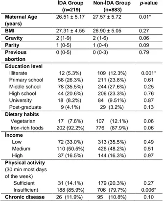
among women included in the study was19.8%. The mean age in the anemic group was lower, and the difference between the groups was statistically significant (26.5% vs. 27.7%, p = 0.01). The BMI, gravity, parity and abortion were not different between the groups. When the groups were compared in terms of education level, only illiterates were found to be higher in the non-anemic group (5.3% vs. 12.3%,p = 0.001).There was no difference between the groups in terms of dietary habits, and the rates of those consuming a vegetarian diet and those eating iron-rich foods were similar. There was no difference between the groups in terms of income levels. The weekly rate of attaining adequate physical activity was lower in the anemic group (85.9% vs. 79.7%, p = 0.006). Both groups were found to have similar chronic diseases (Table 1).
Table 1. Obstetric history, sociodemographic status and life
style according to the presence of iron deficiency anemia
İDA Group (n=219) Non-İDA Group (n=883) p-value Maternal Age (years) 26.51 ± 5.17 27.57 ± 5.72 0.01* BMI 27.31 ± 4.55 26.90 ± 5.05 0.27 Gravity 2 (1-9) 2 (1-6) 0.06 Parity 1 (0-5) 1 (0-4) 0.09 Previous abortion 0 (0-5) 0 (0-3) 0.79 Education level Illiterate Primary school Middle school High school University Post-graduate 12 (5.3%) 58 (26.3%) 78 (35.5%) 44 (20.6%) 18 (8.2%) 9 (4.1%) 109 (12.3%) 211 (23.8%) 244 (27.6%) 206 (23.3%) 84 (9.51%) 29 (3.2%) 0.001* 0.61 0.25 0.76 0.87 0.13 Dietary habits Vegetarian Iron-rich foods 17 (7.8%) 202 (92.2%) 107 (12.1%) 776 (87.9%) 0.06 0.06 İncome Low Medium High 72 (33.0%) 110 (50.5%) 37 (16.5%) 313 (35.5%) 426 (48.2%) 144 (16.3%) 0.49 0.51 0.97 Physical activity
(30 min most days of the week) Sufficient Insufficient 31 (14.1%) 188 (85.9%) 179 (20.3%) 706 (79.7%) 0.27 0.006* Chronic disease 26 (11.9%) 95 (10.8%) 0.10
Values are given as mean ± standard deviation, median (min-max) and %
* p <0.05 indicates statistical significance. IDA: identify iron deficiency anemia BMI: Body Mass Index
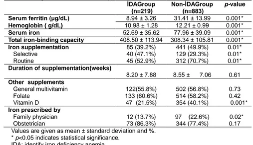
Serum ferritin, hemoglobin, serum iron concentrations and total iron-binding capacity were lower in the anemic group and the difference between the groups was statistically significant. Of the women included in the study, 47.7% (n = 526) were receiving iron supplementation. The iron-use rate was 39.2% in the anemic group and 49.9% in the non-anemic group (p = 0.01). The selective use of iron was higher in the anemic group, while the routine use of iron was higher in the non-anemic group (47.1% vs. 29.3%, p = 0.01; 52.9% vs. 70.7%, p = 0.01, respectively).The mean duration of iron supplementation was 8.2 weeks in the anemic group and 8.5 weeks in the non-anemic group, and the difference between the groups was not statistically significant. Evaluation of micronutrient supplementation other than iron revealed that 58.7% (n = 647) took folate, 56.6% (n = 624) general multivitamins and 30% (n = 401) vitamin D supplements. Only the vitamin D supplementation was higher in the non-anemic group (40.1% vs. 21.5%, p = 0.01).Iron supplementation was prescribed by a family physician in 18% of the pregnant women and by an obstetrician in 82%.The selective iron supplementation rate was higher with family physicians (64% vs. 36%, p = 0.007), whereas with obstetricians, there was no difference between selective and routine iron use(52.75% vs. 47.25%, p = 0.27). Although in the non-anemic group the iron supplementation prescription rate by family physicians was higher (22.6% vs 13.7%, p = 0.02), there was no difference between the groups in terms of iron supplementation initiation by obstetricians (Table 2).
Weight gain during pregnancy was similar in both groups.The interpregnancy interval was longer in the anemic group, and the difference
232
between the groups was statistically significant(48 vs. 34 months, p = 0.01). There was no difference between the groups in terms of birth weight, term birth and preterm birth. The SGA
rate was similar between the groups, while the LGA rate was higher in the non-anemic group (6.8% vs. 6.2%, p = 0.81; 2.8% vs. 9.2%, p = 0.01, respectively).
Table 2. Status of serum hemoglobin and ferritin and supplementation with iron and other micronutrients. İDAGroup (n=219) Non-İDAGroup (n=883) p-value Serum ferritin (μg/dL) 8.94 ± 3.26 31.41 ± 13.99 0.001* Hemoglobin ( g/dL) 10.98 ± 1.28 12.21 ± 0.99 0.001* Serum iron 52.69 ± 35.62 77.96 ± 39.09 0.001*
Total iron-binding capacity 408.50 ± 113.94 308.34 ± 105.81 0.001*
Iron supplementation Selective Routine 85 (39.2%) 40 (47.1%) 45 (52.9%) 441 (49.9%) 129 (29.3%) 312 (70.7%) 0.01* 0.01* 0.01* Duration of supplementation(weeks) 8.20 ± 7.88 8.55 ± 7.06 0.61 Other supplements General multivitamin Folate Vitamin D 122(55.8%) 133 (60.6%) 47 (21.5%) 502 (56.8%) 514 (58.2%) 354 (40.1%) 0.73 0.42 0.001* Iron prescribed by Family physician Obstetrician 12 (13.7%) 73 (86.3%) 97 (22.6%) 344 (77.4%) 0.02* 0.17 Values are given as mean ± standard deviation and %.
* p<0.05 indicates statistical significance. IDA: identify iron deficiency anemia
When the complications of pregnancy were compared, the incidence of pre-eclampsia was higher in the anemic group than in the non-anemic group (11.4% vs. 4.6%, p = 0.01). There was no difference between the groups in terms of GDM and postpartum hemorrhage (Table 3).
Table 3. Obstetrical outcomes according to the presence of
irondeficiencyanemia IDA Group (n=219) Non-IDA Group (n=883) p-value Weight gain during
pregnancy (kg)
7 (2-30) 8 (2-35) 0.53
Interpregnancy interval (months)
48 (5-204) 34 (5-216) 0.01*
Gestational age at birth <37 weeks ≥37weeks 25(11.4%) 194(88.6%) 116 (13.1%) 767(86.9%) 0.37 0.67 Newborn weight (g) 3159 ± 736 3111 ± 646 0.87 SGA GA 15(6.8%) 6 (2.8%) 55 (6.2%) 81 (9.2%) 0.81 0.01* Pregnancy complications Pre-eclampsia Gestational diabetes 25 (11.4%) 24 (11.0%) 41 (4.6%) 86 (9.7%) 0.01* 0.64 Values are given as mean ± standard deviation, median (min-max) and %.
* p<0.05 indicates statistical significance. IDA: identify iron deficiency anemia SGA: Small for gestational age LGA: Large for gestational age
DISCUSSION
Findings from the study showed that IDA was found in 19.8% of the pregnant population
included in the study and the mean age of the anemic group was lower. Routine iron use was more effective in preventing IDA than selective iron use. Longer interpregnancy intervals increased the risk of IDA. In terms of adverse perinatal events, IDA was associated only with pre-eclampsia.
According to the Global Prevalence Of Anemia report published by the World Health Organization (WHO),in 2011 over 800 million children and women were affected by anemia, with half of them suffering from IDA 15.The IDA seen in pregnancy is less than 20% in developed countries such as the USA and European nations, while it is over 60% in developing countries where income levels are low 16. In the present study, IDA in pregnancy was observed 19.7% and this rate is similar to rate of İDA in developed countries.
By way of achieving their goals for 2025, WHO has made some recommendations for reducing the incidence of anemia by 50%. One of these is to integrate iron supplementation into other
233
measures already being taken16. Oral ironsupplementation plays a critical role in the prevention of IDA in pregnancy. Many studies have suggested that oral iron supplementation used to treat IDA is effective in protecting both maternal and fetal health 17. The relationship between maternal/fetal outcome and iron deficiency is in fact dependent on the week of gestation at which the anemia is detected and the degree of the anemia. In their prospective study, Bencaiova and Breymann 18 found no relationship between mild anemia and adverse pregnancy outcome, and even maintained that mild anemia in pregnant womenmay result in non-excessive placental hyperplasia leading to fetal nutritional support and a higher incidence of macrosomia. The present study also found no difference between the anemic and non-anemic groups in terms of GDM, preterm delivery and SGA; however a higher rate of LGA was seen in the nonanemic pregnancies. The pre-eclampsia rate was higher in the anemic group. The reason for this, as suggested by Gambling et al.19, was thought to be that ID induces maternal and fetal stress and increases corticotropin-releasing hormone (CRH), cortisol production and oxidative damage, and that this process results in endothelial damage. Kemper et al. reported that diets low in iron-rich foods and a short interpregnancy interval were associated with IDA in pregnancy 20. In the present study, no difference was found in terms of IDA between women with vegetarian dietary habits and women who consumed iron-rich foods. Furthermore, the IDA rate increased when the interpregnancy interval was longer. The main reason for this was considered to be that these women had more menstrual blood loss and that the IDA was the result of the increasing iron requirement to compensate for chronic blood loss.
Today, WHO recommends the routine daily use of 30-60 mg elemental iron throughout pregnancy for prevention of IDA. This recommendationis based on the Cochrane Review published in 2012, which shows that daily iron supplementation has a positive effect on adverse perinatal outcomes 21. According to this report, women who routinely receive iron supplementation throughout their pregnancies have a reduced risk of giving birth to infants with LBW, maternal anemia at term, and maternal ID at term. However, some studies
have suggested that routine iron
supplementation may be associated with SGA and hypertension disorder 22,23. Therefore, instead of routine iron supplementation, they recommend selective supplementation. In this study, routine iron supplementation was applied more frequently in the non-anemic group. In the anemic group, selective supplementation was preferred as opposed to routine iron supplementation.
Folate deficiency was the second most common cause of anemia during pregnancy, especially in cases impaired by intestinal malabsorption coexisting with ID 24. The desired levels of hemoglobin and ferritin cannot be reached in one third of pregnancies despite adequate iron supplementation 25. Studies conducted by WHO in western Pacific nations found that folate supplementation together with iron reduced IDA rates by 9% to 57% 26. In this study, no difference in IDA was found between the group using iron supplementation with folate and the group using it without folate. Vitamin D deficiency and ID have long been observed together 27, 28 and the mechanisms responsible for this are understood to be the decreased levels of erythropoietin resulting from vitamin D deficiency and the blocked iron absorption resulting from the decreased circulating levels
234
of hepcidin occurring with vitamin Dsupplementation 29,30. In addition to iron supplementation, the use of vitamin D was observed in the non-anemic group.
The present study had some limitations. Only those patients using oral iron supplementation were included in the study, while those using IV iron were excluded. Diagnosis of IDA was based on serum ferritin levels and the methods or indices used for IDA diagnosis were not applied. The strength of this work, conducted as a prospective, multicenter study to determine the frequency of IDA, was that it included a large sample of participating patients and high follow-up rates.
CONCLUSIONS
In the East Marmara region the rate of IDA in pregnancy is close to developed countries and was found 19.8%. However, both the duration and quantity of iron supplementation in non-anemic group was higher than non-anemic group, which proves the need of iron supplementation strategy in pregnancy. The fight against IDA in pregnancy has become a public health issue that cannot be solved by obstetricians alone. A national screening program should be developed in order to compile more accurate data. Subsequently, this data should be used in determining the strategies to be applied to combat IDA in pregnancy and gestation. This should be considered a fundamental national goal for ensuring healthy mothers and healthy future generations.
Conflict of Interest
The authors declare no conflicts of interest.
References
1. Stevens GA, Finucane MM, De-Regil LM, Paciorek CJ, Flaxman SR, Branca F et al. Global, regional, and national trends in haemoglobin concentration and
prevalence of total and severe anaemia in children and pregnant and non-pregnant women for 1995–2011: A systematic analysis of population-representative data. The Lancet Global Health,1(1), e16-e25.
2. WHO, Unicef. UNU. (2001). Iron deficiency anaemia: assessment, prevention and control, a guide for
programme managers. Geneva: World Health
Organization.
3. Stoltzfus RJ, Mullany L, Black RE. Iron deficiency anaemia. In: Ezzati M, Lopez AD, Rodgers A, Murray CJL, editors. Comparative quantification of health risks: Global and regional burden of disease attributable to selected major risk factors. Geneva: World Health Organization; 2004,1: 163–209.
4. Clark SF. Iron deficiency anemia. Nutr Clin Pract 2008;23 (2):128–41.
5. Scholl TO. Maternal iron status: relation to fetal growth, length of gestation, and iron endowment of the neonate. Nutr Rev 2011; 69 (suppl-1): 23–9.
6. Balarajan Y, Ramakrishnan U, Ozaltin E, et al. Anaemia in lowincome and middle-income countries. Lancet 2011;378.9809: 2123–35.
7. de Sa SA, Willner E, Duraes Pereira TA, et al. Anemia in pregnancy: Impact on weight and in the development of anemia in newborn. Nutr Hosp 2015;32 (5):2071–9. 8. Pasricha SR, Flecknoe-Brown SC, Allen KJ, Gibson
PR, McMahon LP, Olynyk JK, et al. Diagnosis and management of iron deficiency anaemia: A clinical update. Med J Aust 2010;193 (9) :525–32.
9. Pfeiffer CM, Looker AC. Laboratory methodologies for indicators of iron status: strengths, limitations, and analytical challenges. Am J Clin Nutr 2017; 106 (supply-6) : 1606–14.
10. Mei Z, Cogswell ME, Parvanta I, Lynch S, Beard JL, Stoltzfus RJ, Grummer-Strawn LM. Hemoglobin and ferritin are currently the most efficient indicators of population response to iron interventions: An analysis of nine randomized controlled trials. J Nutr 2005;135(8):1974–80.
11. World Health Organization. (2011). Serum ferritin concentrations for the assessment of iron status and
iron deficiency in populations. No.
WHO/NMH/NHD/MNM/11.2. World Health
Organization.
12. American College of Obstetricians and
Gynecologists.Exercise during pregnancy and the postpartum period. Clinical obstetrics and gynecology, 2003, 46.2: 496.
13. World Health Organization. (1999). Definition, diagnosis and classification of diabetes mellitus and its complications : report of a WHO consultation. Part 1,
235
Diagnosis and classification of diabetes mellitus. WorldHealth Organization.
14. Gardosi J, Mongelli M, Wilcox M, Chang A. An adjustable Fetal Weight Standard. Ultrasound Obstet Gynecol 1995;6(3) :168–74.
15. . World Health Organization. (2015). The global prevalence of anaemia in 2011.
16. Pinho-Pompeu M, Surita FG, Pastore DA, Paulino DSM, Pinto E, Silva JL. Anemia in pregnant adolescents: Impact of treatment on perinatal outcomes. J Matern Fetal Neonatal Med 2017;30(10): 1158-62.
17. Bencaiova G, Breymann C. Mild Anemia and Pregnancy Outcome in a Swiss Collective. Journal of Pregnancy 2014; 2014:307535.
18. Gambling L, Kennedy C, McArdle HJ. Iron and copper in fetal development. Seminars in Cell and Developmental Biology 2011;22(6):637–44.
19. Kemper AR, Fan T, Grossman DC, Phipps MG. Gaps in evidence regarding iron deficiency anemia in pregnant women and young children: Summary of US Preventive Services Task Force recommendations. Am J Clin Nutr 2017; 106(suppl-6): 1555-58.
20. World Health Organization. Guideline, W. H. O. (2012). Daily iron and folic acid supplementation in pregnant women. Geneva: World Health Organization, 27. 21. Pena-Rosas JP, De-Regil LM, Dowswell T, Viteri FE.
Daily oral iron supplementation during pregnancy. Cochrane Database Syst Rev 2012; 12: CD004736. 22. Ziaei S, Norrozi M, Faghihzadeh S, Jafarbegloo E. A
randomised placebocontrolled trial to determine the effect of iron supplementation on pregnancy outcome in pregnant women with haemoglobin or = 13.2 g/dl. BJOG 2007; 114(6):684–8.
23. Shastri L, Mishra PE, Dwarkanath P, Thomas T, Duggan C, Bosch R, et al. Association of oral iron supplementation with birth outcomes in non-anaemic South Indian pregnant women. Eur J Clin Nutr 2015;69(5):609–13.
24. Frenkel EP, Yardley DA. Clinical and laboratory features and sequelae of deficiency of folic acid (folate) and vitamin B12 (cobalamin) in pregnancy and
gynecology. Hematol Oncol Clin North Am
2000;14(5):1079-1100.
25. Ramakrishnan U, Grant FK, Goldenberg T, Bui V, Imdad A, Bhutta ZA. Effect of multiple micronutrient supplementation on pregnancy and infant outcomes: A systematic review. Paediatric and Perinatal Epidemiology 2012; 26(suppl.1): 153–67.
26. Weekly iron and folic acid supplementation programmes for women of reproductive age: An analysis of best programme practices. Manila, World Health Organization Regional Office for the Western Pacific, 2011.
27. lanco-Rojo R, Perez-Granados AM, Toxqui L, Zazo P, de la Piedra C, Vaquero MP. Relationship between vitamin D deficiency, bone remodelling and iron status in iron-deficient young women consuming iron-fortified food. Eur J Nutr 2013;52(2):695–703.
28. Sim JJ, Lac PT, Liu IL, Meguerditchian SO, Kumar VA, Kujubu DA, Rasgon SA. Vitamin D deficiency and anemia: A cross-sectional study. Ann Hematol 2010;89(5):447–52.
29. Alon DB, Chaimovitz C, Dvilansky A, Lugassy G, Douvdevani A, Shany S, Nathan I. Novel role of 1,25(OH)(2)D(3) in induction of erythroid progenitor cell proliferation. Exp Hematol 2002;30(5):403–9.
30. Bacchetta J, Zaritsky JJ, Sea JL, Chun RF, Lisse TS, Zavala K, Nayak A, Wesseling-Perry K, Westerman M, Hollis BW, et al. Suppression of iron-regulatory hepcidin by vitamin D. J Am Soc Nephrol2014;25(3):564–72.

