doi:10.30569.adiyamansaglik.537624
Bu eser, Creative Commons Atıf-GayriTicari 4.0 Uluslararası Lisansı ile lisanslanmıştır. Telif Hakkı © 2020 Adıyaman Üniversitesi Rektörlüğü
Research Article/Özgün Araştırma
Evaluation of the diagnostic value of platelet indices in acute ischemic
cerebrovascular event
Akut ı̇skemik serebrovasküler olayda trombosit ı̇ndekslerinin tanısal değerinin
ı̇ncelenmesi
Mustafa BOĞAN1 , Yılmaz SAFİ2 , Mehmet Murat OKTAY3 , Mustafa SABAK4 ,
Hasan GÜMÜŞBOĞA5
1Emergency Department, School of Medicine, Düzce University, 81620, Düzce-Turkey 2Emergency Department of Necip Fazıl City Hospital, 46050, Kahramanmaraş-Turkey 3Vocational High School, Hasan Kalyoncu University, 27000, Gaziantep-Turkey 4Emergency Department of Nizip State Hospital, 27705, Gaziantep-Turkey 5Emergency Department of Şehitkamil State Hospital, 27500, Gaziantep-Turkey
Atıf gösterme/Cite this article as: Boğan M, Safi Y, Oktay MM, Sabak M, Gümüşboğa H. Evaluation of the diagnostic value of platelet indices in acute ischemic cerebrovascular event. ADYÜ Sağlık Bilimleri Derg. 2020;6(1):106-110. doi:10.30569.adiyamansaglik.537624
Abstract
Aim: The majority of acute strokes (cerebrovascular event) are ischemic. The aim of this study was to investigate the diagnostic value of Platelet Count (PC), Mean Platelet Volume (MPV) and the MPV/PC ratio in acute ischemic cerebrovascular event (CVE).
Materials and Methods: Patients were searched on the electronic records of hospital by ICD codes and epicrises. The patients who detected were divided in two group that group 1 (CVE group) and group 2 (non-CVE group).
Results: The PC value in the group 1 was determined as (250.67±93.16×10³/L) and in the group 2 as
(226.53±57.86×10³/L). The mean PC value of the
group 1 was found to be higher (p<0,05). MPV values and the MPV/PC ratios showed no significant difference between the two groups (p>0,05).
Conclusion: MPV and the MPV/PC ratio, which are not distinguished parameters for CVE.
Keywords: Platelet Count; Mean Platelet Volume; Stroke.
Öz
Amaç: Akut inmelerin (serebrovasküler olay) çoğu iskemiktir. Bu çalışmanın amacı akut iskemik serebrovasküler olaylarda (SVO) Platelet Sayısı (PC), Ortalama Platelet Hacmi (MPV) ve MPV/PC oranının tanısal değerini araştırmaktır.
Gereç ve Yöntem: Hastalar, hastanenin elektronik kayıt sistenmi üzerinden ICD kodları ve epikrizleri ile tarandı. Tespit edilen hastalar grup 1 (SVO’lu grup) ve grup 2 (SVO’lu olmayan grup) olmak üzere iki gruba ayrıldı.
Bulgular: Hastalık grubundaki PC değeri (250,67±93,16×10³/L) iken kontrol grubunda
(226,53±57.86×10³/L) belirlenmiştir. Hastalık
grubunun ortalama PC değeri daha yüksek bulunmuştur (p<0,05). MPV değerleri ve MPV/PC oranları iki grup arasında anlamlı fark göstermemiştir (p>0,05).
Sonuç: MPV ve MPV/PC oranı SVO için ayırt edici parametreler değildir.
Anahtar Kelimeler: Platelet sayısı; Ortalama Platelet Hacmi; İnme.
Yazışma Adresi/Address for Correspondence: MD. Ph. Mustafa BOĞAN,Emergency Department, School of Medicine, Düzce University, 81620, Düzce-Turkey, E-mail: [email protected]
Geliş Tarihi/Received:08.03.2019 Kabul Tarihi/Accepted:10.04.2020 Yayım Tarihi/Published online:23.04.2020
https://dergipark.org.tr/tr/pub/adiyamansaglik
107
Introduction
The majority of acute cerebrovascular events (87%) are ischemic. Ischemia develops due to deterioration in cerebral perfusion as a result of thromboembolism. The clinical symptoms observed depend on the size of the area affected by these pathophysiological changes. In addition, deteriorated perfusion causes secondary damage due to oxygen and glucose deficiency at cellular level.1 Platelets
play an active role in the formation, destabilization and thrombosis of atherosclerotic plaques, which comprise the etiology of acute ischemic cerebrovascular events (CVE).2 Increased mean platelet volume (MPV), which is known to be associated with platelet activity, is a risk factor for thromboembolic events.3,4 There is evidence that the platelet count (PC) and MPV values are independent risk factors in acute CVE.5 A negative correlation of PC with MPV in inflammatory events is known. Based on this inverse relationship, there are studies that discuss the diagnostic value of the MPV/PC ratio in thromboembolic pathologies such as myocardial infarction, sepsis, deep vein thrombosis and acute CVE.5- 8
The aim of this study was to investigate the diagnostic value of PC, MPV and the MPV/PC ratio in acute CVE.
Materials and Methods
This study was carried out retrospectively with the approval of the ethics committee of Hasan Kalyoncu University (Decision no: 2018/40; Date: 04/12/2018), in a state hospital in the province of Gaziantep using the electronic hospital records.
On the electronic hospital records; we searched patients who had ICD codes below;
G45 Transient cerebral ischemic attacks and related syndromes
G46 Vascular syndromes of brain in cerebrovascular diseases
G81 Hemiplegia and hemiparesis
G82 Paraplegia (paraparesis) and quadriplegia (quadriparesis)
H81 Disorders of vestibular function
I63 Cerebral infarction
I67 Other cerebrovascular diseases R42 Dizziness and giddiness
R47 Speech disturbances, not elsewhere classified
R55 Syncope and collapse.
Then; we looked the epicrises of these patients. The patients who were older than 16 years old; who had presented to the Emergency Department (ED) between June 1, 2018 and November 30, 2018; who had symptoms such as altered state of consciousness, speech disorder, sensory-motor deficits, persistent dizziness, syncope; who were thought to have acute CVE in the differential diagnosis and for whom diffusion-weighted magnetic resonance imaging (DWI) was carried out; were included in the study. 231 patients were detected.
Patients with hematological, oncological and inflammatory connective tissue disease that may deteriorate the platelet counts and functions and activities, patients who used drugs that would alter the platelet count and function, and patients with hemorrhagic CVE were excluded from the study (13 patients).
The DWI results of the patients were evaluated by an emergency medicine physician and a radiologist. All cases were divided into two groups: DWI-positive cases as the "group 1" and negative cases as the "group 2". The DWI results, PC, MPV and MPV/PC values of both groups were recorded in pre-prepared forms and analyzed statistically.
Laboratory examination
After blood sampling in the EDTA blood tubes for platelet count and MPV measurements, the analysis of these values was performed using the electrical impedance method (Beckman Coulter LH 780). The normal reference values for the parameters measured in our study were as follows: PC (150-400×103/L), MPV (7.4–10.4fL).
The normal distribution of the data was tested with the Shaphiro-Wilk test; the Student's t-test was used to compare the normally distributed variables in 2
108
independent groups, and the Mann–Whitney U test was used to compare the non- normally distributed variables in 2 independent groups. The relationship between the categorical variables was analyzed using the Chi-squared test. The mean±standard deviation for numerical variables and the number and percentage values for categorical variables were given as descriptive statistics. The SPSS Windows version 13.0 package program was used for the statistical analysis and a p<0.05 was considered statistically significant.
Results
Between June 1, 2018 and November 30, 2018 231 patients who were performed DWI were detected. 13 of them were excluded the study (Hematological diseases in 2 patients, oncological diseases in 5 patients, inflammatory connective tissue disease in 1 patient, drug using (that would alter the
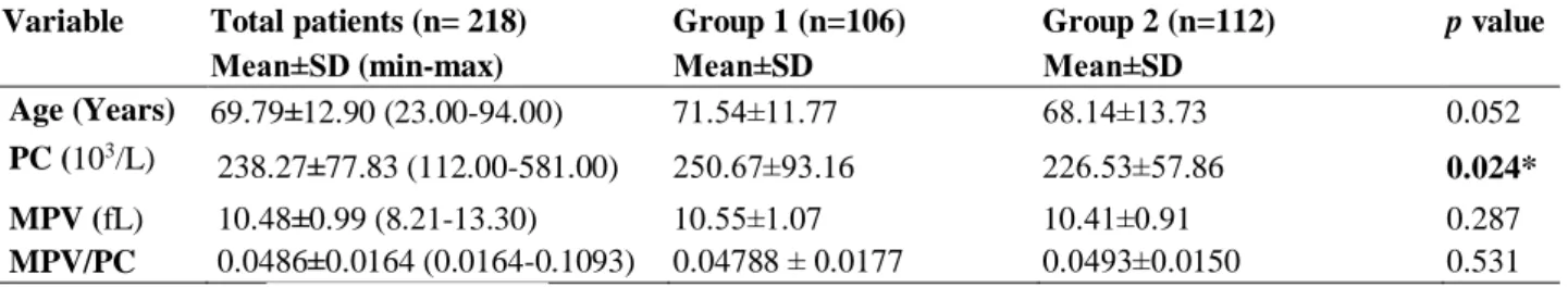
platelet count and functions) in 5 patients were determined). A total of 218 cases (111 males (50.9%), 107 females (49.1%)) were included in the study. Acute CVE was detected in DWI in 106 (48.6%) cases, while 112 (51.4%) cases were included in the group 2. The mean age of all cases included in the study was calculated as 69,79±12,90 years. The descriptive statistics have been displayed in Table 1.
No significant difference was observed between the group 1 and the group 2 in terms of the ages of the patients. The PC value in the group 1 was determined as (250.67±93.16×10³/L) and in the group 2 as
(226.53±57.86×10³/L). The mean PC value of
the group 1 was found to be higher (p<0,05) (Table 1). MPV values and the MPV/PC ratios showed no significant difference between the two groups (p>0,05).
Table 1. Evaluation of changes in platelet indices.
Variable Total patients (n= 218) Group 1 (n=106) Group 2 (n=112) p value
Mean±SD (min-max) Mean±SD Mean±SD
Age (Years) 69.79±12.90 (23.00-94.00) 71.54±11.77 68.14±13.73 0.052 PC (103/L)
238.27±77.83 (112.00-581.00) 250.67±93.16 226.53±57.86 0.024* MPV (fL) 10.48±0.99 (8.21-13.30) 10.55±1.07 10.41±0.91 0.287 MPV/PC 0.0486±0.0164 (0.0164-0.1093) 0.04788 ± 0.0177 0.0493±0.0150 0.531 SD: Standard Deviation, *significant at <0.05 level.
Discussion
Platelets play an active role in the formation, destabilization and thrombosis of atherosclerotic lesions.2 There are studies demonstrating that a high PC values is an important risk factor for acute CVE.9,10 Despite the evidence that elevated PC values pave the way for thrombotic events, no correlation between PC and thrombotic events was found in a study examining 3413 deaths due to thrombotic etiology.11,12 Thrombotic
events have also been shown to be limited to the venous system when PC value is high.13 However, in our study, the PC values of the group 1 were higher than that of the group 2. The PC alone is not a parameter indicating platelet activity; we don't have enough evidence to use PC as a diagnostic criterion in cases with suspicious acute CVE, when its value is measured as >250 K/l.
MPV is one of the most frequently used parameters in determining platelet size and activity.14 The increase in MPV values is known to be a risk factor for thrombotic events.15 Although Cho et al.14 found no
significant difference between the MPV values of the groups in their study, increased MPV values in CVE cases were measured in many studies.7,16-18 Also; Lök et al.19 found no correlation between different acute ischemic stroke subtypes with regard to MPV values. In our study, no significant difference was found between the MPV values of the groups. Elsayed et al. found a higher MPV/PC ratio in CVE cases.3 The evidences that the
increase in the MPV/PC ratio is a risk factor for CVE.3,4,20 Quan et al. found that high MPV/PC value is an independent risk factor large artery atherosclerosis stroke.5 But; there was no significant difference in the MPV/PC ratios between the groups in our study.
109
In previous studies; healthy volunteers were used as control group.3,16-18 But the main difference that differentiates our study from other studies is that the two patient groups. Although this may seem complicated, we think that it is more significant in terms of analyzing the MPV/PC ratio being used as a differential parameter. As a result, compared to other studies, we have obtained different results; the MPV value and the MPV/PC ratio were parameters that showed variations in many clinical conditions, and there was no significant difference between the group 1 and the group 2.
Limitations
The most important limitation of our study is its retrospective design. In addition, the size of the lesions observed in the patients' DWI and their prognosis were unknown.
Conclusion
Increased platelet activity is an important risk factor for thrombosis-related diseases. In our study, the PC value of patients with CVE was higher than the patients without CVE, but we don't have enough evidence to use PC as a diagnostic criterion of CVE. However, MPV and the MPV/PC ratio, which are not distinguished parameters for CVE.
Ethics Committee Approval
The study was approved by Ethics Committee (Decision no: 2018/40; Date: 04/12/2018) and continued in accordance with the Helsinki Principles Declaration.
Informed consent Not required.
Author Contributions
MB, MS: Conceptualization, Methodology, Software. MB, MS, YS: Data curation, Writing- Original draft preparation. MB, YS: Visualization, Investigation. MMO: Supervision. MS, HG: Validation. MB, MS, MMO: Writing- Reviewing and Editing. Conflict of Interest
The authors declared no conflict of interest.
Financial Disclosure
The authors declared no financial support. References
1. Go S, Worman DJ. “Stroke Syndromes” in Tintinalli JE, Stapczynski J, Ma OJ, et al (Editors). Tintinalli’s Emergency Medicine. 7th edition, The McGrew-Hill Companies, China, 2011: 1142–56
2. Coppinger JA, Cagney G, Toomey S, et al. Characterization of the proteins released from activated platelets leads to localization of novel platelet proteins in human atherosclerotic lesions. Blood. 2004; 103(6):2096-104.
3. Elsayed AM, Mohamed GA. Mean platelet volume and mean platelet volume/platelet count ratio as a risk stratification tool in the assessment of severity of acute ischemic stroke. Alexandria
Journal of Medicine. 2017; 53(1), 67-70.
4. D'erasmo E, Aliberti G, Celi FS, Romagnoli E, Vecci E, Mazzuoli GF. Platelet count mean platelet volume and their relation to prognosis in cerebral infarction. J Intern Med. 1990; 227(1): 11-4.
5. Quan W, Chen Z, Yang X, et al. Mean platelet volume/platelet count ratio as a predictor of 90-day outcome in large artery atherosclerosis stroke patients. Int J Neurosci. 2017; 127(11): 1019-27.
6. Ates S, Oksuz H, Dogu B, Bozkus F, Ucmak H, Yanıt F. Can mean platelet volume and mean platelet volume/platelet count ratio be used as a diagnostic marker for sepsis and systemic inflammatory response syndrome? Saudi Med J. 2015; 36(10): 1186.
7. Han JS, Park TS, Cho SY, Joh JH, Ahn HJ. Increased mean platelet volume and mean platelet volume/platelet count ratio in Korean patients with deep vein thrombosis. Platelets. 2012; 24(8): 590–3.
8. Bolat I, Akgul O, Cakmak HA, et al. The prognostic value of admission mean platelet volume to platelet count ratio in patients with ST-segment elevation myocardial infarction undergoing primary percutaneous coronary intervention.
Kardiol Pol. 2016; 74(4): 346-55.
9. Mayda-Domaç F, Mısırlı H, Yılmaz M. Prognostic role of mean platelet volume and platelet count in ischemic and hemorrhagic stroke. J Stroke Cerebrovasc Dis. 2010;19(1): 66-72.
10. Du J, Wang Q, He B, et al. Association of mean platelet volume and platelet count with the development and prognosis of ischemic and hemorrhagic stroke. Int J Lab Hematol. 2016; 38(3): 233-9.
11. Regev A, Stark P, Blickstein D, Lahav M. Thrombotic complications in essential thrombocythemia with relatively low platelet counts. Am J Hematol. 1997; 56(3): 168-72.
12. van der Bom JG, Heckbert SR, Lumley T, et al. Platelet count and the risk for thrombosis and death in the elderly. J Thromb
Haemost. 2009;7(3): 399-405.
13. Griesshammer M, Bangerter M, Sauer T, Wennauer R, Bergmann L, Heimpel H. Aetiology and clinical significance of thrombocytosis: analysis of 732 patients with an elevated platelet count. J Intern Med. 1999; 245(3): 295-300.
14. Cho SY, Jeon YL, Choi S K, Suh JT, Lee HJ, Park TS. Mean platelet volume in Korean patients with acute ischemic stroke: a gender difference. Platelet. 2013; 24(1): 75-6.
15. Braekkan SK, Mathiesen EB, Njølstad I, Wilsgaard T, Størmer J, Hansen JB. Mean platelet volume is a risk factor for venous thromboembolism: the Tromsø study. J Thromb Haemost. 2010; 8(1): 157-62.
16. Arikanoglu A, Yucel Y, Acar A, Cevik MU, Akil E, Varol S. The relationship of the mean platelet volume and C-reactive protein levels with mortality in ischemic stroke patients. Eur
Rev Med Pharmacol Sci. 2013; 17(13): 1774-7
17. Balcik ÖS, Bilen S, Ulusoy E, et al. Thrombopoietin and mean platelet volume in patients with ischemic stroke. Clin Appl
Thromb Hemost. 2013; 19(1): 92-5.
18. Nadar SK, Lip GY, Blann AD. Platelet morphology, soluble P selectin and platelet P-selectin in acute ischaemic stroke. Thromb Haemost. 2004; 92(06) :1342-8.
19. Lok U, Gulacti U, Ekmekci B, Bulut T, Celik M. Predictive and prognostic role of mean platelet volume in patients with first-ever acute ischemic stroke. Neurosciences (Riyadh).
2017;22(2):119–126.
20. Guenancia C, Hachet O, Stamboul K, et al. Incremental predictive value of mean platelet volume/platelet count ratio in
110
in-hospital stroke after acute myocardial infarction. Platelet. 2017; 28(1): 54-9.