https://doi.org/10.1007/s00276-019-02301-z
ORIGINAL ARTICLE
Morphometric development of the tongue in fetal cadavers
Ahmet Dursun1 · Yadigar Kastamonı1 · Demet Kacaroglu2 · Neslihan Yuzbasıoglu3 · Tolga Ertekın4
Received: 25 March 2019 / Accepted: 3 August 2019 © Springer-Verlag France SAS, part of Springer Nature 2019
Abstract
Purpose The tongue is a specific organ for the sense of taste. It consists of the striated muscle and mucous membrane. Fur-thermore, it helps the functions of speech, chewing, and swallowing. In this study, we aimed to reveal some morphometric properties of the tongue in fetal cadavers.
Methods The study was conducted on a total of 45 fetal tongues (25 male tongues, 20 female tongues) aged between 17 and 40 weeks. The fetuses were divided into three groups as trimester II, trimester III, and full term. For each tongue, the length, width, area, free tongue length, and the terminal sulcus angle were measured using Image J program. The free tongue length/tongue length ratio was examined.
Results The obtained data were compared according to the trimester groups and genders. It was determined that the tongue length, width, area, and free tongue length increased during the trimesters and that there was no significant difference in the terminal sulcus angle and the free tongue length/tongue length ratio between the trimesters. No significant difference was found in all parameters between the genders.
Conclusions This study presented significant data on morphometric development of the tongue. These data are thought to be useful for determining the anomaly and variations of the tongue.
Keywords Tongue development · Morphometry · Fetus tongue · Tongue length · Tongue width
Introduction
The tongue, which is located at the base of the oral cavity, is an organ composed of the striated muscle. In addition to speech, it has functions such as chewing, swallowing, respi-ration, control of secretions, suckling, and the sensation of taste [12, 18]. The tongue, which is wrapped by a mucous membrane, consists of three parts: the radix, corpus, and apex linguae. On the posterior side of the dorsum linguae, there is a “V”-shaped groove called the terminal sulcus with
the forward-looking opening. In the middle of the terminal sulcus, there is a blind-ended opening called the foramen caecum. The foramen caecum is the embryonic remnant of the upper end of the thyroglossal duct [24].
The frenulum of the tongue is a fold located in the mid-dle part of the mucosa covering the inferior surface of the tongue. With this fold, the tongue is connected to the base of the mouth [24]. The term-free tongue length is defined as the length of the tongue from the insertion of the lingual frenu-lum into the base of the tongue to the tip of the tongue [14]. Tongue development begins in the fourth and fifth weeks of the intrauterine period. Unlike the maxillary and man-dibular structures, the tongue differentiates from the occipi-tal myotome. Subsequently formed primary tongue cells migrate to the oral cavity. This process has a significant effect on the shaping of the oral cavity [2, 9, 23]. Knowing the typical development of the tongue facilitates the diag-nosis of tongue-related oral cavity pathologies and tongue anomalies in the intrauterine period.
The tongue, which continues to develop during the post-natal period, grows faster than maxillofacial structures [9]. This plays an essential role in the development of the * Ahmet Dursun
1 Department of Anatomy, Faculty of Medicine, Suleyman Demirel University, 32260 Isparta, Turkey
2 Department of Medical Biology, Faculty of Medicine, Suleyman Demirel University, 32260 Isparta, Turkey 3 Department of Anatomy, Faculty of Medicine, Istanbul
Medipol University, 34815 Istanbul, Turkey 4 Department of Anatomy, Faculty of Medicine,
Afyonkarahisar Health Sciences University, 03200 Afyonkarahisar, Turkey
mandibular arch by causing muscular stimulation [11, 16]. Significant reduction in the size of the tongue causes the down and forward growth of the mandible to slow down. Disorders in the transverse development of the mandibular arch in aglossia cases and lower lip retraction and microg-nathia in hypoglossia cases are examples of this condition [11]. Pierre Robin sequence is a congenital anomaly char-acterized by mandibular hypoplasia, glossoptosis, and cleft palate. The small jaw may cause airway obstruction by caus-ing the tongue to fall backward into the hypopharynx [7]. In Down syndrome, the rapid development of the tongue compresses the pharynx and causes mandibular prognathism [9]. Macroglossia is significantly correlated to mandibular prognathism [28].
Investigating the development of the tongue in fetal cadavers and revealing standard parameters will guide the early diagnosis and treatment of congenital anomalies in studies on the tongue. In the present study, some morpho-metric properties of the tongue in fetal cadavers and changes that occurred in these properties with development were examined. Most of the studies on the tongue have been car-ried out using imaging methods [1, 16]. Although there are histological studies carried out on fetal tongues [2, 9, 26], there are few morphometric studies on tongue development [23]. This study aimed to investigate some morphometric properties of the tongue in fetal cadavers and the effect of development on these morphometric properties.
Materials and methods
The study was carried out in the Laboratory of Süleyman Demirel University, Faculty of Medicine, Department of Anatomy on 45 fetuses (25 males, 20 females), which had no external anomaly and pathology, were aged between 17 and 40 gestational weeks, and which were provided from Isparta Maternity and Children’s Hospital with the permission of their families between the years 1996 and 2014. The causes of death in fetal cadavers are unknown. All fetal cadavers were fixed by arterial injection of 10 L of 10% (v/v) formal-dehyde solution into the water and stored in a pool of 10 L of 10% (v/v) formaldehyde solution. Although there was no study on tissue contraction of formaldehyde in the human tongue, in a study on the cervix, the median shrinkage was found to be zero in both distal and proximal anteroposte-rior diameters in 8% formalin fixation. The median shrink-age in the longitudinal direction was 3.0% and 2.4% in the transverse direction [3]. When rat liver tissues were fixed in 1.3 M formaldehyde solution and examined by an acceler-ated video camera, 1 × 1 × 8 cm rat liver strips shrunk to only about 3% at room temperature [6]. Although formaldehyde has a shrinkage effect on tissues, it can be understood from
the publications that this rate is 3%. The 3% effect should be considered in the evaluation of the data in the publication.
The gestational age of fetuses was determined accord-ing to the crown rump length (CRL), bi-parietal diameter (BPD), head circumference (HC), and femur length (FU). In the fetal period, fetuses were evaluated by being divided into three groups: fetuses between 17 and 25 weeks were deter-mined to be trimester II, fetuses between 26 and 37 weeks were determined to be trimester III, and fetuses between 38 and 40 weeks were determined to be full term.
First, the general parameters of each fetus (CRL, BPD, HC, and FL) were determined. Afterward, the mandible of the fetuses was dissected from the joints and removed from the fetuses with fat, muscle, and skin. The tongue of the fetuses was ablated together with the root of the tongue using a scalpel. The extracted tongues were placed on a flat surface close to the intraoral position. The photographs of the removed tongues were taken next to a ruler. The photo-graphs were recorded with Olympus SP-600UZ camera with 12.0 megapixels, 15 × optical zoom. The taken photographs were transferred to the Image J program, and the following parameters were measured (Fig. 1).
Tongue length: a distance from the tip of the tongue to the foramen caecum.
Tongue width: the measured widest distance of the dor-sum of the tongue.
Tongue area: an area of the region in front of the terminal sulcus.
Free tongue length: the length of the tongue from the insertion of the lingual frenulum into the base of the tongue to the tip of the tongue.
Male: the angle between the lines drawn to the front edge of the place, where the palatoglossal arch ends in the tongue, by considering the foramen caecum as the center.
Statistical analysis was performed using SPSS Inc. SPSS for Windows 17.0 statistical package program. According to gestational age (trimester), the parameters’ arithmetic means and standard deviations were determined. The parametric values presented according to trimesters were displayed as arithmetic mean ± standard deviation. Other parameters except the tongue area showed normal distribution. We used nonparametric analysis methods, because the number of data was less than 30 in the inter-group comparison. The Kruskal–Wallis test was used for the comparison of param-eters between the trimester groups, and the Mann–Whitney
U test was used for the comparison between the genders.
Pearson’s correlation analysis was used in the correlation analysis of the tongue length and tongue width with gesta-tional age, because the data were normally distributed.
Results
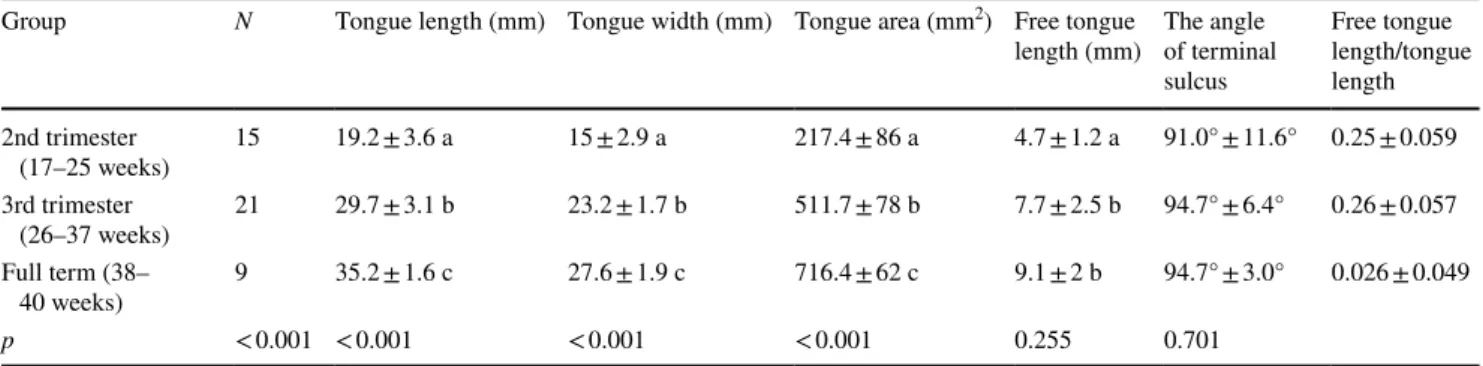
The tongue length, width, area, free tongue length, the ter-minal sulcus angle, and free tongue length/tongue length ratio, and the maximum, minimum, and mean values, and standard deviations of these parameters are shown in Table 1. We think that the reason for the high standard deviation of the tongue area is due to the use of two factors
in the field calculation. The mean values of these param-eters according to trimesters are presented in Table 2. It was determined that among these parameters, the length, width, area, and free tongue length increased during tri-mesters, and there was a statistically significant difference between the trimesters (p ˂ 0.05; Table 2). No significant difference was found between the trimesters in terms of the
Fig. 1 Measured parameters related to tongue
Table 1 Minimum, maximum, mean and standard deviation values of measured parameters related to tongue
Parameters N Minimum (mm) Maximum (mm) Mean ± std. deviation (mm)
Tongue length 45 14.68 37.78 27.38 ± 6.87
Tongue width 45 10.31 30.26 21.39 ± 5.35
Tongue area 45 107.89 768.40 454.59 ± 201.73
Free tongue length 45 2.73 13.80 7.02 ± 2.45
Free tongue length/tongue length 45 0.16 0.40 0.26 ± 0.055 The angle of terminal sulcus 45 68.58 115.03 93.36 ± 8.32
Table 2 Comparison of measured parameters related to tongue according to trimesters
In paired comparisons between groups (trimester), each group was represented by letters. If the letters were the same, there was no statistically significant difference between the groups (p > 0.05) and if the letters were different, there was statistically significant difference between the groups (p < 0.05)
Group N Tongue length (mm) Tongue width (mm) Tongue area (mm2) Free tongue
length (mm) The angle of terminal sulcus Free tongue length/tongue length 2nd trimester (17–25 weeks) 15 19.2 ± 3.6 a 15 ± 2.9 a 217.4 ± 86 a 4.7 ± 1.2 a 91.0° ± 11.6° 0.25 ± 0.059 3rd trimester (26–37 weeks) 21 29.7 ± 3.1 b 23.2 ± 1.7 b 511.7 ± 78 b 7.7 ± 2.5 b 94.7° ± 6.4° 0.26 ± 0.057 Full term (38– 40 weeks) 9 35.2 ± 1.6 c 27.6 ± 1.9 c 716.4 ± 62 c 9.1 ± 2 b 94.7° ± 3.0° 0.026 ± 0.049 p < 0.001 < 0.001 < 0.001 < 0.001 0.255 0.701
terminal sulcus angle and the free tongue length/tongue length ratio (p > 0.05; Table 2).
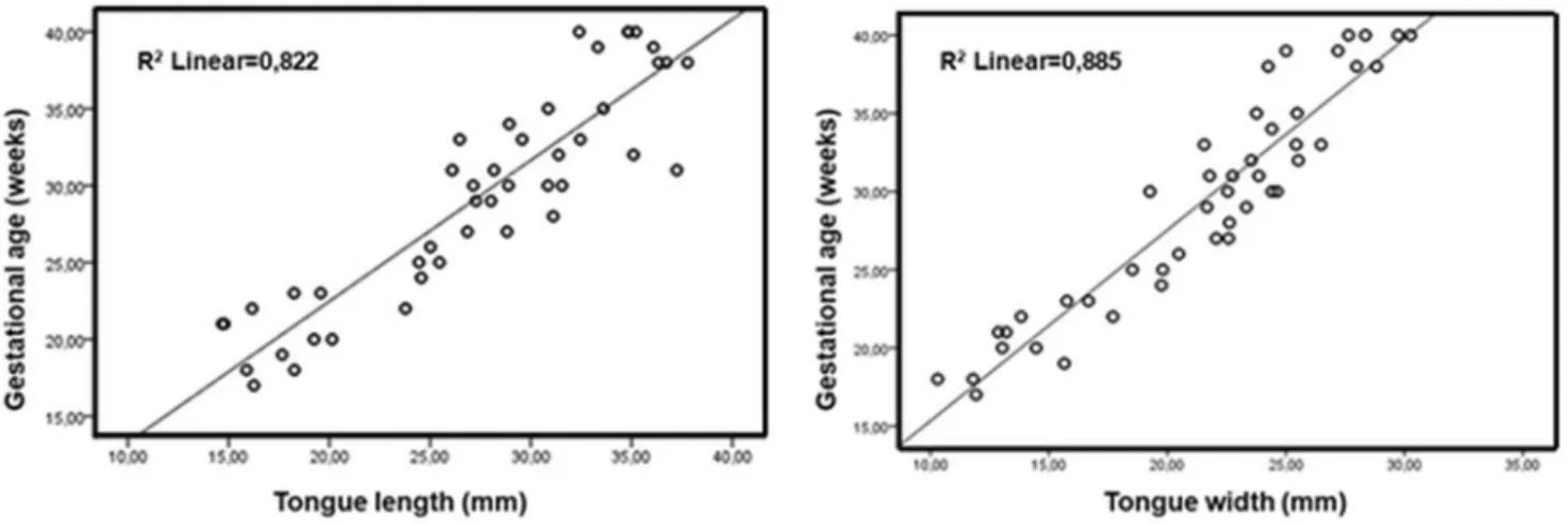
When the correlation between the tongue length (r = 0.907) and tongue width (r = 0.941) was examined according to gestational age, a very strong positive correla-tion was found (Fig. 2).
When among the tongue-related parameters, the length, width, area, free tongue length, the terminal sulcus angle, and the free tongue length/tongue length ratio were com-pared between the genders, the mean values of males were higher in all parameters except for the free tongue length/ tongue length ratio, but there was no significant difference between genders in any parameters (p > 0.05; Table 3).
Discussion
An abnormal tongue size can be associated with more than 25 syndromes and diseases [8, 13, 20–22]. Macroglossia is considered to be rare among children [15]. However, accord-ing to a study conducted on tongue disorders in a child popu-lation, macroglossia is one of the most common diagnoses (24%) [10]. It is difficult to determine the accurate incidence of macroglossia because of its multiple causes. Various syn-dromes include macroglossia, such as Down syndrome (1 per 700 live births) [4, 19] and the Beckwith–Wiedemann
syndrome (0.07 per 1000 live births) [27, 28], gangliosi-dosis, athyrotic hypothyroidism sequence, trisomy 4p, and autosomal dominant macroglossia [5, 25]. The enlarged tongue can be observed occasionally in cases such as tri-somy 21, Hurler syndrome, Robinow syndrome, and Scheie syndrome. The small tongue is often observed in the oro-mandibular extremity hypogenesis spectrum and hypoglos-sia–hypodactylia syndrome, and rarely in other syndromes such as Arthrogryposis syndrome type II, Freeman–Sheldon syndrome, Lenz-Majewski hyperostosis syndrome, Moebius sequence, Pallister–Hall syndrome, and Short rib-Polydac-tyly Majewski type [5]. Knowledge of typical tongue param-eters may be helpful in detecting these syndromes when abnormalities are encountered in tongue measurements to be performed during the intrauterine period. This may be the main step for further investigations.
Aguiar et al. [2] found a positive correlation between gestational age and tongue length and circumference in the macroscopic analysis of 55 tongues aged between 23 and 40 weeks: R2 = 0.527; p < 0.001. In the present study, a
strong correlation was determined between the tongue length and gestational age: R2 = 0.822; p < 0.001 (Fig. 2). In the
study in which Bronshtein et al. [5] studied 80 fetuses aged between 13 and 18 weeks using transvaginal ultrasonogra-phy, a linear relationship between gestational age and lingual width was found: R2 = 0.83. In the present study, a linear
Fig. 2 Graphs of correlation of tongue length and width with gestational age
Table 3 Comparison of measured parameters related to tongue according to gender
Gender N Tongue length (mm) Tongue width (mm) Tongue area (mm2) Free tongue
length (mm) The angle of terminal sulcus Free tongue length/tongue length Male 25 28.15 ± 6.27 21.86 ± 4.84 466.43 ± 183.40 7.20 ± 2.54 95.11° ± 7.60° 0.254 ± 0.060 Female 20 26.41 ± 7.59 20.79 ± 5.99 439.79 ± 226.58 6.77 ± 2.37 91.33° ± 8.85° 0.256 ± 0.049
correlation was determined between the tongue width and gestational age: R2 = 0.885; p < 0.001 (Fig. 2).
Using regression equations, Siebert [23] defined the rela-tionship between the weight, length, width, and thickness of 83 healthy tongues, aged between 25 weeks and 10.5 years, with various body and head circumference measurements. In this study, Siebert [23] could not find any difference between genders in the tongue dimension. In contrast, Liegeois et al. [17] examined the tongue volume of 70 volunteers (35 males and 35 females) aged between 20 and 37 years by the mag-netic resonance imaging method and determined that the tongue volume was different between genders. In the present study, no significant difference was found between genders in terms of the measured parameters (Table 3).
The frenulum of the tongue is demonstrated to be shaped according to changes in the embryonic process of the tongue [9]. In the present study, the mean value of the measurement which was made from the frenulum of the tongue to the apex of the tongue and is called the free tongue length was found to be 7.02 ± 2.45 mm. This value was higher in males, but there was no statistically significant difference between males and females. The free tongue length increased during the trimesters, but the free tongue length to tongue length ratio was not different between trimesters. Knowing the dis-tance from the distal of the frenulum of the tongue to the tip of the tongue will be useful for the early diagnosis of pathologies such as ankyloglossia, hypertrophic frenulum linguae, and short frenulum.
Conclusions
The tongue is a vital organ that plays a critical role in the development of adjacent maxillofacial structures such as oral and nasal cavities, pharynx and maxilla, and abnormal tongue development is associated with congenital maxillofa-cial anomalies. In the present study, the typical development of the tongue was defined on fetal tongues, which had no congenital anomaly morphometrically. It is believed that the data obtained from this study will be useful in the prenatal diagnosis of cases such as developmental disorders of the tongue and congenital syndromes.
Author contributions AD: project development, data acquisition, man-uscript draft, and manman-uscript review. YK: data acquisition, manman-uscript draft, manuscript review. DK: data acquisition, manuscript review. NY: data analysis, manuscript editing. TE: data analysis, manuscript editing.
Funding This research received no specific grant from any funding agency in the public, commercial, or not-for-profit sectors.
Conflict of interest The authors declare that they have no conflict of interest.
Ethical approval This is a retrospective study. Ethics committee approval is not required. This article does not contain any studies with human participants or animals performed by any of the authors.
References
1. Achiron R, Ben Arie A, Gabbay U, Mashiach S, Rotstein Z, Lipitz S (1997) Development of the fetal tongue between 14 and 26 weeks of gestation: in utero ultrasonographic measurements. Ultrasound Obstet Gynecol 9:39–41
2. Aguiar LS, Juliano GR, Silveira LAM, Oliveira MS, Torquato BGS, Juliano GR, Araujo MF, Pereira SAL, Teixeira VPA, Fer-raz MLF (2018) Tongue development in stillborns autopsied at different gestational ages. J Pediatr (Rio J) 94:616–623 3. Boonstra H, Oosterhuis JW, Oosterhuis AM, Fleuren GJ (1983)
Cervical tissue shrinkage by formaldehyde fixation, paraffin wax embedding, section cutting and mounting. Virchows Archiv A 402:195–201
4. Boyd D, Ouick A, Murray C (2004) The Down syndrome patient in dental practice, part ll: clinical. N Z Dent J 100:4–9 5. Bronshtein M, Zimmer EZ, Tzidony D, Hajos J, Jaeger M,
Blazer S (1998) Transvaginal sonographic measurement of fetal lingual width in early pregnancy. Prenat Diagn 18:577–580 6. Fox CH, Jhonson FB, Whiting J, Roller PP (1985)
Formalde-hyde fixation. J Histochem Cytochem 33:845–853
7. Gunlemez A, Işken T (2008) Pierre Robin Sekansı: Yenidoğan döneminde hava yolu açıklığının ve beslenmenin sağlanması. Çocuk Dergisi 8:235–242
8. Gupta OP (1971) Congenital macroglossia. Arch Otolaryngol 93:378–383
9. Hong SJ, Cha BG, Kim YS, Lee SK, Chi JG (2015) Tongue growth during prenatal development in Korean fetuses and embryos. J Pathol Transl Med 49:497–510
10. Horn C, Thaker HM, Tampakopoulou DA, Serres LMD, Keller JL, Haddad J Jr (2001) Tongue lesions in the pediatric popula-tion. Otolaryngol Head Neck Surg 124:164–169
11. Hutchinson EF, Kieser JA, Kramer B (2014) Morphometric growth relationships of the immature human mandible and tongue. Eur J Oral Sci 122:181–189
12. Iskander A, Sanders I (2003) Morphological comparison between neonatal and adult human tongues. Ann Otol Rhinol Laryngol 112:768–776
13. Kotlow LA (1999) Ankyloglossia (tongue-tie): a diagnostic and treatment quandary. Quintessence Int 30:259–262
14. Lam AM, Vavilala MS (2000) Macroglossia: compartment syndrome of the tongue? Anesthesiol J Am Soc Anesthesiol 92:1832
15. Lauder R, Muhl ZF (1991) Estimation of tongue volume from magnetic resonance imaging. Angle Orthod 61:175–184 16. Liegeois F, Albert A, Limme M (2010) Comparison between
tongue volume from magnetic resonance images and tongue area from profile cephalograms. Eur J Orthod 32:381–386
17. Mangold AR, Torgerson RR, Rogers RS III (2016) Diseases of the tongue. Clin Dermatol 34:458–469
18. Moura CP, Cunha LM, Vilarinho H, Cunha MJ, Freitas D, Palha M, Pueschel SM, Pais-Clemente M (2008) Voice parameters in children with Down syndrome. J Voice 22:34–42
19. Petersen MB, Hansen M, Djernes BW (1987) Full trisomy 22 in a newborn infant. Ann Genet 30:101–104
20. Reynoso MC, Hernandez A, Soto F, Garcia-Cruz O, Martinez Y, Martinez R, Cantu JM (1986) Autosomal dominant macroglossia in two unrelated families. Hum Genet 74:200–205
21. Rice JP, Stephen HC (1985) A case report of lingual lymphangi-oma presenting as recurrent massive tongue enlargement. Clin Pediatr 24:47–50
22. Siebert JR (1985) A morphometric study of normal and abnormal fetal to childhood tongue size. Arch Oral Biol 30:433–440 23. Standring S (2008) Gray’s anatomy: the anatomical basis of
clini-cal practice. Elsevier Churchill Livingstone, New York
24. Topouzelis N, Iliopoulos C, Kolokitha OE (2011) Macroglossia. Int Dent J 61:63–69
25. Tozak Yıldız H, Ozdamar S (2009) İnsan fetuslarında dil papillalarının gelişiminin taramalı elektron mikroskobunda incelenmesi. Sağlık Bilimleri Dergisi 18:129–137
26. Van Lierde KM, Mortier G, Huysman E, Vermeersch H (2010) Long-term impact of tongue reduction on speech intelligibil-ity, articulation and oromyofunctional behaviour in a child with
Beckwith–Wiedemann syndrome. Int J Pediatr Otorhinolaryngol 74:309–318
27. Ylmaz M, Mercan H, Karaman E, Kaytaz A (2009) Tongue reduc-tion in Beckwith–Wiedemann syndrome with CO2 laser. J Crani-ofac Surg 20:1202–1203
28. Yoo E, Murakami S, Takada K, Fuchihatal H, Sakuda M (1996) Tongue volume in human female adults with mandibular progna-thism. J Dent Res 75:1957–1962
Publisher’s Note Springer Nature remains neutral with regard to jurisdictional claims in published maps and institutional affiliations.