Combined Vibrational Spectroscopic and Quantum Chemical
Investigations of 1,8-diaminooctane
Akif Özbay1*, Aysun Gözütok2
1Gazi University, Faculty of Science, Department of Physics, Ankara, Turkey 2Selçuk University, Faculty of Science, Department of Physics, Konya, Turkey
Received: 06 December 2018 Accepted: 18 January 2019 DOI: 10.18466/cbayarfbe.492861
Abstract
This paper contains the molecular parameters, vibrational properties and some theoretical calculations of 1,8-diaminooctane. Bond angles, bond lengths, vibrational properties, dipole moments, frontier molecular orbitals and molecular electrostatic potential of 1,8-diaminooctane were performed with using density
functional theory calculations with B3LYP/6-311++G(d,p) level of theory. Vibrational properties were
interpreted with the by using scaled quantum mechanical force field. This study enables us to figure out the vibrational and structural properties and some electronic properties of the 1,8-diaminooctane by means of the theoretical and experimental studied methods.
Keywords: 1,8-diaminooctane, density functional theory, theoretical calculations, molecular electrostatic potential.
1. Introduction
This molecule concerns of the monoalkylamines which are organic compounds including a primary aliphatic amine group. An alkane-α and ω-diamine in which the two amino groups are separated by eight methylene groups [1]. The 1,8-diaminooctane is used as cross link effects while the synthesis of various molecular carbon nanotubes and macrocycle molecules [2]. Aliphatic diamines are important compounds that are widely used in coordination chemistry, biochemistry and polymer chemistry. This molecule has a fundamental carbon chain structure. So, it is used like synthesis molecule. This molecule is used as the ligand in the formation of
Hofmann type host-guest compounds. Molecular
formula of 1,8-diaminooctane is C8H20N2 and be found in solid phase.
FT-IR spectra of 1,8-diaminooctane was reported by Kasap and Özbay [3]. In that study, this molecule had been purchased from Sigma-Aldrich and used without further purification. IR spectra of 1,8-diaminooctane had been recorded 400-4000 cm-1 with Perkin-Elmer Spectrum One FT-IR spectrometer.
In our study, the molecular structure (bond angles, bond lengths, dipole moments) and vibrational frequencies of 1,8-diaminooctane have been calculated by density functional theory (DFT) with B3LYP method and 6-311++G(d,p) level of theory.
2. Materials and Methods
In this study, 1,8-diaminooctane molecule was drawn in three dimensions using the molecular drawing program GaussView 3.0 [4]. The geometrical parameters of this structure were automatically entered as input data in the Gaussian 03W package program [5]. Then, 1,8-diaminooctane molecule were optimized by means of the B3LYP/6-311++G(d,p) level of theory. Bond angles, bond lengths and vibrational frequencies of
1,8-diaminooctane molecule have been calculated
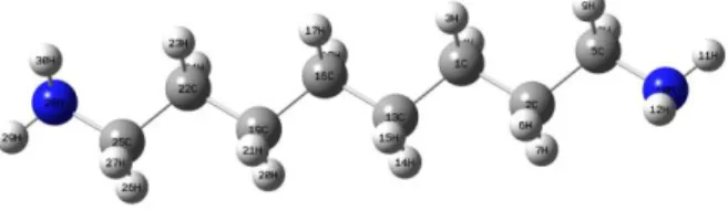
DFT/B3LYP with 6-311++G(d,p). The vibrational frequencies of the optimized geometry were corrected by multiplying with scaling factors [6] for comparing the experimental values. The optimized geometrical structure of the title molecule is shown in Figure 1.
Figure 1. Molecular structure and atomic numbering of 1,8-diaminooctane.
Since there is not published experimental data on the bond lengths and bond angles of the 1,8-diaminooctane, the calculated geometrical parameters of this molecule were compared with the experimental values of the geometrical parameters of the 1,6-diaminohexane molecule as similar molecule performed by Meng and
Doi:10.18466/cbayarfbe.492861 A. Özbay Lin [7].
The potential energy distribution (PED) was calculated via using the scaled quantum mechanics (SQM) program [8] and the fundamental vibrational modes were assigned with their PED values.
Addition to these properties, HOMO-LUMO energy gap and MEP have been calculated by DFT/B3LYP with same basis set.
3. Results and Discussion
Geometrical parameters
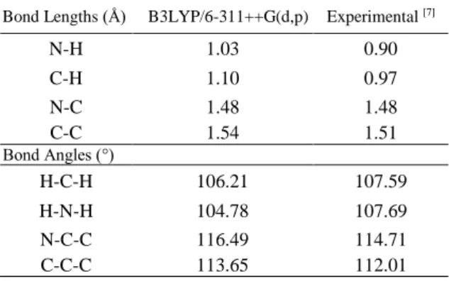
The experimental values and the calculated geometrical parameters of 1,8-diaminooctane were compared in Table 1.
In the B3LYP/6-311++G(d,p) level of theory, the N-C-C (116.49°) bond angle is greater than the C-C-C (113.65°), H-N-H (104.78°) and H-C-H (106.21°). In general, the N-C-C bond angles are greater than the C-C-C, H-C-H and H-N-H bond angles. This is also consistent with the experimental results.
In the B3LYP/6-311++G(d,p) level of theory, the C-C (1.54 Å) bond length is greater than the N-C (1.48 Å), C-H (1.10 Å) and N-C-H (1.03 Å). In general, the C-C bond lengths are greater than the N-C, C-H and N-H bond lengths. This is also consistent with the experimental results.
Our calculated the geometrical parameters were found to be very compatible with compared experimental the geometrical parameters.
Table 1. Experimental and calculated parameters of 1,8-diaminooctane.
Bond Lengths (Å) B3LYP/6-311++G(d,p) Experimental [7]
N-H 1.03 0.90 C-H 1.10 0.97 N-C 1.48 1.48 C-C 1.54 1.51 Bond Angles (°) H-C-H 106.21 107.59 H-N-H 104.78 107.69 N-C-C 116.49 114.71 C-C-C 113.65 112.01
1,8-diaminooctane molecule has 30 atoms in which the C and N atoms are planar and the H atoms are out of plane. This molecule belongs to the point group C2h. Since this molecule is a non-linear molecule, it has 84 normal vibrational modes.
Vibrational Assignment
The vibrational frequencies and IR vibrational intensity are given in Table 2. The experimental and calculated FT-IR and FT-Raman spectra for 1,8-diaminooctane are given in Figures 2-4. Also, the corresponding data are given in Table 2.
Figure 2. Experimental FT-IR spectra of 1,8-diaminooctane.
Figure 3. Theoretical FT-IR spectra of 1,8-diaminooctane.
Figure 4. Experimental and theoretical FT-Raman spectra of 1,8-diaminooctane.
Table 2. The vibration frequencies of 1,8-diaminooctane molecule. Freqa. 𝑰 𝑰𝑹 𝒃 FT-IR [3] PED (%) 1 34 0.137 2 49 0.081 3 62 0.658 4 82 0.281 5 108 2.070 6 130 0.528 7 134 0.485 8 152 0.304 9 165 0.071 10 210 0.854 11 230 7.822 12 254 9.932 13 260 19.49 Wavenumber (cm-1) Wavenumber (cm-1) T ra n smit ta n ce ( %) In fr ar ed I n te n si ty
14 368 4.383 15 393 0.601 16 475 0.247 17 498 2.918 18 515 6.845 19 709 1.826 20 715 0.027 21 732 0.715 22 765 5.648 23 797 47.03 24 802 81.22 25 836 9.548 860 m, br HCH(37%) 26 904 0.755 27 930 2.610 28 954 1.229 951 vw HCH(53%) 29 975 2.719 30 987 1.514 31 998 6.143 1005 vw CCC(27%)+HNH(10%)+HCH(10%) 32 1023 0.280 33 1028 0.515 34 1031 0.283 35 1040 3.126 36 1047 11.54 1052 vw CCH(13%)+HNH(13%) 37 1068 2.193 1075 vw CCH(20%)+HNH(18%) 38 1109 1.554 1089 vw CCC(40%)+HNH(13%)+HCH(10%) 39 1174 0.160 40 1190 0.432 41 1212 0.101 1218 vw CCH(48%) 42 1231 0.105 43 1252 0.181 44 1270 0.519 45 1282 0.233 46 1291 0.122 47 1296 0.042 48 1299 0.242 1305 vw HCH(30%)+HNH(18%) 49 1325 0.572 1338 vw HCH(26%)+HNH(21%) 50 1342 0.229 51 1348 3.970 52 1353 0.379 53 1356 1.059 1367 vw HCH(25%)+HNH(18/%) 54 1380 6.998 1390 w, sh HCH(36%) 55 1434 0.107 56 1437 0.020 1440 vw HCH(40%) 57 1439 0.005 58 1442 0.048 59 1449 0.272 60 1457 0.083 61 1464 3.048 1466 s HCH(32%)+HNH(28%) 62 1471 1.840 1487 m HCH(42%)+HNH(21%) 63 1604 14.86 1584 s HCH(57%) 64 1609 11.58 65 2841 31.22 2856 vs CH(65%) 66 2892 1.911 67 2893 0.368 68 2896 4.347 69 2897 0.423 70 2901 0.201 71 2908 39.41 72 2910 9.590 73 2913 3.190 74 2919 35.355 75 2920 3.950 2926 vs CH(87%) 76 2932 5.181 77 2936 12.24 78 2947 5.435 79 2958 0.661 80 2960 100.0 81 3376 0.627 82 3380 0.365 83 3452 0.231 3321 s NH(79%) 84 3456 0.309 3381 s NH(79%)
vs:very strong, m: medium, s:strong, w:weak, vw:very weak,ν: stretching, : in plane bending aObtained from the wavenumbers calculated at DFT using scaling factor 0.967. bRelative absorption intensities normalized with the highest peak absorption equal to 100, cFT-IR values were taken Ref. [3]
dPotential energy distribution (PED) calculated at the B3LYP/6-311++G(d,p) level of theory, Only contributions ≥10% are listed,
The NH stretching vibrations appear at 3500-3300 cm-1 [9,10]. For 1,8-diaminooctane N-H stretching vibrations are observed at 3381 cm-1 and 3321 cm-1 experimentally (FT-IR spectra) [3] and assigned at 3456 cm-1 and 3452 cm-1 (DFT/B3LYP with 6-311++G(d,p)) according to our calculation. It can be seen that in Table 2, experimental and theoretical results are very consistent with each other.
The asymmetric and symmetric stretching modes of the CH2 group usually occur in the region from 2800 to 3000 cm-1[11]. In FT-IR spectra for 1,8-diaminooctane C-H stretching vibrations are observed at 2856 cm-1 and 2926 cm-1 experimentally [3]. These vibrations calculated at 2841 cm-1 and 2920 cm-1 by B3LYP/6-311++G(d,p) calculation (Table 2). The theoretical results are very good agreement with experimental ones.
The H-C-H in-plane bending vibrations characterize in the region 1300-1500 cm-1[12]. Experimentally, the in-plane H-C-H bending modes were measured at 1305, 1338, 1367, 1390, 1440, 1466, 1487 and 1584 cm-1 in FT-IR spectra [3]. These vibrations calculated at 1299, 1325, 1356, 1380, 1437, 1464, 1471 and 1604 cm-1 by means of B3LYP/6-311++G (d,p) (Table 2). It can be seen that in Table 2, experimental and theoretical results are very consistent with each other.
The C-C-H in-plane bending vibrations appear in the region 1300-1000 cm-1 [12]. Experimentally, the in-plane C-C-H bending modes were measured at 1052, 1075, 1089 and 1218 cm-1 in FT-IR spectra [3]. These vibrations calculated at 1047, 1068, 1109 and 1212 cm-1 by means of B3LYP/6-311++G (d,p) (Table 2). It can be seen that in Table 2, experimental and theoretical results are very compatible.
All aromatic C-H stretching vibrations were found to be weak. This reason may be attributed to decrease in the dipole moment because of decrease the negative charge on the C atom [13]. This decrease is due to the withdrawing of electrons from the carbon atom of the molecule due to the reduction of inductive effect, resulting in an increase in the chain length of the molecule [13].
HOMO-LUMO analysis
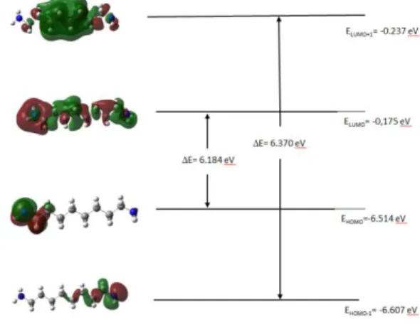
The highest occupied molecular orbital (HOMO), the lowest unoccupied molecular orbital (LUMO) and HOMO-LUMO energy gaps have been performed with using DFT at B3LYP/6-311++G(d,p) level. They are very important parameters in quantum chemistry. HOMO defines the ability to donate an electron and LUMO defines the ability to obtain an electron. The plots of HOMO, HOMO-1, LUMO, LUMO+1 for 1,8-diaminooctane were given in Figure 5. The HOMO-LUMO energy gap (∆𝐸) value of 1,8-diaminooctane calculated as 6.18 eV. This value for Urea 6.72 eV in the
Doi:10.18466/cbayarfbe.492861 A. Özbay literature [14]. So, 1,8-diaminooctane is very soft
molecule and very stable than Urea molecule.
Figure 5. Frontier molecular orbitals of 1,8-diaminooctane.
Besides, dipole moment values of 1,8-diaminooctane calculated by DFT and these values are given in Table 3. Table 3. The dipole moment (field-independent basis, Debye) values of 1,8-diaminooctane.
𝝁𝒙 𝝁𝒚 𝝁𝒛 𝝁𝒕𝒐𝒕
-1.21 1.62 1.07 2.29
MEPS of 1,8-diaminooctane
The molecular electrostatic potential (MEP) maps of 1,8-diaminooctane’s ground state are shown in Figure 6. Using the B3LYP/6-311++G(d,p) level of theory, the MEP maps were drawn. The MEP can be seen as reactivity maps on organic molecules, showing the most likely regions where point charges can approach the molecule. The MEP is used for researching reactivity regions. In MEP maps, red color shows negative, blue color shows positive areas. In Figure 6 right side picture, MEP in 1,8-diaminooctane is mainly over Nitrogen atoms.
Figure 6. The MEP maps of 1,8-diaminooctane
4. 4. Conclusion
In this work, the geometrical parameters, vibrational frequencies, dipole moments, HOMO-LUMO and MEP of 1,8-diaminooctane were performed with using Gaussian 03W program. In that all theoretical calculations, we used B3LYP/6-311++G(d,p) level of theory. The calculated geometrical structural and vibrational spectral data of 1,8-diaminooctane with DFT methods are good consistent with available experimental data.
Author’s Contributions
Akif Özbay: Drafted and wrote the manuscript, performed the experiment and result analysis.
Aysun Gözütok: Assisted in analytical analysis on the structure, supervised the experiment’s progress, result interpretation and helped in manuscript preparation. Ethics
There are no ethical issues after the publication of this manuscript.
5. References
1. Chemical Entities of Biological Interest (ChEBI) UK, http://www.ebi.ac.uk/chebi/searchId.do?chebiId=CHEBI:73112 (accessed at 01.06. 2018).
2. Sigma Aldrich Chemical Company, https://www.sigmaaldrich.com/catalog/product/aldrich/d22401?la ng=en®ion=TR (accessed at 01.06. 2018).
3. Kasap, E, Özbay, A. 1997. Infrared spectroscopic study of the Hofmann -daon-type clathrates : M( 1,8-diaminooctane)Ni(CN)4.G(M = Co, Ni or Cd ; G = Aromatic Guest Molecules). Journal of Inclusion Phenomena and Molecular Recognition in Chemistry; 28: 335-347.
4. Frisch, A, Dennington, RD, Keith, TA, Nielsen, AB, Holder, AJ. 2003. GaussView 3.0, Gaussian, Inc., Wallingford CT.
5. Frisch, MJ, Trucks, GW, Schlegel, HB, Scuseria GE, Robb, MA, Cheeseman, JR, Zakrzewski, VG, Montgomery, JA, Stratmann, Jr RE, Burant, JC, Dapprich, S, Milliam, JM, Daniels, AD, Kudin, KN, Strain, MC, Farkas, O, Tomasi, J, Barone, V, Cossi, M, Cammi, R, Mennucci, B, Pomelli, C, Adamo, C, Clifford, S, Ochterski, J, Petersson, GA, Ayala, PY, Cui, Q, Morokuma, K, Malick, DK, Rabuck, AD, Raghavachari, K, Foresman, JB, Cioslowski, J, Ortiz, JV, Baboul, AG, Stefanov, BB, Liu, G, Liashenko, A, Piskorz, P, Komaromi, I, Gomperts, R, Martin, RL, Fox, DJ, Keith, T, Al-Laham, MA, Peng, CY, Nanayakkara, A, Challacombe, M, Gill, PMW, Johnson, B, Chen, W, Wong, MW, Andres, JL, Gonzalez, C, Head-Gordon, M, Replogle, ES, Pople, JA. Gaussian, Inc., Wallingford CT, 2004.
6. Scaling factor, https://cccbdb.nist.gov/vibscalejust.asp (accessed at 01.06. 2018).
7. Meng, XG, Lin, ZD. 2005. Catena- Poly[[tetraaquacadmium(II)-µ-hexane-1,6-diamine-κ2N:N’] terephthalate dihydrate]. Acta Crystallographica E; 61: 263-264.
8. Pulay, P, Baker, J, Wolinski, K. 2013. Green Acres Road Suite A Fayettevile, Arkansas, 72703, USA.
9. Spire, A, Barthes, M, Kellouai, H, De Nunzio G. 2000. Far-infrared spectra of acetanilide. Physica D:Nonlinear Phenomena; 137: 392 – 396.
10. Panicker, CY, Varghese, HT, Thansani, T. 2009. Spectroscopic studies and Hartree-Fock ab initio calculations of a substituted amide of pyrazine-2-carboxylic acid-C16H18ClN3O. Turkish Journal of Chemistry; 33: 633 – 646.
11. Sajan, D, Binoy, J, Pradeep, B, Venkatakrishnan, K, Kartha, VB, Joe, IH, Jayakumar, VS. 2004. NIR-FT Raman and infrared spectra and ab initio computations of glycinium oxalate.
Spectrochimica Acta Part A: Molecular and Biomolecular Spectroscopy; 60: 173–180.
12. Thilagavathi, G, Arivazhagan, M. 2010. Density functional theory calculation and vibrational spectroscopy study of 2-amino-4,6-dimethyl pyrimidine (ADMP). Spectrochimica Acta Part A: Molecular and Biomolecular Spectroscopy; 79: 389 – 395. 13. Shanmugam, R, Sathyanarayana, D. 1984. Experimental (FT-IR
and FT-Raman), electronic structure and DFT studies on 1-methoxynaphthalene. Spectrochimica Acta Part A: Molecular and Biomolecular Spectroscopy; 40: 757 – 761.
14. Tesch, MF, Golnak, R, Ehrhard, F, Schön, D, Xiao, J, Atak K, Bande, A, Aziz, EF. 2016. Analysis of the Electronic structure of aqueous urea and its derivatives: a systematic soft X-Ray-TD-DFT approach. Chemistry; 22: 12040.