Angel Wings Syndrom in Swans (Cygnus cygnus and Cygnus atratus)
[1]Mustafa ARICAN
1,a Kurtuluş PARLAK
1,bMustafa YALÇIN
1,c[1] This study was presented poster in the 16th National Veterinary Surgery Congress and 2nd International Veterinary Surgery
Congress, September 20-23, 2018, Bafra, KKTC
1 Department of Surgery, Faculty of Veterinary Medicine, The University of Selcuk, TR-42003 Konya - TURKEY
a ORCID: 0000-0001-8180-135X; b ORCID: 0000-0002-8656-037X; c ORCID: 0000-0002-5479-1165
Article ID: KVFD-2019-21995 Received: 07.02.2019 Accepted: 02.06.2019 Published Online: 03.06.2019
How to Cite This Article
Arıcan M, Parlak K, Yalçın M: Angel wings syndrom in swans (Cygnus cygnus and Cygnus atratus): A retrospective study. Kafkas Univ Vet Fak Derg, 25
(6): 873-877, 2019. DOI: 10.9775/kvfd.2018.21995 Abstract
In the present study 6 male swans two black (Cygnus atratus) and four white (Cygnus cygnus), weighing about 5.5-7.5 kg, with bilateral wing sagging and turning out of the wing tips belonging to Parks and Gardens Directorate of Metropolitan Municipality were used as materials. The swans were being fed on daily ration (pellet feed) as well as food residues such as bread, bagels, chips, etc. thrown by visitors. The clinical examination revealed that developing feathers and the tips of the wings of swans were stuck up from its normal position. The wings of the swans were bandaged adjacent to their bodies for one week and their rations were arranged. Angel wings have a high chance of being treated if the problem is diagnosed early.
Keywords: Swan, Angel wings, X-ray
Kuğularda (Cygnus cygnus ve Cygnus atratus) Melek Kanat Sendromu
Öz
Sunulan çalışmada, Büyükşehir Belediyesi Park ve Bahçeler Müdürlüğüne ait, yaklaşık 5.5-7.5 kg ağırlığında, kanatları bilateral olarak ventrale doğru sarkmış ve kanat uçları dorsale doğru dönmüş 6 erkek kuğunun [iki siyah (Cygnus atratus) ve dört beyaz (Cygnus cygnus)] tanı ve tadavi süreci ele alındı. Kuğuların günlük rasyonda (pelet yemi) ve ayrıca parkı gezenler tarafından (ekmek, simit, cips vb.) beslendiği öğrenildi. Klinik muayenede kuğuların kanatlarında yeni tüy gelişiminin olmadığı ve uç kısımlarında deformasyonlar bulunduğu görüldü. Kuğuların kanatlarını kullanamadıkları ve abdomenden uzak pozisyonda tuttukları dikkat çekti. Kuğuların rasyonları düzenlenerek, kanatları 7 gün süre ile abdominal duvara bandajlanarak hareketleri kısıtlandı. Melek kanatları sendromu erken teşhis edilirse, tedavi edilme şansının yüksek olduğu belirlendi.
Anahtar sözcükler: Kuğu, Melek Kanatları, X-ışını
INTRODUCTION
“Angel wing” (AW) is a deformity commonly found in
ducks, geese, swans and other waterfowl [1]. There has
been little scientific study done on the condition, yet most wildlife and waterfowl experts agree the overwhelming cause of angel wing is an unhealthily-high protein and/or carbohydrate-based diet. The disorder causes the last joint in one or both wings to unnaturally twist outward rather than lying flat against the bird’s body. The resulting image is named in 20 different ways, depending on whether these primary flight flaps are twisted (angel wing, aircraft wing etc.) or hanging (low wing, sling wing etc.). But AW is the most common and accepted name for this
deformity [1-4]. AW has widely been reported in domesticated
birds raised in the hunting grounds as compared to wild birds. The vast majority of the cases have been reported in waterfowl however other species including parrots, rattles,
toys, fishermen and tournaments are also predisposed [1,3].
The etiology of AW is unknown however some of the documented predisposing factors include nutritional deficiency or imbalance especially vitamins elevated concentration of dietary protein, increased levels of contaminants (polychlorinated biphenyls, polychlorinated dibenzo-p-dioxins and polychlorinated dibenzofurans and perhaps a genetic predisposition due to relatives after a
bottle neck event [1,5,6]. Kear [1] reported that inappropriate
nutrition, high protein diet and lack of exercise were the
İletişim (Correspondence)
+90 332 2233591main causes of AW in wild-water birds. If the AW is noticed early, the wing can be brought to its normal position for 3-5 days to improve. However, in cases of adulthood, the carpus can be traumatized leaving amputation as a is the best solution [7,8].
Swan with AW problem in same herd was not observed very often. In the present study, 6 swans with AW were considered with clinical, radiological and laboratory analysis to be shared with clinicians.
MATERIAL and METHODS
In the present study 6 male swans [(two black [(Cygnus atratus) and four white (Cygnus cygnus)], weighing about 5.5-7.5 kg, with bilateral wing sagging and turning out of the wing tips belonging to Parks and Gardens Directorate of Metro-politan Municipality were used as materials. As a result of the anamnesis, the swans (ages between 4-6 month) were being fed on daily ration (pellet feed) as well as food residues such as bread, bagels, chips, etc. thrown by visitors.
Clinic and Radiological Examination
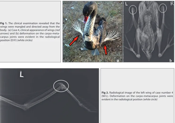
The clinical examination revealed that developing feathers and the tips of the wings were stuck up from its normal position. The wings were mangled and directed away
from the body. The wing joints (A. carpo-phalangeal) were twisted, deformed and no longer flat and smooth (Fig.
1-a,b). The radiological examination revealed a deformation
on the distal tip of carpo-metacarpus and bending on the dorsolateral side of the primary fl ight fl aps in the wings
(Fig. 2). The wings were bilaterally shaped in 5 cases. In
the black swans the wing twist, twist was advanced in dorsolateral direction. In white swans, primary fl ight fl aps were found to be pendulous in the ventral direction.
Laboratory Analysis
Blood (4 mL) was collected from brachial wing vena (v.
brachialis) in all cases for hematological and biochemical
analysis. In all the affected birds, significant findings of hematologic analysis were including leukocytosis consisting of lymphocytosis, granulocytosis and mono-cytosis. Additionally, a slight increased MCHC and marginally low MCH values were also recorded. However, red blood cell count remained normal in all birds (Table 1).
As a result of biochemical analysis, sodium value was found above or above the upper limit at all of the cases, again calcium value was found to be well below the lower limit in all cases (Table 2).
Daily feed analysis of swans has been analyzed in laboratory in animal Nutrition Department (Table 3).
Fig 2. Radiological image of the left wing of case number 4
(M/L). Deformation on the carpo-metacarpus joints were evident in the radiological position (white circle)
Fig 1. The clinical examination revealed that the
wings were mangled and directed away from the body. (a) Case 4, clinical appearance of wings (red
arrows) and (b) deformation on the
carpo-meta-carpus joints were evident in the radiological position (D/V) (white circles)
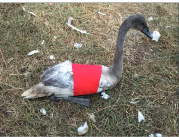
Treatment
For the treatment, the wings of the swans were bandaged adjacent to their bodies for one week (7 days) and their rations were arranged (Fig. 3). In addition to feeding balanced protein, carbohydrate, fat, vitamins, minerals, green vegetables (lettuce, etc.) and clean drinking water.
RESULTS
It was determined that the success rate of the applied treatment was high in early diagnosed cases (Fig. 4). In 4 swans determined to be in the early stage of the AW, bandage application and regulation of the ration prevented the wing degeneration and corrected wing position. No improvement was observed in the wings of 2 swans which were determined to be in the advanced stage.
Table 1. Hematological outcomes of cases
Hematological Values Reference Values Case 1 Case 2 Case 3 Case 4 Case 5 Case 6
WBC (x109/L) 6.3-22 36.71 38.39 41.25 37.03 33.6 31.85 Lym. (x109/L) 0.9-9.77 11.41 11.59 11.79 11.14 10.95 10.73 Mon. (x109/L) 0.05-1.39 3.63 3.76 3.87 4.77 3.12 4.20 Gra. (x109/L) 3.33-14.6 21.67 23.4 25.59 21.12 19.53 16.92 RBC (x1012/L) 1.96-2.9 2.36 2.59 2.74 2.8 2.05 2.22 MCV (fl) 164-200 123.5 121.2 130.1 127 125.6 117.2 Hct - 29.1 31.3 35.6 35.5 25.7 26.0 MCH (pg) 52.9-65.5 50.0 49.4 52.9 50.7 54.1 46.8 MCHC (g/dL) 29-36.5 40.5 40.8 40.7 40.0 43.1 40.0 RDW - 6.7 7.4 8.1 8.5 6.6 7.2 Hb (g/dL) 11-16.5 11.8 12.8 14.5 14.2 11.1 10.4 THR (m/mm3) - 29 429 432 475 530 330 MPV (fl) - 7.3 6.7 6.7 6.4 6.6 6.7
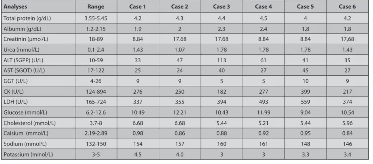
Table 2. Results of biochemical analysis of swans
Analyses Range Case 1 Case 2 Case 3 Case 4 Case 5 Case 6
Total protein (g/dL) 3.55-5.45 4.2 4.3 4.4 4.5 4 4.2
Albumin (g/dL) 1.2-2.15 1.9 2 2.3 2.4 1.8 1.8
Creatinin (µmol/L) 18-89 8.84 17.68 17.68 8.84 8.84 17,68
Urea (mmol/L) 0.1-2.4 1.43 1.07 1.78 1.78 1.78 1.43
ALT (SGPP) (U/L) 10-59 33 47 113 61 41 35
AST (SGOT) (U/L) 17-122 25 24 40 27 45 27
GGT (U/L) 4-26 9 9 5 5 10 9 CK (U/L) 124-894 276 250 182 277 399 217 LDH (U/L) 165-724 337 355 394 493 559 374 Glucose (mmol/L) 6.2-12.6 10.49 12.21 10.43 11.99 9.04 10.54 Cholesterol (mmol/L) 3.7-8 6.68 6.68 5.44 5.21 5.44 5.96 Calsium (mmol/L) 2.19-2.89 0.98 0.86 0.88 0.92 0.95 0.84 Sodium (mmol/L) 132-150 154 157 160 161 148 146 Potassium (mmol/L) 3-5 4.5 4.0 3 3 3.3 3.4
Table 3. Feed analysis results
Physical Analysis Result
Appearance Normal
Color Normal
Smell Normal
Foreign body None
Chemical Analysis Natural State In Dry Matter
Dry matter, % 92.19 ---Raw ash, % 12.51 13.57 Raw fat, % 4.43 4.80 Raw cellulose, % 7.04 7.64 Raw protein, % 17.2 18.46 ME, kcal/kg 2546 2762
However, the progression of the problem was stopped by the regulation of the ration. Among all the aff ected birds, clinical improvement was seen in 4 white swans as compared to 2 black swans with no improvement. The partial and complete wing amputation is recommended for these two cases. But it was not accepted.
DISCUSSION
Angel Wings or Slipped Wing is more commonly observed in swans and geese and is to a lesser extent reported
in ducks [1,4,5,7,8]. The deformed wing developed during
growth, results in one or both wings sticking out from the body leaving the bird unable to fl y. The left wing is more commonly aff ected than the right wing. In our cases, the frequency of bilateral appearance was very obvious. This condition becomes apparent while the fl ight feathers are growing, with the weight of the primary feathers appearing
to be too great for the carpal joint muscles, leading to the dropping wing tip. The primary fl ight feathers may become damaged. In this study, deformities wings have been permanent since the long-standing of events for in
the wrong nutritional feeding [8,9]. The recommendations
for blood tests in waterfowl are similar to those commonly performed in other birds and include the following: complete blood count, advanced serum chemistry panel
with bile acids, serum protein electrophoresis [10].
If the treated patients are very young, the condition can sometimes be minimized by splinting and repositioning the affected wing while feeding them a proper diet for optimal growth. Even then, a full recovery is not guaranteed. For rehabilitators, it can be emotionally taxing to see birds denied the chance for a full and productive life because people didn’t know about the dangers of improperly feeding them. But, as the swans evaluated in this study were adults, there was no desired improvement. Although a mild improvement was seen in the swans
that were in acute phase of the disease [9,11,12]. Male birds
seem to be more prone to this condition than females. Although it looks odd, it is not painful to the bird. It can occur with one wing or both. Radiological examination showed deformation of the carpometacarpal joints in all
cases [11]. The principles of positioning the avian patient
for radiography are the same as for other species. At least two views, 90° to each other, are suggested. Positioning may be maintained by taping the patient directly to the cassette or to a radiolucent plexiglas board. Masking tape is usually sufficient in the chemically immobilized patient and has the advantage of not pulling out feathers when
it is removed [13]. It was shown that in adult wild birds the
disease is incurable and usually leads to an early death as aff ected birds are rendered eff ectively or totally fl ightless. However, adult swan has been adversely affecting the solution of the problem. Among all the affected birds, clinical improvement was seen in 4 white swans as compared to 2 black swans with no improvement. These results brought to mind the question that the propensity of the swan might be diff erent between the swan species, even though it is in the same age range in the course of the problem. The partial and complete wing amputation is recommended for these two cases. But it was not accepted.
In young birds wrapping the wing and binding it against the bird›s fl ank for a few days and feeding more natural diet can reverse the damage. If diet is the primary issue, reducing the protein by adding wheat to the birds› feed may be recommended. A diet that provides sufficient amounts of vitamin D (the «sunshine» vitamin), vitamin E
and manganese may also be indicated [6,14].
Another interesting fact of this study is that it appears in a swan colony living in the same place. AW respond well if
treated early [12,13]. In the case of late-onset and long-onset
Fig 3. Bandage application of the swans were bandaged adjacent to
their bodies for one week
Fig 4. Case No. 1 after the bandage is removed, on the 7th day of treatment
treatment, the treatment will often fail. These phenomena must be rehabilitated in certain regions. Because these birds cannot fly and move fast.
REFERENCES
1. Kear J: Notes on the nutrition of young waterfowl, with special
reference to slipped wing. In, Duplaix-Hall N (Ed): International Zoological Year Book. 13: 97–100; London: Zoological Society, 1973.
2. Drew ML, Kreeger TJ: Skeletal abnormalities in wings of free-flying
juvenile White Pelicans (Pelecanus erythrorhynchos) in Minnesota. J Wildl
Dis, 22, 447-449,1986.
3. Pitman RL, Balance LT, Bost CA: Incidence of wing deformities
(Angel Wing) among Masked Boobiesat Clipperton Island: Life history
consequences and insight into Etiology. Wilson J Ornithol, 124 (3): 597-602, 2012. DOI: 10.1676/11-208.1
4. Zsivanovits P, Monks DJ, Forbes NA: Bilateral valgus deformity of
the distal wings (Angel Wing) in a Northern Goshawk (Accipiter gentilis). J
Avian Med Surg, 20 (1): 21-26, 2006. DOI: 10.1647/1082-6742
5. Min-Jung L, Shen-Chang C, Ko-Hua T, Wei-Chih L, Che-Lun C, Tzu-Tai L: Effect of T-2 toxin and antioxidants on angel wing incidence and
severity in White Roman geese. J Appl Anim Res, 46 (1): 340-348, 2018. DOI: 10.1080/09712119.2017.1301257
6. Thompson HM, Fernandes A, Rose M, White S, Blackburn A: Possible
chemical causes of skeletal deformities in Grey Heron nestlings (Ardea
cinerea) in North Nottinghamshire. Chemosphere, 65, 400-409, 2006.
DOI: 10.1016/j.chemosphere.2006.02.007
7. Gardner GR, Brown C, Funk FF, Bolin S, Webb R, Bolin S: Selected
topics in captive swan medicine and surgery. Exotic DVM, 5 (4): 33-38, 2003.
8. Flinchum GB: Exotic pet care: Swans. Exotic DVM, 2 (1): 36-38, 2000. 9. Harcourt-Brown N: Development of the skeleton and feathers of
dusky parrots (Pionus fuscus) in relation to their behaviour. Vet Rec, 154, 2-48, 2004. DOI: 10.1136/vr.154.2.42
10. Wood KA, Newth JL, Hilton GM, Rees EC: Has winter body condition
varied with population size in a long-distance migrant, the Bewick’s Swan
(Cygnus columbianus bewickii)? Eur J Wildl Res, 64:38, 2018. DOI: 10.1007/
s10344-018-1200-3
11. Azmanis PN, Wernick MB, Hatt JM: Avian luxations: Occurrence,
diagnosis and treatment. Vet Q, 34 (1): 11-21, 2014, DOI: 10.1080/ 01652176. 2014.905731
12. Wimsatt J, Dressen P, Dennison C, Turner AS: Ultrasound therapy
for the prevention and correction of contractures and bone mineral loss associated with wing bandaging in the domestic pigeon (Columba livia). J
Zoo Wildl Med, 31, 190-195, 2000. DOI: 10.1638/1042-7260(2000)031
13. Helmer P: Advances in diagnostic imaging. In, Harrison GJ,
Lightfoot TL (Eds): Clinical Avian Medicine. 25, 653-659, Amazon, USA, 2005.
14. Lin MJ, Chang SC, Lin TY, Cheng YS, Lee YP, Fan YK: Factors affecting
the incidence of angel wing in White Roman Geese: Stocking density and genetic selection. Asian Australas J Anim Sci, 29 (6): 901-907, 2016. DOI: 10.5713/ajas.15.0456