Research Article e-ISSN: 2528-858X doi: 10.19159/tutad.425732
Cholinesterase, α-Glucosidase, α-Amylase, and Tyrosinase Inhibitory
Effects and Antioxidant Activity of Veronica officinalis Extracts
Nuraniye ERUYGUR1, Esra UÇAR2*
1Selçuk University Faculty of Pharmacy, Department of Pharmacognosy, Konya, TURKEY 2Cumhuriyet University, Sivas Vocational School, Department of Crop and Animal Production, Sivas, TURKEY
Received: 21.05.2018 Accepted: 19.10.2018
ORCID ID (by author order)
orcid.org/0000-0002-4674-7009; orcid.org/0000-0001-6327-4779 *Corresponding Author: [email protected]
Abstract: In this study, inhibition ability of Veronica officinalis extracts against Alzheimer’s disease-related enzymes
acetylcholinesterase (AChE) and butyrylcholinesterase (BuChE), diabetes mellitus related enzymes glucosidase and α-amylase and antioxidant of Veronica officinalis were investigated. To the best of our knowledge, there are no previous studies on the enzyme inhibition activities of the V. officinalis extracts. For this aim, V. officinalis extracted with methanol and water by maceration method and their antioxidant activities were evaluated by DPPH (2,2-Diphenyl-1-picrylhydrazyl), ABTS [2,2′-Azino-bis-(3-ethylbenzothiazoline-6-sulphonic acid)] radical scavenging assays, total phenol, total flavonoid content, iron chelating and ferric reducing power assays. The enzyme inhibition activity was performed with 96-well plate method. According to the obtained results; the plant extracts have good antioxidant effects. In the same time, the water extract have stronger inhibition activity against AChE, BuChE and α-glucosidase, α-amylase, and tyrosinase than methanol extract. The experimental findings indicated that the water extract exerted the higher α-glucosidase, α-amylase and tyrosinase (82.07 ± 2.91, 63.61 ± 3.93 and 47.47 ± 0.53 at 2 mg mL-1, respectively) inhibition than reference drugs. The obtained results
demonstrate that this plant has a significant potential for improving pharmaceutical formulations.
Keywords: Antioxidant, alzheimer’s, diabetes, tyrosinase, Veronica officinalis
1. Introduction
Veronica L. genus belongs to Plantaginaceae
family, and is characterized by 450 species. These species are annual and perennial plants and located in Europe and Turkey. The aerial parts of Veronica species are used for the treatment of cough, rheumatism and have wound-healing properties (Albach and Meudt, 2010; Beara et al., 2015; Mocan et al., 2015; Raclariu et al., 2017). According to the previous data on phytochemical investigations, Veronica species have different constituents such as iridoid glucosides, especially benzoic and cinnamic acid esters of catalpol, some phenylethanoid and flavonoid glycosides (Taskova et al., 2002; Taskova et al., 2006; Harput et al., 2011).
The oxygen radical derivatives and peroxides-like reactive species cause oxidative stress and
reactive free radicals can oxidize proteins, lipids, and nucleic acids. As a result, various diseases such as neurodegenerative, cardiovascular, inflammation are emerging as a result of oxidative stress. (Halliwell, 1987; Emerit et al., 2004; Junqueira et al., 2004; Kiss et al., 2009). Medicinal plants that are rich in terms of polyphenols, can retard the oxidative degradation of lipids. Flavonoids are phenolic components and can prevent some cardiovascular and inflammatory diseases (Havsteen, 2002; Rice-Evans, 2004).
Tyrosinase is a copper-containing proteolytic enzyme that allows o-hydroxylation of monophenols and then oxidation of the o-quinones from the o-diphenols (Zhou et al., 2017). This enzyme is playing an important role in melanin synthesis in mammalian cells and browning in plants and microorganisms. However, excessive production and accumulation of melanin in the
orcid.org/0000-0002-4674-7009 orcid.org/0000-0001-6327-4779
ORCID ID (By author order)
İD İD
human skin lead to the formation of hyperpigmentation. Therefore, control the overproduction of melanin by using tyrosinase inhibitory agents is a therapeutical strategy for hyperpigmentation in the skin.
Alzheimer’s disease (AD) is a neurodegenerative chronic disease related to decreasing of brain transmitter choline. Acetylcholinesterase and butyrylcholinesterase are involved in the hydrolysis of acetylcholine to choline and acetic acid. Therefore, cholinesterase inhibitors have been used in the treatment of AD (Ertaş et al., 2014). However, the side effects of cholinesterase inhibitors make it necessary to search for new, potential and alternative cholinesterase inhibitors from natural sources.
Diabetes mellitus is a multifactorial disease leading to severe complications. Therefore, multiple therapeutic approaches are used in the treatment. In diabetic patients, postprandial hyperglycemia occurs after a meal due to glucose absorption from the gastrointestinal tract. Preventing glucose uptake in the intestines and promoting glucose uptake in tissues can control the level of blood glucose in the case of postprandial hyperglycemia, which is common for people with diabetes (Thilagam et al., 2013). When the literature were evaluated, there was a litte study of biological and enzyme activity on V. officinalis. For this reason, we planned to evaluate the V. officinalis for potential inhibitory activities against ɑ -glucosidase, ɑ-amylase, cholinesterase, and tyrosinase as well as its in vitro antioxidant activities.
2. Materials and Methods
2.1. Plant material
This study was conducted in the Laboratories of Faculty of Pharmacy, Cumhuriyet University in Sivas in the 2018 year. Leaves of V. officinalis were collected from Cumhuriyet University Campus.
2.2. Preparation of extracts
The plant material was powdered with a grinder after dried in shade. 10 gr of the leaves were soaked in 50 mL of methanol (Sigma) and water for 24h with intermittent shaking. At the end of extraction, it was filtrated by No. 1 Whatman filter paper. The filtrate was concentrated to dryness under reduced pressure with a rotary evaporator at 40°C and this process was repeated for three times. The residue of plant material then subjected to maceration with water as solvent and after filtering dried under vacou to yield water extract. The obtained extracts were analyzed by GC-MS (Gas Chromatography-Mass Spectrometry). The obtained methanol and
water extracts were used for evaluation of the antioxidant potential and enzyme inhibition activity as well as chemical composition through GC-MS.
2.3. In-vitro antioxidant activity
The experiments were performed in completely randomized design with three replications.
2.3.1. DPPH radical scavenging activity
The free radical scavenging activity of the extracts was conducted according to the method
reported previously (Miser-Salihoglu et al., 2013).
The 150 µL of methanol extract was mixed with 50
µL of 1.0×10⁻³M DPPH solution freshly prepared
in methanol. Methanol was used as a control of the experiment. After incubation for 30 min at room temperature (25°C), the reduction of the DPPH free radical was measured by spectrophotometer at 517
nm. Trolox was used as positive control. The
antioxidant activity as a percentage (%) of inhibition was calculated using the formula given
below (Equality 1):
% Inhibition = [(Absorbance of control – Absorbance of test sample) / Absorbance of control] × 100 (1)
2.3.2. ABTS radical scavenging activity
For determination of ABTS radical scavenging activity of the extracts, followed by the method of Re et al. (1999) with slight modification. The stock solution of ABTS was generated by reacting 7 mM ABTS solution with 2.4 mM of potassium persulfate solution in equal volume for 16h. Working solution was then prepared by diluting the stock ABTS solution with methanol to give an absorbance of 0.7 ± 0.02 units at 734 nm using a spectrophotometer. In each assay, the ABTS solution was prepared freshly. 50 μL of extract was mixed with 150 μL ABTS working solution and left for 10 min in darkness. All the analyses were performed in triplicate and the results expressed as a mean ± standard deviation. Appropriate blanks
(methanol) and standard [BHT (Butyl hydroxy
tolüen)in methanol] were run simultaneously. The
absorbance was measured at 734 nm in a microplate reader (Epoch, USA).
2.3.3. Determination of total phenolics (TPC, total phenolics content)
The spectrophotometric Folin–Ciocalteu (F-C) method was used to measure the TPC of the extracts, as previously described by Clarke et al. (2013). Briefly, 20 μL of extract diluted
appropriately in DMSO (Dimethyl sulfoxide) was
mixed with 100 μL F-C reagent freshly diluted 1/10 with distilled water. After five minutes, the solution
incubated for 30 min at 25°C. The absorbance was measured at 650 nm with a microplate reader
(Epoch, USA). All the analyses were performed in
triplicate and the results expressed as a mean ± standard deviation. Appropriate blanks (DMSO) and standard (gallic acid in DMSO) were run simultaneously, after which the TPC was calculated as milligrams gallic acid equivalents per gram of dry extract.
2.3.4. Estimation of total flavonoids (TFC, total flavonoid concentration)
The aluminum chloride colorimetric method was used for the determination of TFC, as
previously described by Molan and Mahdy (2014),
using catechin as the reference standard. Briefly, 25 μL of test sample solution with a concentration of 1
mg/mL, 100 μL of doubled distillated H2O and 7 μL
of NaNO25% were mixed together in 96-well
plates. After 15 min of incubation at room
temperature, 7 μL of 10% (w/v) AlCl3was added. 5
min later, 50 μL of 1 M NaOH and 60 μL of distilled water were added to each well. Then the absorbance was measured at 490 nm with a spectophotometer. All determinations were carried out in triplicates. The content of total flavonoids was expressed as mg of catechin equivalent per g of dry weight of the extract.
2.3.5. Iron chelating activity
The iron chelating activity of the extracts was determined according to their interaction with the
formation of the ferrozine-Fe2+ complex. A
previously described procedures were used (Chai et
al., 2014). Briefly, a mixture of 0.2 mL of 0.1 mM
FeSO4[Iron (II) sulfate], 0.2 mL of extract, and 0.4
mL of 0.2 mM ferrozine was allowed to react at room temperature. Absorbance was read after 10 min of incubation at 562 nm.
2.3.6. Ferric reducing antioxidant power (FRAP) assay
FRAP assay uses antioxidants as reductants in a redox-linked colorimetric method. The FRAP assay was conducted according to the previously reported
method with slight modification (Wojdylo et al.,
2007; Yang et al., 2011). Each extract was dissolved
in DMSO to prepare a stock solution. The working FRAP reagent was prepared by freshly mixing 0.3 M acetate buffer (pH 3.6), a solution of 10 mM TPTZ (2,4,6-tripyridyl-s-triazine) in 40 mM HCI
and 20 mM FeCI3solution at a ratio of 10:1:1. 30
µL of the sample solution and 270 µL of FRAP working solution were mixed together in 96-well plates and warmed at 37ºC for 4 min. All determinations were performed in triplicate. The absorbance was measured at 593 nm. A standard
calibration curve was prepared using different
concentrations of FeSO4 solution. All solutions
were prepared freshly. Trolox was used as reference. The results were expressed as FRAP value.
2.4. Enzyme inhibitory activity
2.4.1. Acetylcholinesterase (AChE)/
Butyrylcholinesterase (BChE) inhibition assay
A 96-well plate colorimetric method described by Ellman et al. (1961) was used for determining of the AChE inhibitory activity of the plant extracts. The experiments were carried out with three replications. Galanthamine was used a positive control, while the buffer was used as a blank. Briefly, 140 µL of 0.1 mM phosphate buffer (pH 6.8), 20µL of extracts prepared in the buffer from
50 mg mL-1stock sample solution in DMSO, 20µL
AChE from Electric eel (5×10⁻³M)/BuChE from
equine serum (5×10⁻³M) was added in each well of
microtiter plate. After 10 min of incubation, 10 µL of 3 mM DTNB was added. Then the reaction was initiated with adding of 10 µL of 0.71 mM acetylcholine iodide (AChI)/0.2mM Butyrylcholine iodide (BChI), absorbance was measured at 412 nm with a microplate reader (Epoch, USA).
2.4.2. Alpha-glucosidase inhibition activity
Alpha-glucosidase inhibition method was followed by Kumar et al. (2012). Acarbose was used as a positive control, while phosphate buffer was used as negative control in place of the sample. Each concentration was carried out in triplicate. 25 µL of sample solution was mixtured with 25 µL of
α-glucosidase (0.5 U mL-1), then incubated for
about 10 min at 25ºC. After incubation 25 µL of 0.5 mM PNPG was added to each well as substrate and incubated further 30 min at 37ºC. After the incubation period, 100 µL of 0.2 M sodium carbonate was added to terminating the reaction and the absorbance was read at 405 nm.
2.4.3. Alpha-amylase inhibition activity
Alpha-amylase inhibition method was followed by Kumar et al. (2013). Acarbose was used as a positive control, while phosphate buffer (0.02 M PBS, pH 6.9) was used as negative control in place of sample. Each sample was carried out in triplicate with different concentrations. The reaction mixture containing 50 µL of sample solution, 25 µL of ɑ-amylase (0.5 mg/mL) incubated for about 10 min at 25ºC. Then 50 µL of 0.5% starch solution (w/v) prepared freshly was added to each well as substrate and incubated further 10 min at 25ºC. After the incubation period, 100 µL of 1% DNS (3,5-dinitrosalicylic acid), the color reagent was added
as a color reagent and heated at water a bath for 10 min. The absorbance was read at 540 nm.
2.4.4. Tyrosinase enzyme inhibition activity Tyrosinase enzyme inhibition method was followed by Jeong et al. (2009). In 96 well plate, 20 µL of sample solution diluted with buffer, and 100 µL of phosphate buffer were mixed with 20 µL of
tyrosinase (250 U mL-1) in each well and incubated
for about 10 min at 25ºC. Then 20 µL of 3 mM L-tyrosine was added as a substrate and incubated further 30 min at 25ºC. After the incubation period, the absorbance was read at 492 nm. Kojic acid was used as a positive control, while phosphate buffer (100 mM PBS, pH= 6.8) was used as a negative control in place of sample. Each sample was carried out in triplicate with different concentrations.
2.5. Statistical analysis
Data obtained from in vitro antioxidant and enzyme inhibition activity were expressed as the mean ± standard deviation (SD). The % inhibitory activity of the extract and reference compounds were calculated trough extract dose-response curve by GraphPad Software (San Diego, CA, USA).
3. Results and Discussion
3.1. Chemical composition of the extracts
The chemical compositions of water and methanol extracts obtained from leaves of V.
officinalis have been determined (Table 1). When
methanol and water extracts were compared in terms of content, methanol extract was found to
have a higher component content. While the most
Table 1. Chemical components of water and methanol extracts of Veronica officinalis
Components Retention time (min) Water(%) Methanol(%)
2-Furanmethanol (CAS) 6.743 3.81
1,2-Cyclopentanedione 8.941 1.51
4-Hydroxy-6-methyl2H-pyran-2-one 15.424 1.30
4H-Pyran-4-one, 2,3-dihydro-3,5-dihydroxy-6-methyl- (CAS) 16.865 1.28
1,4:3,6-Dianhydro-.alpha.-d-glucopyranose 18.565 6.24 1,4-Benzenediol (CAS) 20.865 9.30 2-Methoxy-4-vinylphenol 22.502 2.41 Phenol, 2,6-dimethoxy- 24.121 1.86 Phenol, 2,6-dimethoxy- 24.184 5.77 Cyclopropane, nonyl-1-Tridecanol 28.241 4.72 Phenol, 2,4-bis(1,1-dimethylethyl)- 29.316 3.23 2,6-Dimethyl-3-(methoxymethyl)-p-benzoquinone 30.695 3.41 (-)-Caryophyllene oxide 31.205 1.32 1,5-Cycloundecadiene, 8,8-dimethyl-9-methylene- 33.196 3.82 Cyclododecane 33.448 4.70 Neophytadıene 36.206 1.62 2-Pentadecanone, 6,10,14-trimethyl 36.326 1.18
6-Octen-1-ol, 3,7-dimethyl-, propanoate 36.961 1.09
(4S,5R)-5-Hydroxycaryophyll-8(13)-ene-4,12-epoxide 37.333 0.99
Hexadecanoic acid, methyl ester (CAS) 37.705 0.85
(+) spathulenol 38.151 1.65
Hexadecanoic acid (CAS) 38.426 6.60
Aromadendrene oxide-(2) 38.603 0.93 3,4'-Difluoro-4-Methoxybıphenyl 39.215 1.43 (3E,5E,8E)-3,7,11-Trimethyl-1,3,5,8,10-dodecapentaene 39.381 0.56 2,2,6-Trimethyl-1-(2-methyl-cyclobut-2-enyl)-hepta-4,6-dien-3-one 40.383 3.74 1-(3'-methyl-2'-cyclobutenyl)-2,2,6-trimethyl-4,6-heptadien-3-one 40.726 3.10 Cyclododecyne 41.167 1.27 Isoxeniolide-A 45.372 0.71
Isosteviol methyl ester 45.904 0.88
Lactic Acid Di-TMS 47.060 1.26
Silane, Trimethyl 5-Methyl-2- 1-Methylethyl Phenoxy - 55.168 0.64
Total 18.04 65.14
abundant components are 1,4:3,6-dianhydro-alpha-d-glucopyranose (6.24%) and 1,4-benzenediol (9.30%) for water extract, are Hexadecanoic acid (6.60%) and Phenol, 2,6-dimethoxy- (5.77%) for methanol extract (Table 1).
3.2. In-vitro antioxidant activity
Natural antioxidants present in plants can be inhibited or prevent reactive oxygen species, by the way, protect the human body from oxidative stress. In this study, we evaluated the antioxidant activities
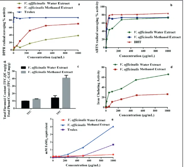
of water and methanol extracts of V. officinalis leaves using DPPH and ABTS radical scavenging assays. In both assays, the decrease in the absorbance of free radical causes by the natural antioxidants present in the extract which can be scavenged radical by hydrogen donation (Supritha and Radha, 2018). According to our results which were shown in Figure 1, the methanol extract of V.
officinalis leaves showed a higher percentage of
DPPH radical scavenging activity in lower concentration in comparison to water extract. However, in ABTS radical scavenging, both the methanol and water extract showed the same effect which was comparable with the positive control of BHT.
Phenolic compounds are primarily responsible for the antioxidant activity of plants. In our study, the total phenolic contents of the water and methanol extracts of V. officinalis are shown in Figure 1, which were found as 14.26 and 40.93 mg
GAE/g (GAE: Gallic acid equivalent) dry weight of
extract, respectively. Total phenolic contents of both of the extracts showed higher values than those reported in three Veronica species that was presented TPC values expressed as gallic acid
equivalent ranging 17.71-34.40 mg g-1 (Mocan et
al., 2015). The obtained results showed that the difference in the amount of phenolic compounds may be dependent on the geographical environment, collection time, drying and extraction method, as well as other genetic factors.
In our study, the total flavonoid content of water and methanol extracts of V. officinalis was detected
as 9.98 and 12.57 mg CE g-1, respectively. Our
results revealed that higher concentrations of TFC as compared to those reported in tree Veronica species that their TFC were expressed as quercetin
equivalents ranged from 2.52 to 6.60 mg g-1(Mocan
et al., 2015). The variations in the content of flavonoids may be due to the growing habitat, investigated plant parts and extraction solvent differences.
Figure 1. The in-vitro antioxidant activity of methanol and water extracts of V. Officinalis. a) DPPH radical
scavenging activity, b) ABTS radical scavenging activity, c) total phenol and total flavonoid contents, d) iron
Antioxidant activity of herbal extracts resulting from various compounds by different kinds of antioxidant mechanisms such as radical scavenging, delaying peroxidation, iron chelating etc. Therefore, it was suggested that the antioxidant capacity was not evaluated by using a single test. In our study, total phenol and flavonoid content, iron chelating and ferric reducing antioxidant power assays which were widely used were conducted for determining the antioxidant potential of water and methanol extracts of V. officinalis. When compared each other, the V. officinalis water extract exhibited relatively low levels of DPPH scavenging activity, however in ABTS radical scavenging assay, there is not much difference between the water and methanol extract. Both scavenged DPPH and ABTS radical in a concentration-dependent manner (Figure 1).
3.3. In-vitro enzyme inhibition activity
The water and methanol extracts of V. officinalis were tested to evaluate their tyrosinase, AChE and BuChE enzyme inhibition effects as well as starch breakdown enzyme inhibition abilities against α-amylase and α-glucosidase. Prevention of the absorption of glucose and control of blood sugar by using these two enzyme inhibitors in reducing the postprandial blood glucose level in diabetic patients is an effective treatment approach in the treatment of diabetes nowadays. According to our results, the water extract showed stronger amylase, α-glucosidase, AChE BuChE and tyrosinase inhibition activities than the methanol extract at the
concentration of 2 mg mL-1 (Table 2). From the
results it can be say that the enzyme inhibition activity of water extract may be attributed to the
water-soluble polar compounds which are perhaps
Table 2. Enzyme inhibitory activities (%) of the water and methanol extracts of Veronica officinalis (in 2 mg
mL-1concentrations)
Extracts Anticholinesterase activityAChE BChE α-glucosidaseAntidiabetic activityα-amylase Tyrosinase Water extract 70.94 ± 2.37 66.89 ± 2.35 82.07 ± 2.91 63.61 ± 3.93 47.47 ± 0.53 Methanol extract 69.65 ± 1.69 65.66 ± 1.94 72.60 ± 1.73 57.45 ± 0.84 26.40 ± 0.57 Galanthamine HBr 93.87± 0.56 89.89 ± 0.01
Acarbose 57.56 ± 0.52 58.40 ± 0.63
Kojic acid 45.55 ± 1.65
playing effective role against enzymes than non-polar compounds dissolved in methanol extract. When the results compared with standard compounds, both water and methanol extract exhibited lower AChE and BuChE inhibitory activity than galanthamine, however, at the α-glucosidase inhibition assay, both of the water and methanol extract demonstrated higher inhibitory activity than reference drug acarbose.
The antityrosinase activity of plant extracts is important in terms of their usage in depigmenting agents for cosmetic formulations. Otherwise, the water extract (47.47%) also demonstrated higher tyrosinase inhibitory activity than the methanol
extract (26.40%) at the concentration of 2 mg mL-1.
Moreover, it was found that the water extract had higher inhibition effect than the positive control (45.55%) at the same concentration (Table 2).
4. Conclusions
In conclusion, water and methanol extract of V.
officinalis were evaluated for in-vitro antioxidant
activity and were tested for their inhibition activity against five different enzymes which are AChE, BuChE, α-glucosidase, α-amylase, and tyrosinase. From the obtained results, it was observed that the
methanol extract showed higher total phenolic content and possess a higher antioxidant activity than water extract. Otherwise, the water extract exhibits higher inhibition effect on all the tested enzymes except for α-glucosidase than methanol extract. However the water extract demonstrated lower inhibitory activity than Galanthamine in AchE and BuchE and higher than the acarbose in α-glucosidase and α-amylase. It could be emphasize that the water extract is possessing potential tyrosinase inhibition activity even stronger than the reference compound- kojic acid. The results demonstrated that the plant may be a good source for developing herbal formulations for the Alzheimer’s disease and diabetes patients.
Acknowledgement
The authors are thankful to the Advanced Technology Application and Research Center, Cumhuriyet University.
References
Albach, D.C., Meudt, H.M., 2010. Phylogeny of veronica in the southern and northern hemispheres based on plastid, nuclear ribosomal and nuclear low-copy DNA. Molecular Phylogenetics and Evolution, 54(2): 457-471.
Beara, I., Živković, J., Lesjak, M., Ristic, J., Savikin, K., Maksimovic, Z., Jankovic, T., 2015. Phenolic profile and anti-inflammatory activity of three Veronica species. Industrial Crops and Products, 63: 276-280. Chai, T., Mohan, M., Ong, H., Wong, F., 2014.
Antioxidant, iron-chelating and anti-glucosidase activities of Typha domingensis Pers (Typhaceae). Tropical Journal of Pharmaceutical Research, 13(1): 67-72.
Clarke, G., Ting, K.N., Wiart, C., Fry, J., 2013. High correlation of 2,2-diphenyl-1-picrylhydrazyl (DPPH) radical scavenging, ferric reducing activity potential and total phenolics content indicates redundancy in use of all three assays to screen for antioxidant activity of extracts of plants from the Malaysian Rainforest. Antioxidants, 2(1): 1-10.
Ellman, G.L., Courtney, KD., Andres, V., Featherstone, R.M., 1961. A new and rapid colorimetric determination of acetylcholinesterase activity. Biochemical Pharmacology, 7(2): 88-90.
Emerit, J., Edeas, M., Bricaire, F., 2004. Neurodegenerative diseases and oxidative stress. Biomedicine & Pharmacotherapy, 58(1): 39-46. Ertaş, A., Gören, A.C., Boğa, M., Yeşil, Y., Kolak, U.,
2014. Essential oil compositions and anticholinesterase activities of two edible plants Tragopogon latifolius var. angustifolius and Lycopsis orientalis. Natural Product Research, 28(17): 1405-1408.
Halliwell, B., 1987. Oxidants and human disease: Some new concepts. Federation of American Societies for Experimental Biology, 1(5): 358-364.
Harput, U.Ş., Genç, Y., Khan, N., Saracoglu, I., 2011. Radical scavenging effects of different Veronica species. Records of Natural Products, 5(2): 100-107. Havsteen, B.H., 2002. The biochemistry and medical
significance of the flavonoids. Pharmacology & Therapeutics, 96(2-3): 67-202.
Jeong, S.H., Ryu, Y.B., Curtis-Long,, M.J., Ryu, H.W., Baek, Y.S., Kang, J.E., Lee, W.S., Park, K.H., 2009. Tyrosinase inhibitory polyphenols from roots of Morus lhou. Journal of Agricultural and Food Chemistry, 57(4): 1195-1203.
Junqueira, V.B., Barros, S.B., Chan, S.S., Rodriques, L., Giavarotti, L., Abud, R.L., Deucher, G.P., 2004. Aging and oxidative stress. Molecular Aspects of Medicine, 25(1-2): 5-16.
Kiss, B., Popa, D.S., Crişan, G., Bojiţǎ, M., Loghin, F., 2009. The evaluation of antioxidant potential of Veronica officinalis and Rosmarinus officinalis extracts by monitoring malondialdehide and glutathione levels in rats. Farmacia, 57(4): 432-441. Kumar, D., Gupta, N., Ghosh, R., Gaonkar, R.H., Pal,
B.C., 2013. α-Glucosidase and α-amylase inhibitory constituent of Carex baccans: Bio-assay guided isolation and quantification by validated RP-HPLC-DAD. Journal of Functional Foods, 5(1): 211-218. Kumar, D., Kumar, H., Vedasiromoni, J.R., Pal, B.C.,
2012. Bio- assay guided isolation of a-glucosidase inhibitory constituents from Hibiscus mutabilis leaves. Phytochemical Analysis, 23(5): 421-425.
Miser-Salihoglu, E., Akaydin, G., Caliskan-Can, E., Yardim-Akaydin, S., 2013. Evalution of antioxidant activity of various herbal folk medicines. Journal of Nutrition Food Sciences, 3(5): 222.
Mocan, A., Vodnar, D.C., Vlase, L., Crișan, O., Gheldiu, A.M., Crișan, G., 2015. Phytochemical characterization of Veronica officinalis L., V. teucrium L. and V. orchidea crantz from Romania and their antioxidant and antimicrobial properties. International Journal of Molecular Science, 16(9): 21109-21127.
Molan, A.L., Mahdy, A.S., 2014. Iraqi medicinal plants: Total flavonoid contents, free-radical scavenging and bacterial beta-glucuronidase inhibition activities. Journal of Dental and Medical Sciences, 13(5): 72-77.
Raclariu, A.C., Mocan, A., Popa, M.O., Vlase, L., Ichim, M.C., Crisan, G., Brysting, A.K, Boer H., 2017. Veronica officinalis product authentication using DNA metabarcoding and HPLC-MS reveals widespread adulteration with Veronica chamaedrys. Frontiers in Pharmacology, 8: 378, doi: 10.3389/fphar.2017.00378.
Re, R., Pellegrini, N., Proteggente, A., Pannala, A., Yang, M., Rice-Evans, C., 1999. Antioxidant activity applying an improved ABTS radical cation decolorization assay. Free Radical Biology & Medicine, 26(9-10): 1231-1237.
Rice-Evans, C., 2004. Flavonoids and isoflavones: absorption, metabolism, and bioactivity. Free Radical Biology & Medicine, 36(7): 827-828. Supritha, P., Radha, K.V., 2018. Estimation of phenolic
compounds present in the plant extracts using high pressure liquid chromatography , antioxidant properties and its antibacterial activity. Indian Journal of Pharmaceutical Education and Research, 52(2): 321-326.
Taskova, R.M., Gotfredsen, C.H., Jensen, S.R., 2006. Chemotaxonomy of Veroniceae and its allies in the Plantaginaceae. Phytochemistry, 67(3): 286-301. Taskova, R., Peev, D., Handjieva, N., 2002. Iridoid
glucosides of the genus Veronica s.l. and their systematic significance. Plant Systematics and Evolution, 231(1): 1-17.
Thilagam, E., Parimaladevi, B., Kumarappan, C., Chandra Mandal, S., 2013. Glucosidase and α-Amylase inhibitory activity of Senna surattensis. JAMS Journal of Acupuncture and Meridian Studies, 6(1): 24-30.
Wojdylo, A., Oszmianski, J., Czemerys, R., 2007. Antioxidant activity and phenolic compounds in 32 selected herbs. Food Chemistry, 105(3): 940-949. Yang, H., Dong, Y., Du, H., Shi, H., Peng, Y., Li, X.,
2011. Antioxidant compounds from propolis collected in Anhui, China. Molecules, 16(4): 3444-3455.
Zhou, J., Tang, Q., Wu, T., Cheng, Z., 2017. Improved TLC bioautographic assay for qualitative and quantitative estimation of tyrosinase inhibitors in natural products. Phytochemical Analysis, 28(2): 115-124.