Experimental Study / Deneysel Çalışma
Effect of levetiracetam on hepatic fibrosis in diabetic rats
Yiğit Uyanıkgil,1,2 Tülay Akman,3 İbrahim Çavuşoğlu,4 Erkan Kısmalı,5 Dilek Taşkıran,6 Oytun Erbaş7
1Department of Histology and Embryology, Medical Faculty of Ege University, İzmir, Turkey 2Cord Blood, Cell-Tissue Research and Application Center, Ege University, İzmir, Turkey 3Department of Medical Oncology, İzmir Tepecik Training and Research Hospital, İzmir, Turkey
4Department of Radiology, Kolan British Hospital, Lefkosa, Republic of Northern Cyprus 5Department of Radiology, Medical Faculty of Ege University, İzmir, Turkey 6Department of Physiology, Medical Faculty of Ege University, İzmir, Turkey 7Department of Physiology, Medical Faculty of İstanbul Bilim University, İstanbul, Turkey
Received: February 11, 2015 Accepted: June 16, 2015
Correspondence: Yiğit Uyanıkgil, MD. Ege Üniversitesi Tıp Fakültesi Histoloji ve Embriyoloji Anabilim Dalı, 35100 Bornova, İzmir, Turkey.
Tel: +90 232 - 390 59 08 e-mail: [email protected] ABSTRACT
Objectives: In our study we investigated the possible effects of levetiracetam (LEV) on hepatic fibrosis in streptozotocin (STZ) induced diabetic rats with histopathology, real-time elastography imaging technique. We also aimed to investigate the effects of LEV on oxidative stress markers.
Materials and methods: Diabetes was induced by intraperitoneal (i.p.) single dose injection of STZ (60 mg/kg). Twenty-one rats were randomly divided into three groups; control group, STZ group treated with 1 mL/kg/day saline (STZ+SP), and STZ group treated with 600 mg/kg/day LEV was administrated by i.p. for four weeks. All rats underwent a real-time elastography (strain-elasticity %). The liver sections are examined by histopathologically (liver fibrosis score). In addition, malondialdehyde, total antioxidant capacity, and glutathione levels were measured in plasma.
Results: Treatment with LEV significantly decreased liver fibrosis score and increased strain measurement % on the real-time elastography in diabetic rats. In addition, treatment with LEV significantly increased total antioxidant capacity, glutathione and malondialdehyde levels decreased in plasma of diabetic rats.
Conclusion: Levetiracetam has potential as a treatment for diabetic liver injury and hepatic fibrosis and can be a good candidate among new treatment options. In addition, real-time elastography is reliable imaging non-invasive technique for detecting hepatic fibrosis.
Keywords: Diabetes mellitus; levetiracetam; oxidative stress; real-time elastography.
Diyabetik sıçanlarda karaciğer fibrozisi üzerine levetirasetam etkisi
ÖZ
Amaç: Bu çalışmada, streptozotocin (STZ) ile indüklenmiş diyabetik sıçanlardaki hepatik fibrozis üzerine levetirasetamın (LEV) pozitif etkilerinin histopatolojik ve gerçek zamanlı elastografik görüntüleme tekniği ile incelenmesi amaçlandı. Yine bu çalışmada LEV’nin oksidatif stres belirteçleri üzerine etkisi araştırıldı.
Gereç ve yöntemler: Diyabetik model, tek doz intraperitoneal (i.p.) STZ (60 mg/kg) enjeksiyonu ile yapıldı. Yirmi bir sıçan rastgele olarak üç gruba ayrıldı; kontrol grubu, STZ uygulaması yapılıp 1 mL/kg/gün salin verilen grup (STZ+SP) ve STZ uygulanıp dört hafta boyunca 600 mg/kg/gün i.p LEV uygulanan grup. Tüm sıçanlar gerçek zamanlı elastografik yöntem ile incelendi (gerilme-elastikiyet yüzdesi). Karaciğerden alınan kesitler histopatolojik olarak değerlendirildi (karaciğer fibroz skoru). Bunlara ek olarak, plazmadan malondialdehit, total antioksidant kapasite ve glutatyon seviyeleri ölçüldü. Bulgular: Levetirasetam ile tedavi edilen diyabetik sıçanlarda karaciğer fibroz skorlamasında anlamlı bir azalma ve gerçek zamanlı elastografik gerilim-elastikiyet yüzdesinde artış saptandı. Bununla birlikte LEV tedavisinde diyabetik sıçanlarda total antioksidant kapasitesinin arttığı, glutatyon ve malondialdehitin plazma seviyelerinin azaldığı görüldü.
Sonuç: Levetirasetam, diyabetik karaciğer hasarı ve karaciğer fibrozisi için yeni tedavi seçenekleri arasında iyi bir aday olabilir. Buna ek olarak gerçek zamanlı elastografi, hepatik fibrozisin tespitinde non-invazif ve güvenilir bir görüntüleme bir tekniğidir.
Type 2 diabetes mellitus and hyperinsulinemia are closely associated with the development of fatty changes in the liver ranging from simple steatosis to steatohepatitis to advance liver disease such as hepatic fibrosis and cirrhosis.[1,2] Despite extensive
studies, the underlying mechanisms of these liver diseases still remain largely undefined. Evidence of oxidative stress with decreased antioxidant defenses has been found to be associated with hepatic fibrosis regardless of etiology and fibrosis progression rate.[3,4]
Although there are few effective therapies used for the treatment of hepatic fibrosis, no drug has been approved as anti-fibrotic agent in humans so far.[5,6] The benefit of the agents reducing
the oxidative stress, inflammation and immune response were determined in the previous studies and to date, only limited antifibrogenic treatment options are available to protect hepatocytes from oxidative stress and reduce inflammatory damage. In the previous studies they have shown that different types of antioxidants could attenuate the complications of diabetes including fatty liver diseases and fibrosis.[7-9]
Levetiracetam (LEV) is a commonly used antiepileptic drug that acts as a histone deacetylase inhibitor and an inhibitor of high-voltage activated N-type calcium channels. The recent evidence shows that the drug has anti-inflammatory and antioxidative effects.[10-12] In a
previous experimental study the neuroprotective effects of LEV by reducing the level of oxidative stress markers and anti-inflammatory cytokines were determined.[10] On account of this results
LEV may be a promising candidate for treating other inflammatory diseases such as hepatic fibrosis.
In this present study we aimed to investigate the possible effects of LEV on hepatic fibrosis in streptozotocin (STZ) induced diabetic rats with histopathology and real-time elastography imaging technique. We also aimed to investigate the effects of LEV on oxidative stress.
MATERIALS AND METHODS
Animals
In this study 21 male Sprague Dawley albino mature rats at eight weeks, weighing 200-220 g, were used. Animals were fed ad libitum and housed
impairs in steel cages having a temperature-controlled environment (22±2 °C) with 12-h light/ dark cycles. The experimental procedures were approved by the Committee for Animal Research of Ege University. All animal studies are strictly conformed to the Animal Experiment Guidelines of the Committee for Human Care.
Experimental protocol
Diabetes was induced by intraperitoneal (i.p) injection of STZ (Sigma-Aldrich, Inc.; Saint Louis, MO, USA) (60 mg/kg in 0.9% NaCl, adjusted to a pH 4.0 with 0.2M sodium citrate) for 14 rats. No drug was administered to the remainder of rats (n=7) (Control group). Diabetes was verified after 24 hours by evaluating blood glucose levels with the use of glucose oxidase reagent strips (Boehringer-Mannheim Corp, Indianapolis, IN). The rats with blood glucose levels 250 mg/dL and higher were included in this study. We waited 21 days for the development of diabetes-related microvascular complications.[13] Then, 14 diabetic rats were
randomly divided into two groups; STZ group treated with 1 mL/kg saline (STZ+SP) (n=7), and STZ group treated with 600 mg/kg/day LEV (Keppra falcon, UCB, Greece) (STZ+LEV) (n=7) was administrated by i.p for four weeks.
Histopathological examination of liver For histological studies, all animals were anesthetized by an i.p. of ketamin (40 mg/kg)/ xylazine (4 mg/kg) and perfused with 200 mL of 4% formaldehyde in 0.1 M phosphate-buffer saline (PBS). Formalin-fixed liver sections (4 μm) were stained with Masson's trichrome. All sections were photographed with Olympus C-5050 digital camera mounted on Olympus BX51 microscope (Olympus optical Co. Ltd., Hatagaya, Shibuya-ku, Tokyo, Japan).
Morphological analysis was assessed by computerized image analysis system (Image-Pro Express 1.4.5, Media Cybernetics, Inc. USA) on 10 microscopic fields per section examined at a magnification of x40, with the observer blind to the study group. Histological lesions (fibrosis) were evaluated according to the following scale: 0= absent; 1= mild (involving 10% per microscopic field); 2= moderate (>11 and 25%); 3= severe (>26 and 50%) or 4= very severe (>50%).
Real-time elastography
Elastography measures mechanically induced deformation (strain) of structures in the B mode image to quantify the elasticity of the tissue. By measuring the tissue strain induced by compression, it is possible to estimate the tissue hardness.
Transabdominal real-time elastography was proposed as a new method for noninvasive staging of liver fibrosis relative tissue elasticity is calculated and displayed as a color overlay of the conventional B-mode image.
Stiffer tissue (low elasticity) structures are displayed in blue, while the more easily deformed tissues (high elasticity) are in red. If there is an increase in liver fibrosis is reduced strain (elasticity), so the color turns blue and strain measured the percent (%) value is reduced.[14,15]
Strain (elasticity) measurement
All groups underwent real-time elastography (EUB-8500, Hitachi Hi-Vision Avius; 9-MHz probe, Japan). The rats were examined in a supine position. An area was chosen where the liver tissue was at least 3 cm thick and was free of large blood vessels. Strain measurements of circular ROI (Region of Interest) were used. ROI was placed 0.5 cm lateral to the portal vein.
The examination was performed with a 9-MHz transducer because, similar to B-mode imaging, higher frequencies allow better analysis of areas close to the transducer, and assessment of real-time elastography is optimized by the manufacturer on superficial tissues. The measurement depth was between 10 and 15 mm (mean, 12 mm)
with a 80-90 mm2 area of measurement (mean,
85 mm2). 5-10 minutes per rats (Figure 1).
Measurement of lipid peroxidation
Lipid peroxidation was determined in plasma samples by measuring malondialdehyde (MDA) levels as thiobarbituric acid reactive substances.[16] Briefly, trichloroacetic acid and
thiobarbituric acid reactive substances (TBARS) reagent were added to the plasma samples, then mixed and incubated at 100 °C for 60 min. After cooling on ice, the samples were centrifuged at 3000 rpm for 20 min and the absorbance of the supernatant was read at 535 nm. Malondialdehyde levels were
calculated from the standard calibration curve using tetraethoxypropane and expressed as μM.
Measurement of plasma total antioxidant capacity (TAC)
Plasma total antioxidant capacity was measured by ferric reducing antioxidant power (FRAP) assay according to Benzie and Strain.[17] Briefly, the
FRAP reagent (sodium-acetate, tripiridiltriazyne in hydrochloric acid and ferric chloride) pre-warmed to 37 °C were mixed with plasma; the absorbance was read after 4 min at 593 nm. A calibration curve was prepared by substituting the sample with freshly prepared of ascorbic acid solution (100-1000 μM).
Measurement of plasma glutathione (GSH) levels
Glutathione content in plasma samples was measured spectrophotometrically according to Ellman’s method.[18] In this method, thiols interact
with 5,5’-dithiobis-(2-nitrobenzoic acid) (DTNB) and form a colored anion with maximum peak at 412 nm. Glutathione levels were calculated from the standard calibration curve and expressed as μM.
Statistical analysis
All quantitative data were analyzed by using non-parametric (Mann-Whitney U) test. Student’s t test was used to evaluate the differences between the groups. Data are presented as mean values ± standard error of the mean (SEM). P values of 0.05 or less were regarded as statistically significant.
RESULTS
Figure 1 shows tissue elasticity distribution displayed as a color overlay of the conventional B-mode image. Stiffer or fibrotic tissue (low elasticity) structures are displayed in blue, while the more easily deformed or low fibrotic tissues (high elasticity) are in red. If there is an increase in liver fibrosis is reduced strain (elasticity), so the color turns blue and strain measured the percent (%) value is reduced.
Figure 2 shows histopathological sections of control rat liver and LEV-treated diabetic rat liver induced by STZ. The liver histopathology of the STZ+SP group showed significantly higher liver fibrosis score when compared with the control group (p<0.001). We also observed notably
reduced liver fibrosis score in the STZ+LEV group when compared with the STZ+SP group (p<0.05). The STZ+SP group showed significantly decreased strain on the real-time elastography when compared with control the group (p<0.001). We also observed notably
increased strain in the STZ+LEV group when compared with the STZ+SP group (p<0.001).
Table 1 shows the results of liver fibrosis score and strain measurement of control rat liver and diabetic rat liver induced STZ.
Figure 2. Histopathological assessment of control rat and levetiracetam treated diabetic rat induced by streptozotocin. The liver sections were stained with Masson’s trichrome. Normal liver tissue were shown in control group (a; x10, b; x20). Perivascular (portal and central area) fibrotic alterations and inflammation were shown in diabetic rats (STZ+SP group) (c; x10, d; x40). Regression of fibrotic alteration and inflammation were shown in levetiracetam treated diabetic rats (LEV+STZ group) compared to STZ+SP group (e; x20, f; x20).
(a) (d) (b) (e) (c) (f)
Figure 1. Tissue elasticity distribution displayed as a color overlay of the conventional B-mode image.
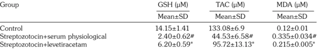
Table 2 shows the results of GSH, TAC, and MDA levels in the plasma of diabetic rats induced STZ. The STZ+SP group showed significantly decreased total TAC, GSH and increased MDA levels when compared with the control group. The STZ+LEV group showed significantly increased total TAC, GSH and decreased MDA levels when compared with the STZ+SP group.
DISCUSSION
In this present study we have shown the protective antioxidant effects of LEV on hepatic fibrosis in STZ induced diabetic rats with histological examination, real-time elastography imaging technique and measurement of oxidative and antioxidative markers. Our result was consistent with the other studies and showed that type 2 DM and hyperglycemia are major risk factors for diabetic hepatic injury.[9]
Pathogenesis of diabetic liver injury comprises two steps. The first step is thought to be insulin resistance increasing the transport of fatty acids from adipose tissue to the liver and causes accumulation of fat in the hepatocytes. In the second step increased oxidative stress and production of inflammatory cytokines lead to the hepatocellular injury and subsequently progress to hepatic fibrosis.[19,20] Pro-inflammatory and
pro-fibrogenic role of oxidative stress products play a critical role in this step.[21] In the previous studies,
increased levels of lipid peroxidation products and oxidative stress markers and decreased levels of antioxidant enzymes are found in nonalcoholic steatohepatitis (NASH) patients.[22,23] In prior
reports the protective effect of antioxidants and stimulation of collagen production and hepatic fibrogenesis by oxidative stress were shown.[3,24]
Oxidative stress-related molecules may act as mediators and trigger the events responsible for the progression of hepatic fibrosis.[4] Reactive oxygen
species (ROS) trigger formation of cytokines and growth factors leading to inflammatory response and eventually liver damage.
Currently, liver biopsy is still the gold standard for determining hepatic fibrosis. In spite of this, it is an invasive and extremely uncomfortable procedure, and sometimes may result with serious complications.[25,26] Besides, significant intra-
and interobserver variability is reported in the pathologic evaluation of the specimens.[27,28] As
a consequence, the evaluation of non-invasive methods for liver fibrosis assessment has become important in recent researches. Real-time elastography is a method used for the detection of focal lesions in the breast, thyroid gland, and prostate gland.[29] In a previous study, they had
shown that real-time elastography can be used for the measurement of liver fibrosis in patients with chronic viral hepatitis.[14] In a recent study,
Lin et al.[30] used real-time elastography as a reliable
imaging technique for quantitative evaluation of Table 1. The results of histopathological examination (Liver fibrosis score) and
real-time elastography (Strain-elasticity) of control rat liver and diabetic rat liver induced by streptozotocin
Group Liver fibrosis score Strain elasticity (%)
Mean±SD Mean±SD
Control 0.8±0.37 83.1±1.8
Streptozotocin+serum physiological 3.6±0.24* 31.8±3.0*
Streptozotocin+levetiracetam 2.4±0.50† 50.1±2.3‡
SD: Standard deviation; * p<0.001, different from control group; † p<0.05, different from streptozotocin+serum physiological group; ‡ p<0.001, different from streptozotocin+serum physiological group.
Table 2. The results of glutathione, total antioxidant capacity, and malondialdehyde levels in the plasma of diabetic rats induced by treptozotocin
Group GSH (μM) TAC (μM) MDA (μM)
Mean±SD Mean±SD Mean±SD
Control 14.15±1.41 133.08±6.9 0.12±0.01
Streptozotocin+serum physiological 2.40±0.62# 44.53±6.58# 0.335±0.034#
Streptozotocin+levetiracetam 6.20±0.59* 95.72±13.13* 0.215±0.005*
GSH: Glutathione; TAC: Total antioxidant capacity; MDA: Malondialdehyde; # p<0.000, different from control group; * p<0.001, different from streptozotocin+serum physiological group.
hepatic fibrosis induced by dimethylnitrosamine (DMN) in rats. They compared the results of real-time elastography [acquire area ratio of low-strain region (% AREA) and liver fibrosis index] with the stage of liver fibrosis and grade of necroinflammatory pathologically. They determined that the values of liver fibrosis index and low-strain region were both increased with liver fibrosis stage. They found a certain correlation between liver fibrosis index and liver fibrosis stage, so as low-strain region and liver fibrosis stage. In this present study, perivascular fibrotic alterations in the liver was determined in diabetic rats histopathologically and lower strain measurement was found by using real-time elastography.
Levetiracetam is an antiepileptic drug which has been proved to have antioxidative
and antiinflammatory efficacy.[10-12] It was
demonstrated that besides the antiepileptic efficiency of the drug LEV has neuroprotective effects associated with the reduction of lipid peroxidation, and the increase of the level of endogenous antioxidants.[11] Levetiracetam also
regulates the influx of calcium into the cells, and modulates membrane depolarization and prevents irreversible cellular damage.[31-33] In
this present study we determined the protective antioxidant effects of LEV on hepatic fibrosis. Significant increases were detected in the serum levels of GSH and TAC while there was a significant decrease in the serum levels of MDA in the LEV+STZ group. Much evidence supports the association between oxidant injury and hepatic fibrogenesis.[34] It was reported that
a well known antioxidant molecule vitamin E, reduced the level of lipid peroxidation molecules in the liver of STZ-diabetic rats and prevented the development of diabetic hepatic injury.[8] Another
well-known antioxidant molecule melatonin was used in a study and significant decrease in an oxidative stress parameter, MDA, in brain, liver and kidney was determined.[8] In another study
by using angiotensin converting enzyme inhibitor to protect liver damage in STZ-induced diabetic rats, they had shown the negative correlation between liver tissue fibrosis and glutathione related antioxidant defenses.[9]
As a result we showed that when compared with histological investigation, real-time elastography is a reliable imaging technique for
detecting hepatic fibrosis. Taken together the mechanism of action of LEV and pathogenesis of diabetic liver injury, LEV has potential as a treatment for diabetic liver injury and hepatic fibrosis and can be a good candidate among new treatment options. Given the scope of the problem more evidence is needed for the effectiveness and safety of this drug and future prospective and randomized studies will be needed to confirm recommendations.
Declaration of conflicting interests
The authors declared no conflicts of interest with respect to the authorship and/or publication of this article.
Funding
The authors received no financial support for the research and/or authorship of this article.
REFERENCES
1. Stone BG, Van Thiel DH. Diabetes mellitus and the liver. Semin Liver Dis 1985;5:8-28.
2. Keeffe EB, Adesman PW, Stenzel P, Palmer RM. Steatosis and cirrhosis in an obese diabetic. Resolution of fatty liver by fasting. Dig Dis Sci 1987;32:441-5. 3. Di Sario A, Candelaresi C, Omenetti A, Benedetti A.
Vitamin E in chronic liver diseases and liver fibrosis. Vitam Horm 2007;76:551-73.
4. Parola M, Robino G. Oxidative stress-related molecules and liver fibrosis. J Hepatol 2001;35:297-306. 5. Svegliati-Baroni G, De Minicis S, Marzioni M. Hepatic
fibrogenesis in response to chronic liver injury: novel insights on the role of cell-to-cell interaction and transition. Liver Int 2008;28:1052-64.
6. Pinzani M, Rombouts K. Liver fibrosis: from the bench to clinical targets. Dig Liver Dis 2004;36:231-42. 7. Wu YG, Xia LL, Lin H, Zhou D, Qian H, Lin
ST. Prevention of early liver injury by breviscapine in streptozotocin-induced diabetic rats. Planta Med 2007;73:433-8.
8. Baydas G, Canatan H, Turkoglu A. Comparative analysis of the protective effects of melatonin and vitamin E on streptozocin-induced diabetes mellitus. J Pineal Res 2002;32:225-30.
9. de Cavanagh EM, Inserra F, Toblli J, Stella I, Fraga CG, Ferder L. Enalapril attenuates oxidative stress in diabetic rats. Hypertension 2001;38:1130-6.
10. Stettner M, Dehmel T, Mausberg AK, Köhne A, Rose CR, Kieseier BC. Levetiracetam exhibits protective properties on rat Schwann cells in vitro. J Peripher Nerv Syst 2011;16:250-60.
11. Ueda Y, Doi T, Takaki M, Nagatomo K, Nakajima A, Willmore LJ. Levetiracetam enhances endogenous antioxidant in the hippocampus of rats: in vivo evaluation by brain microdialysis combined with ESR
spectroscopy. Brain Res 2009;1266:1-7.
12. Gibbs JE, Walker MC, Cock HR. Levetiracetam: antiepileptic properties and protective effects on mitochondrial dysfunction in experimental status epilepticus. Epilepsia 2006;47:469-78.
13. Coppey LJ, Davidson EP, Dunlap JA, Lund DD, Yorek MA. Slowing of motor nerve conduction velocity in streptozotocin-induced diabetic rats is preceded by impaired vasodilation in arterioles that overlie the sciatic nerve. Int J Exp Diabetes Res 2000;1:131-43. 14. Friedrich-Rust M, Ong MF, Herrmann E, Dries V,
Samaras P, Zeuzem S, et al. Real-time elastography for noninvasive assessment of liver fibrosis in chronic viral hepatitis. AJR Am J Roentgenol 2007;188:758-64. 15. Kanamoto M, Shimada M, Ikegami T, Uchiyama H,
Imura S, Morine Y, et al. Real time elastography for noninvasive diagnosis of liver fibrosis. J Hepatobiliary Pancreat Surg 2009;16:463-7.
16. Demougeot C, Marie C, Beley A. Importance of iron location in iron-induced hydroxyl radical production by brain slices. Life Sci 2000;67:399-410.
17. Benzie IF, Strain JJ. Ferric reducing/antioxidant power assay: direct measure of total antioxidant activity of biological fluids and modified version for simultaneous measurement of total antioxidant power and ascorbic acid concentration. Methods Enzymol 1999;299:15-27.
18. Ellman GL. Tissue sulfhydryl groups. Arch Biochem Biophys 1959;82:70-7.
19. Day CP, James OF. Steatohepatitis: a tale of two “hits”? Gastroenterology 1998;114:842-5.
20. Neuschwander-Tetri BA, Caldwell SH. Nonalcoholic steatohepatitis: summary of an AASLD Single Topic Conference. Hepatology 2003;37:1202-19.
21. Clark JM, Diehl AM. Nonalcoholic fatty liver disease: an underrecognized cause of cryptogenic cirrhosis. JAMA 2003;289:3000-4.
22. Maiese K, Morhan SD, Chong ZZ. Oxidative stress biology and cell injury during type 1 and type 2 diabetes mellitus. Curr Neurovasc Res 2007;4:63-71. 23. Leclercq IA, Da Silva Morais A, Schroyen B, Van Hul
N, Geerts A. Insulin resistance in hepatocytes and
sinusoidal liver cells: mechanisms and consequences. J Hepatol 2007;47:142-56.
24. Das KS, Balakrishnan V, Mukherjee S, Vasudevan DM. Evaluation of blood oxidative stress-related parameters in alcoholic liver disease and non-alcoholic fatty liver disease. Scand J Clin Lab Invest 2008;68:323-34. 25. Bravo AA, Sheth SG, Chopra S. Liver biopsy. N Engl
J Med 2001;344:495-500.
26. Castéra L, Nègre I, Samii K, Buffet C. Pain experienced during percutaneous liver biopsy. Hepatology 1999;30:1529-30.
27. Bedossa P, Dargère D, Paradis V. Sampling variability of liver fibrosis in chronic hepatitis C. Hepatology 2003;38:1449-57.
28. Regev A, Berho M, Jeffers LJ, Milikowski C, Molina EG, Pyrsopoulos NT, et al. Sampling error and intraobserver variation in liver biopsy in patients with chronic HCV infection. Am J Gastroenterol 2002;97:2614-8.
29. Giuseppetti GM, Martegani A, Di Cioccio B, Baldassarre S. Elastosonography in the diagnosis of the nodular breast lesions: preliminary report. Radiol Med 2005;110:69-76.
30. Lin SH, Ma JJ, Zhang H, Ding H, Yu Q, Zhu HG, et al. Real-time elastography for quantitative assessment of liver fibrosis in a rat model. Zhonghua Gan Zang Bing Za Zhi 2012;20:386-9. [Abstract]
31. Niespodziany I, Klitgaard H, Margineanu DG. Levetiracetam inhibits the high-voltage-activated Ca(2+) current in pyramidal neurones of rat hippocampal slices. Neurosci Lett 2001;306:5-8. 32. Pisani A, Bonsi P, Martella G, De Persis C, Costa
C, Pisani F, et al. Intracellular calcium increase in epileptiform activity: modulation by levetiracetam and lamotrigine. Epilepsia 2004;45:719-28.
33. Madeja M, Margineanu DG, Gorji A, Siep E, Boerrigter P, Klitgaard H, et al. Reduction of voltage-operated potassium currents by levetiracetam: a novel antiepileptic mechanism of action? Neuropharmacology 2003;45:661-71.
34. Poli G, Parola M. Oxidative damage and fibrogenesis. Free Radic Biol Med 1997;22:287-305.