Lymphocytes
Edited by Erman Salih Istifli and Hasan Basri İla
Although all the features of the immune system have not been fully resolved yet,the knowledge we have gained from studies on lymphocytes, the basic elements of the immune system, is quite lucid. For this reason, the significance of lymphocytes (the cells that are the source of most of the information we have obtained about the human genome, the negative effects of drugs on the genetic system, the development
and behavior of immune system, antigen-antibody association, cytotoxic adaptive immunity, antibody-driven adaptive immunity, cancer and autoimmune diseases) is
clear. Studies on lymphocytes will not only help us develop tools to combat human diseases more effectively in the future, but will also help us understand how evolution
shapes the immune system in living organisms.
Published in London, UK © 2018 IntechOpen © jarun011 / iStock ISBN 978-1-78984-919-6
Ly
m
ph
oc
yte
s
LYMPHOCYTES
Edited by Erman Salih Istifli
and Hasan Basri İla
LYMPHOCYTES
Edited by Erman Salih Istifli
and Hasan Basri İla
Edited by Erman Salih Istifli and Hasan Basri İla
Contributors
Yeunwha Gu, Tatsuaki Tsuruyama, Nashwa Abdelaziz, Ghada ElGohary, Sherif Mohamed, Khaled Alsaleh, Aleksandra Weselucha-Birczynska, Magdalena Pietruszewska, Grażyna Biesiada, Jacek Czepiel, Malwina Birczyńska, Paulina Moskal, Mateusz Kozicki, Emilia Hola, Aleksander Garlicki, Takemi Otsuki, Suni Lee, Naoko Kumagai-Takei, Hidenori Matsuzaki, Nagisa Sada, Kei Yoshitome, Yasumitsu Nishimura, Alamelu Sundaresan, Vivek Mann, Elvis Okoro, Ayodotun Sodipe, Courtney Williams, Patricia Ngantcha, Malavika Bhattacharya
© The Editor(s) and the Author(s) 2019
The rights of the editor(s) and the author(s) have been asserted in accordance with the Copyright, Designs and Patents Act 1988. All rights to the book as a whole are reserved by INTECHOPEN LIMITED. The book as a whole (compilation) cannot be reproduced, distributed or used for commercial or non-commercial purposes without INTECHOPEN LIMITED’s written permission. Enquiries concerning the use of the book should be directed to INTECHOPEN LIMITED rights and permissions department ([email protected]).
Violations are liable to prosecution under the governing Copyright Law.
Individual chapters of this publication are distributed under the terms of the Creative Commons Attribution 3.0 Unported License which permits commercial use, distribution and reproduction of the individual chapters, provided the original author(s) and source publication are appropriately acknowledged. If so indicated, certain images may not be included under the Creative Commons license. In such cases users will need to obtain permission from the license holder to reproduce the material. More details and guidelines concerning content reuse and adaptation can be found at http://www.intechopen.com/copyright-policy.html.
Notice
Statements and opinions expressed in the chapters are these of the individual contributors and not necessarily those of the editors or publisher. No responsibility is accepted for the accuracy of information contained in the published chapters. The publisher assumes no responsibility for any damage or injury to persons or property arising out of the use of any materials, instructions, methods or ideas contained in the book.
First published in London, United Kingdom, 2019 by IntechOpen
IntechOpen is the global imprint of INTECHOPEN LIMITED, registered in England and Wales, registration number: 11086078, The Shard, 25th floor, 32 London Bridge Street
London, SE19SG – United Kingdom Printed in Croatia
British Library Cataloguing-in-Publication Data
A catalogue record for this book is available from the British Library Additional hard copies can be obtained from [email protected] Lymphocytes, Edited by Erman Salih Istifli and Hasan Basri İla p. cm.
Print ISBN 978-1-78984-919-6 Online ISBN 978-1-78984-920-2
http://dx.doi.org/10.5772/intechopen.73716 Edited by Erman Salih Istifli and Hasan Basri İla
Contributors
Yeunwha Gu, Tatsuaki Tsuruyama, Nashwa Abdelaziz, Ghada ElGohary, Sherif Mohamed, Khaled Alsaleh, Aleksandra Weselucha-Birczynska, Magdalena Pietruszewska, Grażyna Biesiada, Jacek Czepiel, Malwina Birczyńska, Paulina Moskal, Mateusz Kozicki, Emilia Hola, Aleksander Garlicki, Takemi Otsuki, Suni Lee, Naoko Kumagai-Takei, Hidenori Matsuzaki, Nagisa Sada, Kei Yoshitome, Yasumitsu Nishimura, Alamelu Sundaresan, Vivek Mann, Elvis Okoro, Ayodotun Sodipe, Courtney Williams, Patricia Ngantcha, Malavika Bhattacharya
© The Editor(s) and the Author(s) 2019
The rights of the editor(s) and the author(s) have been asserted in accordance with the Copyright, Designs and Patents Act 1988. All rights to the book as a whole are reserved by INTECHOPEN LIMITED. The book as a whole (compilation) cannot be reproduced, distributed or used for commercial or non-commercial purposes without INTECHOPEN LIMITED’s written permission. Enquiries concerning the use of the book should be directed to INTECHOPEN LIMITED rights and permissions department ([email protected]).
Violations are liable to prosecution under the governing Copyright Law.
Individual chapters of this publication are distributed under the terms of the Creative Commons Attribution 3.0 Unported License which permits commercial use, distribution and reproduction of the individual chapters, provided the original author(s) and source publication are appropriately acknowledged. If so indicated, certain images may not be included under the Creative Commons license. In such cases users will need to obtain permission from the license holder to reproduce the material. More details and guidelines concerning content reuse and adaptation can be found at http://www.intechopen.com/copyright-policy.html.
Notice
Statements and opinions expressed in the chapters are these of the individual contributors and not necessarily those of the editors or publisher. No responsibility is accepted for the accuracy of information contained in the published chapters. The publisher assumes no responsibility for any damage or injury to persons or property arising out of the use of any materials, instructions, methods or ideas contained in the book.
First published in London, United Kingdom, 2019 by IntechOpen
IntechOpen is the global imprint of INTECHOPEN LIMITED, registered in England and Wales, registration number: 11086078, The Shard, 25th floor, 32 London Bridge Street
London, SE19SG – United Kingdom Printed in Croatia
British Library Cataloguing-in-Publication Data
A catalogue record for this book is available from the British Library Additional hard copies can be obtained from [email protected] Lymphocytes, Edited by Erman Salih Istifli and Hasan Basri İla p. cm.
Print ISBN 978-1-78984-919-6 Online ISBN 978-1-78984-920-2
Selection of our books indexed in the Book Citation Index in Web of Science™ Core Collection (BKCI)
Interested in publishing with us?
Contact [email protected]
Numbers displayed above are based on latest data collected.For more information visit www.intechopen.com
3,900+
Open access books available
151
Countries delivered to Contributors from top 500 universities
12.2%
Our authors are among the
Top 1%
most cited scientists
116,000+
International authors and editors
120M+
DownloadsWe are IntechOpen,
the world’s leading publisher of
Open Access books
Meet the editors
Dr Erman Salih İstifli received his Ph.D. from the Biolo-gy Department of Cukurova University, Institute of Sci-ence and Letter. In his doctoral study, Dr İstifli focused on the elucidation of the genotoxic and cytotoxic effects of a commonly used anticancer agent (antifolate) on human lymphocytes. Dr İstifli, during his period of doc-toral research, joined the molecular cytogenetics group at the Max Planck Institute for Molecular Genetics in Berlin, Germany, and there he focused on investigating the molecular cytogenetic causes of some human rare diseases. During these studies, he contributed experimentally to the identification of four candidate genes (GRIA2, GLRB, NPY1R, and NPY5R) responsible for intelligence and obesity. He was assigned as an expert and rapporteur on eight candidate projects in the Marie-Sklodows-ka Curie-Actions Innovative Training Networks in 2016. He is a published author of several articles in journals covered by the SCI and SCI-E, and has manuscripts in other refereed scientific journals. He currently serves as a referee in several journals covered by the SCI and SCI-E. His studies mainly fall into the field of genetic toxicology.
Prof. Dr. Hasan Basri İla received his Ph.D. from Biology Department of Çukurova University, Insitute of Science and Letter. Prof. Dr. Hasan Basri İla, during his doctor-al study, investigated the effects of a commonly used antibiotic on chromosome aberration and micronucleus formation by in vivo tests. Dr. İla has several publica-tions in the internationally indexed (SCI, SCI-E) journals. He actively took responsibility in 17 national projects as a researcher and/ or project leader. He has been actively giving lectures on biology, cytology, genetics, evolution, organelle genetics and cancer genetics. He has nu-merous poster and/or oral scientific presentations in several international conferences. Dr. Ila’s articles have been cited 442 times.
Preface VII
Chapter 1 Alteration of Various Lymphocytes by Particulate and Fibrous Substances 1
Naoko Kumagai-Takei, Suni Lee, Hidenori Matsuzaki, Nagisa Sada, Kei Yoshitome, Yasumitsu Nishimura and Takemi Otsuki
Chapter 2 Lymphocyte Signaling and Function in Altered Physiological Environments 15
Vivek Mann, Elvis Okoro, Ayodotun Sodipe, Courtney Williams, Patricia Ngantcha and Alamelu Sundaresan
Chapter 3 Signaling Pathway for the Development of Pre-B Cells 27
Tatsuaki Tsuruyama
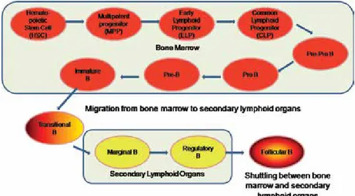
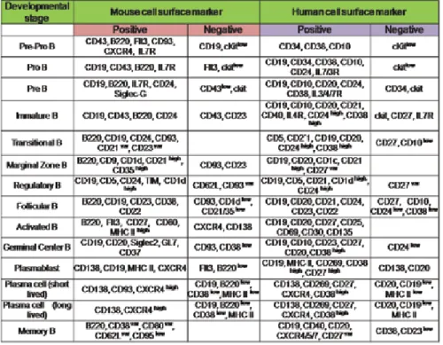
Chapter 4 Understanding B Lymphocyte Development: A Long Way to Go 41
Malavika Bhattacharya
Chapter 5 Prognostic and Therapeutic Implications of Lymphocytes in Hematological Disorders and Solid Malignancies 61
Nashwa Abd El-Aziz, Ghada El Gohary, Sherif Mohamed and Khaled El-Saleh
Chapter 6 Lymphocytes Studied by Raman Microspectroscopy 85
Magdalena Pietruszewska, Grażyna Biesiada, Jacek Czepiel, Malwina Birczyńska, Paulina Moskal, Mateusz Kozicki, Emilia Hola, Aleksander Garlicki and Aleksandra Wesełucha-Birczyńska
Chapter 7 Radiation Protective and Immunopotentiating Effect of Lymphocytes by β-Glucan 101
Preface VII
Chapter 1 Alteration of Various Lymphocytes by Particulate and Fibrous Substances 1
Naoko Kumagai-Takei, Suni Lee, Hidenori Matsuzaki, Nagisa Sada, Kei Yoshitome, Yasumitsu Nishimura and Takemi Otsuki
Chapter 2 Lymphocyte Signaling and Function in Altered Physiological Environments 15
Vivek Mann, Elvis Okoro, Ayodotun Sodipe, Courtney Williams, Patricia Ngantcha and Alamelu Sundaresan
Chapter 3 Signaling Pathway for the Development of Pre-B Cells 27
Tatsuaki Tsuruyama
Chapter 4 Understanding B Lymphocyte Development: A Long Way to Go 41
Malavika Bhattacharya
Chapter 5 Prognostic and Therapeutic Implications of Lymphocytes in Hematological Disorders and Solid Malignancies 61
Nashwa Abd El-Aziz, Ghada El Gohary, Sherif Mohamed and Khaled El-Saleh
Chapter 6 Lymphocytes Studied by Raman Microspectroscopy 85
Magdalena Pietruszewska, Grażyna Biesiada, Jacek Czepiel, Malwina Birczyńska, Paulina Moskal, Mateusz Kozicki, Emilia Hola, Aleksander Garlicki and Aleksandra Wesełucha-Birczyńska
Chapter 7 Radiation Protective and Immunopotentiating Effect of Lymphocytes by β-Glucan 101
Lymphocytes are one of the white blood cell subtypes in the immune system of vertebrates. These cells include natural killer cells (cell-mediated, cytotoxic innate immunity), T cells (for cell-mediated, cytotoxic adaptive immunity), and B cells (for humoral, antibody-driven adaptive immunity). These are the main types of cells found in the "lymph" and are there‐ fore called "lymphocytes". Furthermore, although we emphasize “lymphocytes”, the im‐ mune elements in vertebrates, there is also an immune system in invertebrate organisms that has evolved over approximately 500 million years, which appears to be a precursor form of the immune system of vertebrates.
Lymphocytes have been the main research object of the medical and toxicological sciences for the last six decades. In addition to important roles they play in the immune system, lym‐ phocytes have been the primary test material often employed in pure research-focused stud‐ ies that provide important contributions to our knowledge of biochemistry, molecular biology and genomics. To give a prominent example, the majority of DNA used for sequenc‐ ing of the human genome was isolated from lymphocytes of male donor blood.
Today, the use of lymphocytes for research purposes is increasing, and the variety of tests performed on these cells are also diversifying each day. Lymphocyte activation assays, fluo‐ rescence-based assays for high-throughput screening of small molecules, cytotoxic T lym‐ phocyte assays, cell proliferation assays, blastogenesis assays for T lymphocytes for identifying immunomodulatory drugs of lymphocyte extravasation and sequencing and genotoxicity assays are among the tests that lymphocytes are frequently used for. As can be readily understood, lymphocytes serve in different ways in terms of understanding the cell structure, unknown features of the immune system, genome and chromosomes.
It is clear that lymphocytes will continue to serve as experimental material in the most im‐ portant areas of science in the near future. Although it is impossible to follow simultaneous‐ ly the growing body of knowledge in this area, this book was aimed to present the most pertinent findings and reviews related to the response of the human lymphoid system to various occupational and environmental substances, effects of beta glucan on the radiation protective and immunopotentiating effect of lymphocytes, effects of microgravity on lym‐ phocytes, signaling pathways in the development of pre-B cells, B lymphocyte development, immunotherapeutic approaches against hematologic malignancies and solid tumors as well as the Raman microspectroscopy in lymphocyte studies.
Dr. Erman Salih Istifli
Çukurova University Faculty of Science and Letters Department of Biology Adana, Turkey
Dr. Hasan Basri İla
Çukurova University Adana, Turkey
Chapter 1
Alteration of Various Lymphocytes by Particulate and
Fibrous Substances
Naoko Kumagai-Takei, Suni Lee,
Hidenori Matsuzaki, Nagisa Sada, Kei Yoshitome,
Yasumitsu Nishimura and Takemi Otsuki
Additional information is available at the end of the chapter http://dx.doi.org/10.5772/intechopen.79054
Provisional chapter
© 2016 The Author(s). Licensee InTech. This chapter is distributed under the terms of the Creative Commons Attribution License (http://creativecommons.org/licenses/by/3.0), which permits unrestricted use, distribution, and reproduction in any medium, provided the original work is properly cited.
Alteration of Various Lymphocytes by Particulate and
Fibrous Substances
Naoko Kumagai-Takei, Suni Lee,
Hidenori Matsuzaki, Nagisa Sada, Kei Yoshitome,
Yasumitsu Nishimura and Takemi Otsuki
Additional information is available at the end of the chapter
Abstract
Various occupational and environmental substances alter the cellular and molecular function of the human lymphoid system. For example, silicosis patients who have been chronically exposed to silica particles often complicate with autoimmune diseases such as rheumatoid arthritis and systemic sclerosis. From our investigations, silica particles affect CD4+ responder T cells and regulatory T cells (Tregs), which results in the disrup-tion of autoimmunity. Asbestos fibers are a type of mineral silicate, and patients exposed to asbestos fibers revealed cancers such as mesothelioma and lung cancer. In these cases, asbestos fibers may reduce antitumor immunity. Our results investigating the effect of asbestos on cytotoxic T lymphocyte, natural killer (NK) cells, CD4+ cells, and Tregs revealed a reduction in antitumor immunity. To date, the effects of silica and asbestos on Th17 cells and antigen-presenting cells such as dendritic cells and macrophages remain unclear. Based on these findings, it will be possible to generate earlier detection methods to identify the occurrence of immune alterations in silicosis as well as the appearance of a decreased antitumor immunity in asbestos-exposed populations. Additionally, research efforts should also be directed at discovering and identifying physiological substances from foods, plants, and other sources that can restore the immune status in people exposed to particulate and fibrous substances.
Keywords: silica, asbestos, responder T cell, regulatory T cell, cytotoxic T lymphocyte,
NK cell
© 2018 The Author(s). Licensee IntechOpen. This chapter is distributed under the terms of the Creative Commons Attribution License (http://creativecommons.org/licenses/by/3.0), which permits unrestricted use, distribution, and reproduction in any medium, provided the original work is properly cited.
1. Introduction
There are a variety of lymphocytes including T cells, B cells, and natural killer (NK) cells [1–4]. Additionally, there are other smaller populations of lymphocytes such as natural killer T (NKT) cells. T cells are further divided depending on their surface molecules as well as func-tion and cytokine producfunc-tion in CD4+ and CD8+ T cells. CD8+ cells are designated as cytotoxic T lymphocytes (CTLs), which express T cell receptors (TcRs) and recognize a specific antigen. TcRs on CTLs can bind to the complex of the class I major histocompatibility complex (MHC) molecule and antigen. Thereafter, CTLs can destroy the cells using granzymes and perforins as the attacking molecules. CD4+ T cells include various subpopulations [1–4]. Although CD4+ cells are referred to as T helper (Th) cells, depending on the kind of stimulation, cytokine circumstances surrounding Th cells, naïve Th cells are skewed to Th1, Th2, Th17, and Treg (regulatory T) cells. Th1 and Th2 cells are balanced subpopulations. The proliferation of Th1 cells is triggered by interleukin (IL)-12 and produces IL-2 and interferon (IFN)-γ. The key tran-scription factors are T-bet and the signal transducer and activator of trantran-scription 4 (STAT4). Th1 cells act against intracellular bacteria by activating macrophages. On the other hand, Th2 cells are activated by IL-4 and IL-2, and results in the secretion of IL-4, IL-5, IL-9, IL-10, IL-13 and IL-25 cytokines. The key transcription factors for Th2 are SATA6 and GATA3. IL-4 pro-duced by Th2 cells acts as a positive feedback to restimulate Th2 cells and stimulate B cells to produce immunoglobulin (Ig) E. Therefore, Th2 cells play an important role in the context of allergies and hypersensitivity such as atopic dermatitis and bronchial asthma [1–4].
Additionally, Th17 and Treg cells are also balanced according to the skewing cytokine cir-cumstances surrounding Th naïve cells. Initially, transforming growth factor (TGF)-β stimu-lates naïve Th cells to express two transcription factors, RORγt and forkhead box (Fox) P3. Although IL-6 together with TGF-β causes activation of the STAT3 signaling pathway to suppress FoxP3, IL-2 together with TGF-β facilitates the movement of cells toward the Th17 phenotype. Tregs produce IL-10, TGF-β and IL-35 and act to inhibit effector T cell reactions against foreign as well as non-self/self-antigens. On the other hand, Th17 cells produce IL-17, IL-21 and IL-22 and play an important role in pathogen clearance at the mucosal surface and are implicated to play a role in autoimmune and inflammatory disorders [1–4].
B cells act in humoral immunity to produce Ig/antibodies to attack foreign antigens. Additionally, B cells express B cell receptors (BcRs), which allow for the binding of B cells to a specific antigen in the process involving innate immunity [1–4].
NK cells also act in innate immunity by quickly attacking virally infected cells, as well as tumor cells, after recognition. NK cells do not need MHC presentation. These cells can kill virally infected cells and tumor cells via lytic reactions and apoptosis by releasing granzymes and perforins [1–4]. When the immune system recognizes the presence of foreign substances such as bacteria and viruses, as well as transformed cells derived from self, the human immune system works to attack these non-self substances and facilitates the curtailment of alterations in the human body such as various symptoms resulting from bacterial and viral infections and cancers.
These reactions are physiological. Additionally, certain alterations of the immune system may recognize self-antigens and subsequently cause various autoimmune diseases [1–4].
2. Occupational and environmental substances that affect the human
immune system
There are many environmental and occupational substances which induce alterations of the immune system. For example, isocyanate latex, cement including chromium, and whiten-ing agents includwhiten-ing persulfates and other substances cause occupational hypersensitivity (allergy). Sometimes, patients exposed to these reagents reveal respiratory asthma and contact dermatitis. However, these allergic reactions may be categorized as a range of physiological reactions against foreign antigens [5–11]. On the other hand, some occupational and environ-mental substances cause autoimmune diseases. For example, vinyl chloride, silica dust, and chemicals including trichloroethene and epoxy resins can cause systemic sclerosis, one of the typical generalized autoimmune diseases that occur with certain frequencies in exposed populations. Unlike occupational allergies, autoimmune diseases caused by occupational and environmental substances seem to be the result of disruption of the human immune system caused by these substances [12–15]. Additionally, there are many cancers caused by occupa-tional and environmental substances such as lung cancers due to asbestos, tobacco smoke, certain metals including arsenic, chromium, nickel and beryllium, bladder cancer caused by benzidine, β-naphthylamine and other aromatic hydrocarbons, and others [16–22]. Of course, there are many mechanisms involved in the genesis of these occupational cancers when trig-gered by certain substances, such as DNA damage caused by reactive oxygen species (ROS), formation of DNA adducts, and others. However, it is also possible that certain substances that cause cancers may also affect the human immune system by reducing antitumor immu-nity. If so, it is understandable that a relatively long latency period may exist prior to the occurrence of occupational cancers following initial exposure to carcinogenic agents.
Taken together, it is important to assess how these occupational and environmental sub-stances alter the human immune system. Unlike standardized toxicological investigations, people or workers are usually exposed to these substances in low-dose, chronic, and continu-ous ways. Thus, it is important to assess the cellular and molecular alterations in immune cells derived from exposed populations, as well as to establish continuous and low-dose exposure models using human lymphoid cells exposed in vitro to substances that may cause alterations in autoimmunity or act as carcinogens.
From this viewpoint, we have been investigating the immunological effects of silica par-ticles or asbestos fibers since certain silica-exposed populations such as silicosis patients (SIL) are often complicated with various autoimmune diseases such as rheumatoid arthri-tis (known as Caplan syndrome) [23], systemic sclerosis (known as Erasmus syndrome) [24], systemic lupus erythematosus (SLE) [25], and antineutrophil cytoplasmic antibody (ANCA)-related vasculitis/glomerulonephritis [26–29]. Asbestos fibers are a type of mineral
silicate, while silica is particulate in nature. Additionally, asbestos can cause cancers such as malignant mesothelioma (MM) and lung cancer [30–33]. Silica particles can act to disrupt the regulation of autoimmune tolerance while asbestos fibers can facilitate a decline in antitumor immunity.
3. Silica and disruption of lymphoid cells
Silica exposure causes disruption of autoimmune tolerance. Thus, silicosis patients are often complicated with autoimmune diseases [34, 35].
When considering the effects of B cells and plasma cells, various autoantibodies have been detected in sera derived from silicosis patients. For example, some autoantibodies (AAB) are typically detected in autoimmune diseases such as anti-nuclear antibodies (ANA), anti-topoisomerase I (Sck-70) AAB [36], and anti-CENP-B (centromere) AAB [37]. In addition to these typical AABs, we found anticaspase-8 AAB [38, 39], anti-Fas-AAB [40], and antidesmoglein AAB [41], which is usually detected in skin bullous autoimmune diseases such as pemphigus vulgaris. However, we have not investigated alterations of B cells caused by silica exposure. The production of various AABs from B cells/plasma cells may be caused by alterations of B cells or be dependent on T cells which produce AABs to B cells.
What about effector T cells? There are many alterations that have been detected in effector T cells. CD4+ responder T cells showed an increase in activation markers and an excess of survival mark-ers. For the former, the expression of CD69, a typical early activation cell surface marker for T cells, increased at 5–10 days when peripheral blood mononuclear cells (PBMCs) derived from healthy donors (HD) were cultured in vitro with silica particles [42]. Additionally, soluble IL-2 receptor (sIL-2R) levels in serum derived from silicosis patients showed an increasing tendency compared with those of HD. Then, if we set 1, 2, and 3 as sequential numbering for HD, silicosis, and SSc for autoimmune disruption, this number and the level of serum sIL-2R in these three categories showed a significant positive correlation [43]. Although sIL-2R is considered as a tumor marker for T cell acute lymphoblastic leukemia and T cell malignant lymphoma, the elevation of serum sIL-2R was recently detected in various autoimmune diseases. This increase in serum sIL-2R is the evidence of chronic activation of T cells in certain pathological situations such as autoimmune diseases. Thus, silicosis is also considered as a condition whereby peripheral T cells are activated chronically, and the degree of activation was higher than that in HD, although less than that in autoimmune diseases such as SSc. Moreover, programmed death-1 (PD-1) gene expression in peripheral CD4+ cells derived from silicosis patients was significantly higher than that in HD [44]. Although PD-1 is one of the most important molecules that act in the immune checkpoint system, PD-1 is also a marker of T cell activation. Thus, upregulation of PD-1 in T cells derived from sili-cosis patients also indicates that T cells in silisili-cosis are chronically activated (due to long-term and continuous exposure to silica particles in the body, such as lung fields and related lymph nodes). Considering survival factors, we found that serum from silicosis patients showed significantly higher levels of soluble Fas (sFas) compared with HD and similar levels with SLE patients [45].
Additionally, since sFas is a product due to alternative splicing of the Fas gene, losing 63 bp of the transmembrane domain, mRNA expression of wild-type membrane Fas and sFas tran-scripts were examined. As a result, the ratio of wild-type Fas message divided by soluble-type alternatively spliced message decreased in PBMCs from silicosis patients compared with HD [46]. These findings indicated that T cells in silicosis patients are protected against Fas-ligand-induced apoptosis by increasing the binding of Fas ligand and sFas at extracellular spaces, thereby resulting in the extended survival of T cells in silicosis patients. Additionally, the scenario of Fas, sFas, and Fas ligand was employed for tumor necrosis factor-related apopto-sis-inducing ligand (Trail) receptor, decoy receptor 3 (DcR3), and Trail. DcR3 mRNA expres-sion levels in PBMCs derived from silicosis patients were higher compared with HD [47]. Furthermore, serum DcR3 levels were higher in silicosis patients compared with HD. Hence, in both Fas and Trail systems, T cells in silicosis patients were protected from apoptosis. Taken together, T cells in silicosis patients are chronically activated as well as continuously protected from apoptosis, thereby resulting in the circulation of longer surviving T cells in the periph-eral blood of silicosis patients. If so, rare self-antigen acting T cell clones may also survive longer and be chronically activated. Thus, antigen (non-self or self)-activated T cells increase their volume in silicosis patients.
What about Tregs? It is known that Fas/CD95 expression on the cell surface of Tregs is one marker of Treg activation. Thereafter, Tregs proceed to apoptosis when its role is ceased. Thus, we examined Fas expression in peripheral Treg (CD4+ and FoxP3+) as well as CD4 + FoxP3- T cells derived from HD and silicosis patients. As a result, Fas expression was significantly higher in Tregs from silicosis patients compared with HD [44, 48]. Of course, CD4 + FoxP3- T cells did not express sufficient amounts of Fas in HD or silicosis patients. Then, PBMCs derived from HD or silicosis patients were cultured with agonistic antibody. Tregs from sili-cosis patients showed earlier and higher apoptosis compared with HD because of a greater expression of Fas. Thereafter, PBMCs from HD were cultured with silica particles for 4 days. As a result, CD4 + FoxP3+ cell levels were significantly reduced. However, CD25+ FoxP3- cell levels were not altered, translating Tregs were reduced, and initial CD4 + CD25- cells were activated to reveal CD25 as an activation marker.
Taking together the results of responder T cells and Tregs, an imbalance between responder CD4 cells and Tregs was found, in that the increase of responder T cell levels increased, while Treg cell levels decreased. This tendency is well known in the area of dysregulation of autoim-munity which leads to the occurrence of autoimmune diseases. Thus, silica exposure affects the immune system to create an imbalance between responder T cells and Tregs and sets the foundation for the appearance of autoimmune diseases in silicosis patients.
What about Th17? Unfortunately, we have not investigated the Th17 status in silicosis patients. It was reported that Th17/I-17 is involved in silicosis to facilitate the progression of lung fibrosis found in silicosis [49, 50]. As mentioned earlier, Tregs in silicosis seem to progress toward apoptosis. This may induce an increase in Th17 levels in the peripheral blood of silicosis patients and consequently make these patients more susceptible to autoimmune diseases. Thus, further investigation of Th17 in silicosis is necessary from the viewpoint of efforts to delineate the early processes involved in the disruption of autoimmunity.
4. Asbestos and antitumor immunity
As mentioned earlier, carcinogenic factors among occupational and environmental sub-stances may facilitate a decline in antitumor immunity. We chose asbestos since it is a mineral silicate and possesses the potential to disrupt human lymphocytes in such a way as to make the human body prone to autoimmune diseases.
For the CTLs, we used a mixed lymphocyte reaction (MLR) to assess the asbestos fibers with respect to clonal expansion of the cells examined. The use of chrysotile asbestos resulted in a decreased differentiation and proliferation of CD8+ cells and a decreased production of cytotoxic granules such as granzyme B and perforin [51–53]. These findings were confirmed using peripheral blood CD8+ cells derived from MM patients considered to have a history of exposure to asbestos even though the patients did not remember the exposure. Interestingly, the status of intracellular perforin expression in CD8+ cells from patients with pleural plaque (PP) differed from that of HD and MM patients [51–53]. Perforin expression in CD8+ cells increased somewhat in PP patients. These findings indicated that immunological alterations, especially in CTLs, differed depending on the disease status when examining PP and MM patients exposed to asbestos. This may be dependent on the occurrence of cancer in the body [51–53].
What about NK cells? We tried to expose a human NK cell line, freshly isolated peripheral NK cells derived from HD, as well as NK cells from PP and MM patients to asbestos fibers. The killing activity was reduced in a cell line model subjected to continuous exposure to asbestos, ex vivo activated and expanded freshly isolated NK cells cultured with asbestos fibers, and NK cells derived from patients. Additionally, the most correlated marker of kill-ing activity was the cell surface expression level of NKp46 [54–58]. NKp46 is an NK cell acti-vating receptor that belongs to the natural cytotoxicity receptor (NCR) family. In addition to a reduction in NKp46 expression, phosphorylation was induced of extracellular signal-regulated protein kinases 1 and 2 (ERK1/2) which are involved in the mitogen-activated protein kinase (MAP-K) signaling pathway [54–58]. Moreover, degranulation to produce cytotoxic molecules such as granzyme and perforin was also suppressed by asbestos expo-sure. These results were obtained from a cell line model, an ex vivo culture model, and from the investigation of freshly isolated NK cells derived from PP and MM patients exposed to asbestos [54–58].
Next, the continuous and low-dose exposure of human CD4+ T cells to asbestos fibers was investigated. We employed the human T cell leukemia virus (HTLV)-1 immortalized human polyclonal T cell line MT-2 as the cell line model, an ex vivo clonal expansion model using freshly isolated peripheral blood CD4+ cells derived from HD, and peripheral CD4+ cells derived from PP and MM patients exposed to asbestos. As a result, all models showed that continuous and low-dose exposure to asbestos induced a decline in chemokine receptor CXCR3, a Gαi protein-coupled receptor of the CXC chemokine receptor family [59, 60]. The
reduction in CXCR3 resulted in diminished trafficking of T cells which produce IFNγ to attack tumor cells. In addition to this, CD4+ T cells exposed to asbestos showed potential to produce IFNγ. Both findings suggest a reduction in antitumor immunity [59, 60].
The MT-2 cell line was reported to possess Treg functionality. Thus, Treg suppressive func-tionality in MT-2 cell sublines continuously exposed to asbestos fibers was compared with the original MT-2 cell line not exposed to asbestos. As a result, sublines showed enhanced suppressive function by cell-cell contact in addition to excess production of typical soluble factors such as IL-10 and TGFβ [61]. In addition to these enhanced functions in MT-2 (a model of Tregs) caused by continuous exposure to asbestos, these sublines showed decreased levels of FoxO1 transcription factor [62]. FoxO1 regulates cell cycle progression in a negative fash-ion by inhibiting various cyclins as well as in a positive fashfash-ion by regulating many cyclin-dependent kinase inhibitors (CDK-Is) such as Cip1/p21, Kip1/p27, Kip2/p57, and CDKN2A to 2D such as p16, p15, p18, and p19. As a result of decreased levels of FoxO1, MT-2 sublines showed an enhanced expression of cyclins, especially cyclin D1, and a reduced expression of CDK-Is. Additionally, cell cycle progression was also enhanced compared with the original MT-2 line not exposed to asbestos [63]. Thus, both function and proliferation were enhanced with continuous and low-dose exposure of Tregs to asbestos, which suggest that the antitu-mor immunity controlled by Tregs was reduced [61–63].
Taken together, continuous and low-dose exposure to asbestos caused a reduction in anti-tumor immunity in CTLs, NK cells, CD4+ T cells, and Tregs [64–66]. Thus, it could be con-sidered that people subjected to continuous, low-dose exposure to asbestos are susceptible to the onset of cancers because of a gradual reduction in antitumor immunity. Thereafter, certain localized areas such as lung fields or the pleural cavity may become the locus where fibers remain in the body and chronic stimulations may occur at these locations on the basis of reduced antitumor immunity [64–66]. This may represent the mechanism by which asbestos-induced cancers occur in the long term, after a latency period of approximately 30–40 years, following initial exposure to asbestos fibers.
5. Conclusion
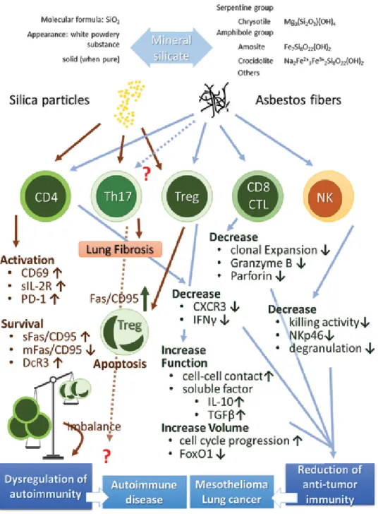
All findings described in this chapter are summarized in Figure 1. In this review, the effects of silica [67–69] and asbestos [64–66] are introduced and discussed. However, people are exposed to many more potentially hazardous occupational and environmental substances, such as various materials in air pollutants and a variety of metals in work environments. Thus, any immunological alterations induced in cells by these and many other substances should be investigated utilizing the methods described earlier to further our understanding of immune responses and cancer.
Additionally, as described earlier, the identification of certain physiologically active materials among various foods, plants, and other sources, which modify and repair the immune state following disruption by various occupational and environmental substances such as silica and asbestos, should lead to the establishment of effective preventive and treatment measures. So far, we have examined immune-neutralizing effects of some extracts from bamboo and some carbohydrates made from starch such as corn. However, we have not obtained enough results yet. In the future, these approaches should be continued to find some substances.
4. Asbestos and antitumor immunity
As mentioned earlier, carcinogenic factors among occupational and environmental sub-stances may facilitate a decline in antitumor immunity. We chose asbestos since it is a mineral silicate and possesses the potential to disrupt human lymphocytes in such a way as to make the human body prone to autoimmune diseases.
For the CTLs, we used a mixed lymphocyte reaction (MLR) to assess the asbestos fibers with respect to clonal expansion of the cells examined. The use of chrysotile asbestos resulted in a decreased differentiation and proliferation of CD8+ cells and a decreased production of cytotoxic granules such as granzyme B and perforin [51–53]. These findings were confirmed using peripheral blood CD8+ cells derived from MM patients considered to have a history of exposure to asbestos even though the patients did not remember the exposure. Interestingly, the status of intracellular perforin expression in CD8+ cells from patients with pleural plaque (PP) differed from that of HD and MM patients [51–53]. Perforin expression in CD8+ cells increased somewhat in PP patients. These findings indicated that immunological alterations, especially in CTLs, differed depending on the disease status when examining PP and MM patients exposed to asbestos. This may be dependent on the occurrence of cancer in the body [51–53].
What about NK cells? We tried to expose a human NK cell line, freshly isolated peripheral NK cells derived from HD, as well as NK cells from PP and MM patients to asbestos fibers. The killing activity was reduced in a cell line model subjected to continuous exposure to asbestos, ex vivo activated and expanded freshly isolated NK cells cultured with asbestos fibers, and NK cells derived from patients. Additionally, the most correlated marker of kill-ing activity was the cell surface expression level of NKp46 [54–58]. NKp46 is an NK cell acti-vating receptor that belongs to the natural cytotoxicity receptor (NCR) family. In addition to a reduction in NKp46 expression, phosphorylation was induced of extracellular signal-regulated protein kinases 1 and 2 (ERK1/2) which are involved in the mitogen-activated protein kinase (MAP-K) signaling pathway [54–58]. Moreover, degranulation to produce cytotoxic molecules such as granzyme and perforin was also suppressed by asbestos expo-sure. These results were obtained from a cell line model, an ex vivo culture model, and from the investigation of freshly isolated NK cells derived from PP and MM patients exposed to asbestos [54–58].
Next, the continuous and low-dose exposure of human CD4+ T cells to asbestos fibers was investigated. We employed the human T cell leukemia virus (HTLV)-1 immortalized human polyclonal T cell line MT-2 as the cell line model, an ex vivo clonal expansion model using freshly isolated peripheral blood CD4+ cells derived from HD, and peripheral CD4+ cells derived from PP and MM patients exposed to asbestos. As a result, all models showed that continuous and low-dose exposure to asbestos induced a decline in chemokine receptor CXCR3, a Gαi protein-coupled receptor of the CXC chemokine receptor family [59, 60]. The
reduction in CXCR3 resulted in diminished trafficking of T cells which produce IFNγ to attack tumor cells. In addition to this, CD4+ T cells exposed to asbestos showed potential to produce IFNγ. Both findings suggest a reduction in antitumor immunity [59, 60].
The MT-2 cell line was reported to possess Treg functionality. Thus, Treg suppressive func-tionality in MT-2 cell sublines continuously exposed to asbestos fibers was compared with the original MT-2 cell line not exposed to asbestos. As a result, sublines showed enhanced suppressive function by cell-cell contact in addition to excess production of typical soluble factors such as IL-10 and TGFβ [61]. In addition to these enhanced functions in MT-2 (a model of Tregs) caused by continuous exposure to asbestos, these sublines showed decreased levels of FoxO1 transcription factor [62]. FoxO1 regulates cell cycle progression in a negative fash-ion by inhibiting various cyclins as well as in a positive fashfash-ion by regulating many cyclin-dependent kinase inhibitors (CDK-Is) such as Cip1/p21, Kip1/p27, Kip2/p57, and CDKN2A to 2D such as p16, p15, p18, and p19. As a result of decreased levels of FoxO1, MT-2 sublines showed an enhanced expression of cyclins, especially cyclin D1, and a reduced expression of CDK-Is. Additionally, cell cycle progression was also enhanced compared with the original MT-2 line not exposed to asbestos [63]. Thus, both function and proliferation were enhanced with continuous and low-dose exposure of Tregs to asbestos, which suggest that the antitu-mor immunity controlled by Tregs was reduced [61–63].
Taken together, continuous and low-dose exposure to asbestos caused a reduction in anti-tumor immunity in CTLs, NK cells, CD4+ T cells, and Tregs [64–66]. Thus, it could be con-sidered that people subjected to continuous, low-dose exposure to asbestos are susceptible to the onset of cancers because of a gradual reduction in antitumor immunity. Thereafter, certain localized areas such as lung fields or the pleural cavity may become the locus where fibers remain in the body and chronic stimulations may occur at these locations on the basis of reduced antitumor immunity [64–66]. This may represent the mechanism by which asbestos-induced cancers occur in the long term, after a latency period of approximately 30–40 years, following initial exposure to asbestos fibers.
5. Conclusion
All findings described in this chapter are summarized in Figure 1. In this review, the effects of silica [67–69] and asbestos [64–66] are introduced and discussed. However, people are exposed to many more potentially hazardous occupational and environmental substances, such as various materials in air pollutants and a variety of metals in work environments. Thus, any immunological alterations induced in cells by these and many other substances should be investigated utilizing the methods described earlier to further our understanding of immune responses and cancer.
Additionally, as described earlier, the identification of certain physiologically active materials among various foods, plants, and other sources, which modify and repair the immune state following disruption by various occupational and environmental substances such as silica and asbestos, should lead to the establishment of effective preventive and treatment measures. So far, we have examined immune-neutralizing effects of some extracts from bamboo and some carbohydrates made from starch such as corn. However, we have not obtained enough results yet. In the future, these approaches should be continued to find some substances.
Figure 1. Summary of effects of silica and asbestos on various lymphocytes. Silica exposure induces the disruption
of autoimmunity resulting in frequent complications such as autoimmune diseases in silicosis patients. Additionally, asbestos exposure causes a reduction in antitumor immunity in CD4+ T cells, Tregs, CTLs, and NK cells. These result in the onset of cancers such as mesothelioma and lung cancer in the long term after a latency period following initial exposure.
Acknowledgements
The authors thank Ms. Tamayo Hatayama, Shoko Yamamoto, Miho Ikeda, and Mikiko Fukuda for their valuable technical assistance.
Conflicts of interest
All authors declare that there are no conflicts of interest.
Author details
Naoko Kumagai-Takei1, Suni Lee1, Hidenori Matsuzaki1,2, Nagisa Sada1,3, Kei Yoshitome1,
Yasumitsu Nishimura1 and Takemi Otsuki1*
*Address all correspondence to: [email protected] 1 Department of Hygiene, Kawasaki Medical School, Kurashiki, Japan
2 Department of Life Science, Faculty of Life and Environmental Science, Prefectural University of Hiroshima, Shobara, Japan
3 Department of Biophysical Chemistry, Graduate School of Medicine, Dentistry and Pharmaceutical Sciences, Okayama University, Okayama, Japan
References
[1] Abbas AK, Kichtman AHH, Pillai S. Cellular and Molecular Immunology. 9th ed. New York, U.S.A.: Elsevier; 2017
[2] Delves PJ, Martin SJ, Burton DR, Roitt IM. Roitt's Essential Immunology (Essentials). 13th ed. Wiley-Blackwell, Hoboken, New Jersey, U.S.A. 2017
[3] Agrawa G. The Lymphocytes LAP. Saarbrücken, Saarland, Germany: LAMBERT Academic Publishing; 2012
[4] Murphy K, Weaver C. Janeway's Immunobiology. Milton Park, Abingdon-on-Thames, Oxfordshire, U.K.: Garland Science, Taylor & Francis Group; 2016
[5] Salvaggio JE. The impact of allergy and immunology on our expanding industrial envi-ronment. The Journal of Allergy and Clinical Immunology. 1990;85:689-699
[6] Charpin D, Vervloet D. Epidemiology of immediate-type allergic reactions to latex. Clinical Reviews in Allergy. 1993;11:385-390
![Figure 1. Heat map of 17 significant genes. Courtesy: [10]. Heat map of predicted miR-21 target genes showing](https://thumb-eu.123doks.com/thumbv2/9libnet/4140609.63140/29.765.203.569.493.804/figure-heat-significant-genes-courtesy-predicted-target-showing.webp)
![Figure 2. Hypothesized pathways of expression and downregulation. Courtesy: [10]. Postulated actions of miR-21 and](https://thumb-eu.123doks.com/thumbv2/9libnet/4140609.63140/30.765.201.567.115.546/figure-hypothesized-pathways-expression-downregulation-courtesy-postulated-actions.webp)
![Figure 2. Hypothesized pathways of expression and downregulation. Courtesy: [10]. Postulated actions of miR-21 and](https://thumb-eu.123doks.com/thumbv2/9libnet/4140609.63140/31.765.158.608.116.347/figure-hypothesized-pathways-expression-downregulation-courtesy-postulated-actions.webp)