IS CRIMEAN-CONGO HEMORRHAGIC FEVER VIRUS TOPOTYPE IMPORTANT IN THE POSSIBILITY OF NOSOCOMIAL TRANSMISSION?
FERITKUŞCU1, SÜHEYLAKÖMÜR1, DILEKYAĞCIÇAĞLAYIK2,3, ASLIHANULU1, AYŞESEZAİNAL1, BEHICEKURTARAN1, YEŞIM TAŞOVA1, HASANSALIHZEKIAKSU1, AYKUTÖZKUL4
1Cukurova University Faculty of Medicine, Department of Infectious Diseases and Clinical Microbiology, Adana, Turkey - 2Public Health Institute of Turkey, National Arbovirus and Viral Zoonoses Reference and Research Laboratory Ankara, Turkey - 3Marmara University Faculty of Medicine, Department of Infectious Diseases and Clinical Microbiology, Istanbul, Turkey - 4Ankara University Faculty of Veterinary Medicine, Department of Virology, Ankara, Turkey
Introduction
Crimean-Congo hemorrhagic fever (CCHF) virus is a RNA virus from the Bunyaviridae family, which belongs to the Nairovirus genus. CCHF is a zoonotic disease that is mainly transmitted by tick bites. Another mode of transmission is contact with blood and secretions of viremic animals and patients(1-3). Epidemics among healthcare workers
(HCW), who take care of CCHF patients have been reported from different countries(4-7).
HCW who have contact with CCHF patients with bleeding in the nose, gingiva, vagina or injec-tion sites carry a serious risk(3). Injuries related with
needle-stick, interventions performed on patients with gastrointestinal bleeding, contact with infected materials belonging to patients without protection increase the risk of contamination(8).
Although the fatality rate related with CCHF is about 30-50%, it ranges between 2% and 80% in different geographical areas(9). It is thought that, the
variety of these rates may have been related to the phylogenetic variation of the virus.
Received November 30, 2016; Accepted March 20, 2017 ABSTRACT
Introduction: Crimean-Congo hemorrhagic fever (CCHF) is a viral zoonotic disease which is mainly transmitted by tick bite.
Contact with blood or body secretions of viremic humans or animals is also among the other modes of transmission. In this study, the serological states in terms of CCHF tested in healthcare workers (HCW) who had had contact with two fatal patients with a diagno-sis of CCHF. The relation of the transmission possibility with the phylogenetic analydiagno-sis of the virus were evaluated.
Materials and methods: CCHF IgM and IgG were investigated with ELISA one month after contact with index cases in HCW.
The contact levels and states of use of personal protective equipment (PPE) were evaluated. Phylogenetic analysis with sequence analysis based on partial sequences of NP coding region was performed for CCHF viruses detected in the index cases.
Results: CCHF IgM and IgG were found negative in any of 20 healthcare workers some of whom had a history of high-risk
contact. The sequence analysis revealed that the viruses found in both patients were identical. Phylogenetic evaluation showed that both viruses have high homology with the viruses which were determined previously in the endemic area in Turkey.
Conclusion: As in our study, detection of virus topotype with sequence analysis in studies about nosocomial transmission will
help to determine if there is a difference between virus subtypes in terms of transmission.
Keywords: Crimean-Congo hemorrhagic fever, nosocomial transmission, Phylogenetic analysis.
The results of nucleic acid sequence analysis show that there are marked genetic differences between the strains isolated in different endemic areas(10).
The CCHF virus has an envelope and genomic RNA composed of 3 segments of negative polarity. The RNA segments that encode for viral nucleocap-sid, membrane glycoprotein precursors (Gn, Gc) and RNA-dependent RNA polymerase are called, S (Small), M (Medium) and L (Large), respectively(11).
As a result of molecular epidemiologic studies on S segment analysis, it was shown that the CCHF viruses detected in Turkey are closely related to the European strains(10).
Adana is one of the non-endemic provinces for CCHF in Turkey. None of the HCWs had given ser-vice to patients with CCHF before. Thus, this study aimed to evaluate if disease transmission and sero-conversion has occurred with use of personal pro-tective equipment (PPE) in HCWs, who had differ-ent levels of contact with two fatal cases and if the phylogenetic properties of the virus isolated from the index cases had an effect on the possibility of transmission.
Materials and methods Index Cases
A 45-year old male patient who presented to the emergency department of our hospital with high fever, impaired general status, thrombocytopenia and increased urea and creatinine values was inter-nalized in the Infectious Diseases Clinic. The patient who developed hematuria and diffuse ecchymoses on the skin was transferred to the Intensive Care Unit (ICU) when his general status was impaired and he was lost on the 4thday of
hos-pitalization. On the next day, the wife of this patient who was 45-years old was also internalized in the Infectious Diseases Clinic from the emergency department. This patient was also transferred to the ICU when she had impaired general status and developed hemorrhages. She was lost on the 4th day of hospitalization. It was learned that these patients slaughtered their animals which became ill and had contact with their blood and secretions. Serum samples were sent to the Public Health Institute of Turkey, National Arbovirus and Viral Zoonoses Reference and Research Laboratory. The real time PCR (RT-PCR) was performed with a RealStar1 CCHFV RT-PCR Kit 1.0 (Altona Diagnostics, Hamburg, Germany).
Health Care Workers
Twenty HCW who had different levels of con-tact with the patients in the Emergency Department (ED), Infectious Diseases Department (IDD) and in ICU were monitored. The ages, gender, occupation, department of work, contact type, if education about the use of PPE was given, PPE types used, if ribavirin prophylaxis was given and development of side effects in the ones who received ribavirin prophylaxis were recorded.
Risk classification by contact level was evalu-ated with the following criteria(12):
• Casual contacts are persons who had remote contact with the ill patient.
• Close contacts are persons who had more than casual contact with the patient. They include persons nursing or serving the patient when he or she was ill, shaking hands with or hugging the patient, handling the patient's laboratory specimens etc.
• High-risk contacts are persons who have had mucous membrane contact with the patient, such as have had a needle stick or other penetrating injury involving contact with the patient's secretions, excretions, blood, tissues, or other body fluids.
Ribavirin treatment was recommended to all HCWs who had close contact and high-risk contact (risk classification 2 and 3). Prophylaxis was initi-ated to the HCWs in 1-3 days after risky contact. Ribavirin prophylaxis was given as explained below: an oral loading dose of 2000 mg on the first day, 1000 mg every 6 hours on the 1-4th days and
500 mg every 6 hours on the 5-10thdays. All HCWs
were tested serologically one month after contact. Serological Procedures
Serum samples of HCWs were sent to the Virology Reference and Research Laboratory at the Public Health Institute of Turkey in cold chain rules. The presence of IgM and IgG antibodies against CCHFV in the serum samples were investi-gated using a commercially available indirect enzyme-linked immunosorbent assay (ELISA) kit (Vector-Best, Novosibirsk, Russia).
Virus Characterization
Viral RNA was isolated from patients’ serum samples using a viral RNA mini kit (Qiagen, Hilden, Germany), according to the manufacturer’s instructions at the laboratory of the Virology Department of the Faculty of Veterinary Medicine, Ankara University.
Synthesis of individual cDNAs, partial ampli-fication of S-segments, and subsequent sequence was submitted to GeneBank and phylogenetic analysis was carried out as described previously(10).
The S-segment sequences representing regional and global viruses were obtained from GenBank records, and used for phylogenetic evaluation. Sequences involved the study were inferred using the neighbor-joining (NJ) method(13). The
evolution-ary distances were computed using the maximum composite likelihood method(14). Phylogenetic
analyses were conducted using MEGA6(15).
Ethical Approval and statistical analysis Written informed consent was obtained from the HCWs who were included in the study and local ethics committee approval was obtained for the study. The descriptive statistics were performed with Statistical Package for the Social Sciences (SPSS) 20.0 (Chicago IL, USA) program.
Results
Index Cases
Serum samples were reported as positive for RT-PCR with cycle threshold (Ct) and the values were found to be 18 and 20 Ct for the husband and wife, respectively.
Health Care Workers
Nine (45%) of 20 HCWs who had different levels of contact with the index cases were female and 11 (55%) were male. The mean age was 24.7 (19-30) years. All HCWs had received education about
PPE. The rates of use of PPE before contact with patients were as follows: gloves: 75% (15/20), mask: 30% (6/20), gown: 15% (3/20) and safety goggles: 5% (1/20). Ribavirin treatment was initiat-ed to 10 individuals who acceptinitiat-ed to receive minitiat-ed- med-ication after risk assessment was performed. No severe side effect necessitating drug discontinuation developed in the individuals who used ribavirin. CCHF IgM/IgG levels were found to be negative at all HCWs in serological evaluation performed one month after contact.
Detailed information related with the age, gen-der, occupation of the healthcare workers, the unit they worked in, contact types, usage of PPEs including gloves, masks, gowns and safety goggles, risk classification and if they received ribavirin pro-phylaxis are shown in Table 1.
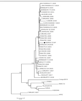
Figure 1: Phylogenetic analysis (Neighbor Joining-NJ) of the virus detected in patients. The percentage of replicate trees in which the associated taxa clustered together in the bootstrap test (1000 replicates) is shown next to the branches(28). The tree is drawn to scale, with branch lengths in the same units as those of the evolutionary distances used to infer the phyloge-netic tree. The evolutionary distances were computed using the Jukes-Cantor method(29)and are in the units of the number of base substitutions per site. The analysis involved 43 nucleotide sequences. There were a total of 440 positions in the final dataset. Evolutionary analyses were conducted in MEGA6(15). Black diamond indicates the CCHF virus detected in this study.
Table 1: Demographic characteristiscs, type and risk
classification of exposure and usage of PPEs of HCWs and whether ribavirin prophylaxis was administered or not.
F: Female, M: Male, PE: Physical Examination, ICU: Intensive Care Unit, ED: Emergency Department, IDD: Infectious Diseases Department
Virus Characterization
S-segment based pylogenetic analysis of the CCHFV (GeneBank Access No: KU695267) detected in this study revealed lack of variation among CCHF viruses reported in this study or viruses defined before. Nucleotide sequences homology was detected as 99% between viruses reported in this study and those reported previously (Figure 1).
Discussion
Nosocomial infection is an important mode of transmission in CCHF. Transmission to healthcare personnel was reported in 1976 in Pakistan for the first time and fatal cases have been identified since then(6). After the first report, nosocomial CCHF
cases have been reported as epidemics or sporadic cases in endemic areas(5, 7, 16-18). The most risky state
in nosocomial infection is injury by needle stick and CCHF was shown to have developed with a rate of 33% after needle injuries at a nosocomial epidemic which developed in Tygerberg hospital in South Africa(7). Infection may occur via the
aerosolization of infected secretions and mucosal contact, however airborne transmission has not been reported(3). In our study, CCHF developed in
none of 20 HCWs including the one whose skin had disrupted integrity and who had high-risk contact with an infected patient’s blood.
In previous molecular studies conducted with CCHF virus, it was found that the virus was divided in 7 clades according to genome sequence. These phylogenetic analyses, which are generally realized based on the S-RNA and L-RNA sequences, showed that 7 clades were related with geographi-cal logeographi-calizations. M-RNA segment sequence-based phylogenetic topology of the CCHF virus is differ-ent from the one which is based on the S-RNA and L-RNA segments. These analyses show that M-RNA segment reassortment events are more com-mon compared to the S- and L-RNA segments(19).
Reassortment events in this M-RNA segment are thought to be efficient in the increase in the virus’ virulence(20-22). Gn and glycoproteins encoded
by Gc M-RNA are responsible for attachment of the virus and induction of the antibodies which neutral-ize the virus(19, 23). It is expected that the
characteris-tics of attachment of the virus to the host cell are different in viruses related with different M-RNA sequence and thus the possibility of transmission of the virus is different in different genetic clades.
In a study in which the total rates of usage of PPE were evaluated in 190 HCWs who took care of patients with CCHF in a regional hospital in an endemic area in terms of CCHF in our country, the rate of usage of gowns was found to be 93.7%, the rate of usage of gloves was found to be 77.4% and the rate of usage of masks was found to be 38.9% and CCHF IgG was found to be positive in only 1 (0,53%) HCW(24).
A low level of nosocomial infection is an expected finding in this hospital which has substan-tially high compliance rates in terms of usage of PPE. Since our hospital was not located in an endemic area in terms of CCHF, the sensitivity of the HCW about this disease was low and thus the rates of usage of PPE was rather low except for gloves. However, it was interesting that clinical dis-ease and seroconversion did not develop (similar to the above-mentioned study) at the HCWs who had high-risk contact with CCHF patients despite com-pliance to the usage of PPE was low. This suggest-ed that the topotypes of the CCHF virus in different geographical regions might be different in terms of contagiousness.
A case series reported from Russia includes findings which support this view. In this study, it was reported that CCHF developed in 8 HCWs who had very low risk contact with a fatal index case and CCHF might have potentially been transmitted by airborne infection in two HCWs(4). However,
phylogenetic evaluation by viral sequencing could not be performed in this study reported from Russia. Thus, viral sequence analysis should be per-formed in studies in different regions to clarify if the modes and possibility of nosocomial transmis-sion in HCWs depending on virus topotype.
Although the efficacy of ribavirin prophylaxis used after risky contact with CCHF is still contro-versial, its use is recommended by World Health organization (WHO) in high risk states. In studies conducted with animal models, findings supported that ribavirin was efficient in preventing CCHF(25, 26).
However, its efficacy in humans has not yet been clearly elucidated(27).
In our study, 10 of the HCWs who had contact with CCHF received ribavirin, while 10 did not. There was no progression to a clinically apparent infection and no seroconversion in both groups. We think that lack of infection in the high-risk group, despite the presence of individuals who did not receive ribavirin, might be related to the virus hav-ing a phylogenetically low virulence.
The most important limitation of our study is stating that the possibility of transmission of the virus isolated was low based on indirect data. In addition, another factor, which decreased the possi-bility of transmission of the disease, might be the PPEs used by the HCWs (though the rates of usage were low). Despite all these limitations, our study has an important place in the literature, because it includes pioneering data about this subject and rec-ommends that phylogenetic analysis should be absolutely performed for the viruses isolated in studies related with nosocomial CCHF infection.
In conclusion, HCW who takes care of patients with a diagnosis of CCHF carries a high-risk and use of PPE is critical in prophylaxis. Standard, contact and droplet precautions are usual-ly considered sufficient when giving routine care to patients with CCHF, but airborne infection isolation precautions are recommended during procedures, which there is a possibility of formation of aerosols(17). Appropriate use of PPE appears to be a
significant factor in preventing nosocomial CCHF infection, but different genetic subtypes of the virus are thought to be important in terms of transmis-sion. This is related to the genetic variance between the viruses in different regions and thus with the variance in the virulence. The latter and the proper-ty of attachment to host cells appear to be signifi-cant factors for low or high possibility of transmis-sion. Phylogenetic analysis of the virus in studies in which nosocomial CCHF infection is evaluated will provide significant data in elucidating if the possi-bility of infection is different for different virus topotypes.
References
1) Charrel R, Attoui H, Butenko A, et al. Tick‐borne virus diseases of human interest in Europe. Clin Microbiol Infect 2004; 10(12): 1040-55.
2) Whitehouse CA. Crimean-Congo hemorrhagic fever. Antivir Res 2004; 64(3): 145-60.
3) Ergönül Ö. Crimean-Congo haemorrhagic fever. Lancet Infect Dis 2006; 6(4): 203-14.
4) Pshenichnaya NY, Nenadskaya SA. Probable Crimean-Congo hemorrhagic fever virus transmission occurred after aerosol-generating medical procedures in Russia: nosocomial cluster. Int J Infect Dis 2015; 33: 120-2. 5) Suleiman MNEH, Muscat-Baron J, Harries J, et al.
Congo/Crimean haemorrhagic fever in Dubai: an out-break at the Rashid Hospital. Lancet 1980; 316(8201): 939-41.
6) Burney M, Ghafoor A, Saleen M, et al. Nosocomial outbreak of viral hemorrhagic fever caused by Crimean Hemorrhagic fever-Congo virus in Pakistan, January 1976. Am J Trop Med Hyg 1980; 29(5): 941-7. 7) Van Eeden P, Joubert J, Van de Wal B. A nosocomial
outbreak of Crimean-Congo haemorrhagic fever at Tygerberg Hospital. Part I. Clinical features. S Afr Med J 1985; 68(10): 711-7.
8) Vorou R, Pierroutsakos IN, Maltezou HC. Crimean-Congo hemorrhagic fever. Curr Opin Infect Dis 2007; 20(5): 495-500.
9) Öncü S. Crimean-Congo hemorrhagic fever: an overview. Virol Sin 2013; 28(4): 193-201.
10) Ozkaya E, Dincer E, Carhan A, et al. Molecular epi-demiology of Crimean-Congo hemorrhagic fever virus in Turkey: occurrence of local topotype. Virus Res 2010; 149(1): 64-70.
11) Haferkamp S, Fernando L, Schwarz TF, et al. Intracellular localization of Crimean-Congo hemor-rhagic fever (CCHF) virus glycoproteins. Virol J 2005; 2(42): 31-8.
12) Centers for Disease Control (CDC). Management of patients with suspected viral hemorrhagic fever. MMWR Morb Mortal Wkly Rep 1988; 37: Suppl 3: 1-16.
13) Saitou N, Nei M. The neighbor-joining method: a new method for reconstructing phylogenetic trees. Mol Biol Evol 1987; 4(4): 406-25.
14) Tamura K, Nei M, Kumar S. Prospects for inferring very large phylogenies by using the neighbor-joining method. Proc Natl Acad Sci USA 2004; 101(30): 11030-5.
15) Tamura K, Stecher G, Peterson D, et al. MEGA6: mol-ecular evolutionary genetics analysis version 6.0. Mol Biol Evol 30(12):2725-9.
16) Tütüncü EE, Gurbuz Y, Ozturk B, et al. Crimean Congo haemorrhagic fever, precautions and ribavirin prophy-laxis: a case report. Scand J Infect Dis 2009; 41(5): 378-80.
17) Celikbas AK, Dokuzoğuz B, Baykam N, et al. Crimean-Congo hemorrhagic fever among health care workers, Turkey. Emerg Infect Dis 2014; 20(3): 477. 18. Guner R, Hasanoglu I, Tasyaran MA, et al. Is ribavirin
prophylaxis effective for nosocomial transmission of crimean-congo hemorrhagic Fever? Vector Borne Zoonotic Dis 2014; 14(8): 601-5.
19) Morikawa S, Saijo M, Kurane I. Recent progress in molecular biology of Crimean–Congo hemorrhagic fever. Comp. Immunol Microbiol Infec Dis 2007; 30(5): 375-89.
20) Papa A, Papadimitriou E, Boźović B, et al. Genetic characterization of the M RNA segment of a Balkan Crimean‐Congo hemorrhagic fever virus strain. J Med Virol 2005; 75(3): 466-9.
21) Ozdarendeli A, Aydin K, Tonbak S, et al. Genetic analysis of the M RNA segment of Crimean-Congo hemorrhagic fever virus strains in Turkey. Arch Virol 2008; 153(1): 37-44.
22) Papa A, Papadimitriou E, Christova I. The Bulgarian vaccine Crimean-Congo haemorrhagic fever virus strain. Scand J Infect Dis 2011; 43(3): 225-9.
23) Ahmed AA, McFalls JM, Hoffmann C, et al. Presence of broadly reactive and group-specific neutralizing epi-topes on newly described isolates of Crimean-Congo
hemorrhagic fever virus. J Gen Virol 2005; 86(12): 3327-36.
24) Gozel MG, Dokmetas I, Oztop AY, et al.. Recommended precaution procedures protect health-care workers from Crimean-Congo hemorrhagic fever virus. Int J Infect Dis 2013; 17(11): e1046-e50. 25) Tignor GH, Hanham CA. Ribavirin efficacy in an in
vivo model of Crimean-Congo hemorrhagic fever virus (CCHF) infection. Antivir Res 1993;22(4):309-25. 26) Bente DA, Alimonti JB, Shieh W-J, et al. Pathogenesis
and immune response of Crimean-Congo hemorrhagic fever virus in a STAT-1 knockout mouse model. J Virol 2010; 84(21): 11089-100.
27) Leblebicioglu H, Ozaras R, Irmak H, et al.. Crimean-Congo hemorrhagic fever in Turkey: current status and future challenges. Antivir Res 2016; 126: 21-34. 28) Felsenstein J. Confidence limits on phylogenies: an
approach using the bootstrap. Evolution 1985: 783-91. 29) Jukes T.H., Cantor C.R. Evolution of protein molecules
In: Munro HN,editor. Mammalian Protein Metabolism, Academic Press, New York,1969, pp. 21-132.
_________
Corresponding author FERITKUŞCU
Cukurova University Faculty of Medicine, Department of Infectious Diseases and Clinical Microbiology, Saricam
Adana (Turkey)