Brief Review of Right Aortic Arch with Aberrant
Left Subclavian Artery
Didem Melis Oztas, MD
1Muzaffer Umutlu, MD
2Melike Ertan, MD
3Metin Onur Beyaz, MD
4Serdar Badem, MD
5Ibrahim Erdinc, MD
6Mustafa Ozer Ulukan, MD
4Orcun Unal, MD
7Cenk Conkbayir, MD
8Ufuk Alpagut, MD
3Murat Ugurlucan, MD
41Department of Cardiovascular Surgery, Bagcilar Training and Research Hospital, Istanbul, Turkey
2Department of Radiology, Istanbul University Istanbul Medical Faculty, Istanbul, Turkey
3Department of Cardiovascular Surgery, Istanbul University Istanbul Medical Faculty, Istanbul, Turkey
4Department of Cardiovascular Surgery, Istanbul Medipol University Medical Faculty, Istanbul, Turkey
5Department of Cardiovascular Surgery, Bursa City Hospital, Bursa, Turkey 6Department of Cardiovascular Surgery, Izmir Bozyaka Training and
Research Hospital, Izmir, Turkey
7Department of Cardiovascular Surgery, Yedikule Chest Diseases and Thoracic Surgery Training and Research Hospital, Istanbul, Turkey 8Department of Cardiology, Near East University, Nicosia, Cyprus
AORTA 2019;7:179–180.
Address for correspondence Murat Ugurlucan, MD, TEM Avrupa Otoyolu Goztepe Cikisi, No: 1, 34214 Bagcilar, Istanbul, Turkey (e-mail: [email protected]).
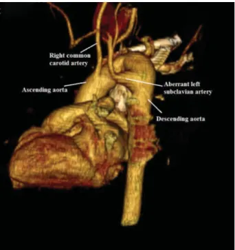
Right aortic arch (RAA) is a rare malformation and is reported at a range of 0.04 to 0.1% in autopsy series.1The anomaly occurs in embryonic life due to the persistence of the right-fourth aortic arch, while regressing the left-fourth arch between the left common carotid artery and the left subclavian artery.2 There are three types of the right-sided aortic arch as follows: Type I involves right aortic arch with mirror image branching, Type II involves right aortic arch with aberrant left subclavian artery, and Type III involves right-sided aortic arch with isolated left subclavian artery communicating with the pul-monary artery.3
Right aortic arch is generally an asymptomatic malfor-mation and diagnosed incidentally. In the Type II form, in which the left subclavian artery is aberrant (►Video 1), patients may present to the clinic with the symptoms occurring secondary to trachea or esophagus compression or aneurysm or dissection of the vessels.1Dysphagia and
dyspnea are usually the symptoms at the infant period, whereas atherosclerotic changes, dissection, or aneurysm may be seen in adulthood.1,3,4
Video 1
Computed tomography angiography video shows right aortic arch with aberrant left subclavian artery. Online content including video sequences viewable at: https:// www.thieme-connect.com/products/ejournals/html/ 10.1055/s-0039-3401999.
Computed tomography angiography is a valuable tool for the diagnosis because of the high resolution and the
Keywords
►
subclavian artery
►
aberrant
►
aortic arch
Abstract
Development anomalies of the aortic arch and its major branches are rare congenital
cardiovascular disorders. In this article, we present aberrant left subclavian artery
associated with right aortic arch.
received July 13, 2018
accepted after revision November 2, 2019
DOIhttps://doi.org/ 10.1055/s-0039-3401999. ISSN 2325-4637.
Copyright © 2019 by Thieme Medical Publishers, Inc., 333 Seventh Avenue, New York, NY 10001, USA.
Tel: +1(212) 760-0888. THIEME
Images in Aortic Disease
179
Published online: 2020-02-17speed of scanning (►Figs. 1 and 2). Also, magnetic reso-nance imaging is another option which may be used for diagnosis.5
The complications of this pathology include aneurysm formation and dissection which may be secondary to ath-erosclerosis in latter ages, as well as recurrent lower respi-ratory tract infections, and growth retardation in the early years of childhood3; hence, these patients should be fol-lowed-up lifelong.
In conclusion, the symptoms are the most important determinants for the treatment of the patients with RAA. Careful follow-up is necessary for the prevention of fatal complications.
Funding
None.
Conflict of Interest
None.
Acknowledgments
None.
References
1 Barr JG, Sepehripour AH, Jarral OA, et al. A review of the surgical management of right-sided aortic arch aneurysms. Interact Car-diovasc Thorac Surg 2016;23(01):156–162
2 Salanitri J. MR angiography of aberrant left subclavian artery arising from right-sided thoracic aortic arch. Br J Radiol 2005;78 (934):961–966
3 Bhatt TC, Muralidharan CG, Singh G, Jain NK. Kommerell’s diver-ticulum: A rare aortic arch anomaly. Med J Armed Forces India 2016;72(01, Suppl 1):S80–S83
4 Mubarak MY, Kamarul AT, Noordini MD. Right-sided aortic arch with aberrant left subclavian artery from Kommerell’s diverticu-lum. Iran J Radiol 2011;8(02):103–106
5 Türkvatan A, Büyükbayraktar FG, Olçer T, Cumhur T. Multidetector computed tomographic angiography of aberrant subclavian arter-ies. Vasc Med 2009;14(01):5–11
Fig. 1 Right aortic arch with aberrant left subclavian artery.
Fig. 2 3D computed tomography angiography image. Right aortic arch with aberrant left subclavian artery. 3D, three-dimensional.
AORTA Vol. 7 No. 6/2019