Introduction
Helicobacter pylori infection is one of the most common chronic infections worldwide and the relationship between H. pylori infection and diabetes patients has been investigated by several authors.[1‑4] The high prevalence of
H. pylori infection among diabetes mellitus and metabolic syndrome patients has been documented in detailed.[5‑9] More
recently, it has been reported that by tracing anti‑H. pylori antibodies in patients with diabetes mellitus and the occurrence of symptoms such as digestive problems in >75% of these patients, it can be concluded that there is a relationship between this bacterium and type 2 diabetes mellitus (T2DM).[10] Furthermore, several
studies reported that the prevalence of H. pylori infection was found to be a
Address for correspondence: Prof. Abdulbari Bener, Department of Biostatistics and Medical Informatics, Cerrahpaşa Faculty of Medicine, Istanbul University Cerrahpaşa and Medipol International School of Medicine, Istanbul Medipol University, 34098 Istanbul, Turkey.
E‑mail: abdulbari.bener@ istanbul.edu.tr
Abstract
Background: Several conducted studies have reported a higher and more frequent Helicobacter pylori infection rate in type 2 diabetes mellitus (T2DM). The aim of this study was to investigate the prevalence and its association between H. pylori infection and T2DM. Materials and Methods: A case and control study was conducted based on 529 T2DM patients and 529 control. H. pylori was assessed by Serum anti‑H. pylori immunoglobulin G (IgG) and IgA. Furthermore, patients were investigated for fasting blood glucose (FBG) levels, glycosylated hemoglobin (HbA1c), serum cholesterol, and other biochemistry parameters. Results: The findings showed a positive significantly higher antibody titer for H. pylori infection (IgA > 250) in diabetic patients (50.7%) compared to controls (38.2%) (P < 0.001). Similarly, H. pylori infection for IgG > 300 titer was higher in T2DM patients (73.5%) compared to controls 61.8%) (P < 0.001). Further, the mean values were statistically significant diabetes with H. pylori infection for IgG > 300 titer and IgA > 250 titer, regarding Vitamin D, HbA1C (P < 0.001), FBG, calcium, creatinine, total cholesterol, LHDL, triglyceride levels, uric acid, bilirubin, thyroid‑stimulating hormone (TSH), and systolic and diastolic blood pressure. The diabetic patients showed higher prevalence rate of symptoms than controls included: hypertension (14.3%), vomiting (15.5%), muscular symptoms (35.2%), bloating/distension (13.2%), abdominal pain (17%), nausea (9.6%), anemia (17%), kidneys (20.8%), chronic bronchitis (14.7%), gastrointestinal (23.8%), and diarrhea (20.4%). Conclusions: The current study revealed that H. pylori infections were significantly higher in diabetic patients compared to controls. Furthermore, T2DM patients infected with H. pylori positive reported a higher prevalence rate of symptoms than H. pylori negative.
Keywords: Diabetes, glycosylated hemoglobin, Helicobacter pylori infection, immunoglobulin, immunoglobulin A, immunoglobulin G
Prevalence of Helicobacter pylori Infection among Type 2 Diabetes
Mellitus
Original Article
Abdulbari Bener1,2,3,
Ahmet Faruk Ağan4,
Abdulla O. A.
A. Al‑hamaq5,
Cem Cahit Barisik3,
Mustafa Öztürk6,
Abdulkadir Ömer6
From the 1Department of
Biostatistics and Medical Informatics, Cerrahpaşa Faculty of Medicine, Istanbul University, Departments of 3Raiology and
Pathology, 4Gastroenterology
and 6Endocrinology, Medipol
International School of Medicine, Istanbul Medipol University, Istanbul, Turkey,
2Department of Evidence
for Population Health Unit, Department of Epidemiology and Health Sciences, The University of Manchester, Manchester, UK, 5Qatar
Diabetic Associations and Qatar Foundation, Doha, Qatar
How to cite this article: Bener A, Ağan AF, Al-hamaq AO, Barisik CC, Öztürk M, Ömer A. Prevalence of Helicobacter pylori infection among type
2 diabetes mellitus. Adv Biomed Res 2020;9:27. Received: 30 November 2019
Revised: 09 March 2020 Accepted: 14 April 2020 Published: 27 July 2020
significantly higher risk in people with diabetes than in controls.[3,5‑11]
The objective of the current study is to determine the prevalence and its association of H. pylori infection with T2DM in the Turkish population.
Materials and Methods
Study designThis case and control study consisted of patients aged between 30 and 70 who visited diabetes, endocrinology, gastroenterology, or outpatient clinics. The sample size was based on matched 529 T2DM patients and 529 controls. Written informed consent was obtained from all individuals prior to
enrolling in the study. Access this article online
Website: www.advbiores.net DOI: 10.4103/abr.abr_248_19
Quick Response Code:
This is an open access journal, and articles are distributed under the terms of the Creative Commons Attribution‑NonCommercial‑ShareAlike 4.0 License, which allows others to remix, tweak, and build upon the work non‑commercially, as long as appropriate credit is given and the new creations are licensed under the identical terms.
Selection of type 2 diabetic mellitus patients
Case patients were considered to have T2DM if they had a history of DM and were currently taking any oral medications for diabetes. DM was defined as individuals with the fasting plasma glucose ≥7.0 mmol/l or 75‑g oral glucose tolerance test with 2‑h plasma glucose ≥11.1 mmol/l or glycosylated hemoglobin (HbA1c) >6.5%[12,13]
and by the International Diabetes Federation.[14]
Selection of controls
The controls aged 30–70 years were identified from a community consisting of a sample of 529 controls who visited the outpatient clinics for any reason other than T2DM and were selected randomly.
Biochemistry data
These individuals were also investigated for fasting blood glucose levels (FBG), HbA1c, serum cholesterol, triglycerides, high‑density lipoprotein (HDL), low‑density lipoprotein (LDL), urea, creatinine, and the presence of other comorbid conditions.
Helicobacter pylori serology
Blood samples were taken from the peripheral veins of the individuals. The serum specimens were obtained from all cases and controls for H. pylori serology test from the participants. Immunoglobulin G (IgG) and IgA classes of anti‑endomysial antibodies (EMAs) were measured with enzyme‑linked immunosorbent assays (ELISAs) (CeliAK EmA human IgG and IgA, generic assays (GA) GmbH, Dahlewitz, Germany). H. pylori was assessed by measuring IgG and IgA among T2DM patients and the control group.[15] A individual was considered to be positive
for H. pylori if IgG and IgA anti‑H. pylori antibody titers were >300 and >250, respectively.[5]
The SPSS computer software (IBM SPSS Statistics for Windows, Version 22.0. Armonk, NY: IBM Corp) was used for the statistical analysis. Student t‑test was used to ascertain the significance of differences between the mean values of two continuous variables. Chi‑square and Fisher exact tests were performed to test for differences in the proportions of categorical variables between two or more groups. Odds ratio and their 95% confidence intervals were calculated using Mantel–Haenszel test. One‑way analysis of variance was employed for the comparison of several group means. The cutoff value for determining significance was chosen as 0.05.
Results
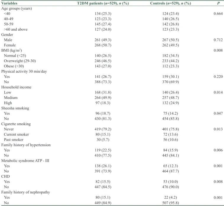
The demographic and clinical characteristics of investigated case patients and controls are shown in Table 1. The findings showed significant differences between T2DM patients compared with controls with respect to their age in years, body mass index (kg/m2), household income, sheesha
smoking, smoking habit, family history of hypertension,
metabolic syndrome, coronary heart disease (CHD), and nephropathy.
In the present study, over 60% of the patients had received H. pylori eradication therapy and about 22% had active infections. Most patients had routine follow‑up as recommended by physicians.
Table 2 gives the magnitude of H. pylori infection among T2DM and controls. A positive antibody titer for H. pylori infection (IgA > 250) was found significantly higher in T2DM (diabetes [50.7%] vs. control [38.2%]), (P < 0.001). Similarly, H. pylori infection for IgG > 300 titer was higher in T2DM patients (73.5%) compared to controls subjects (61.8%) (P < 0.001).
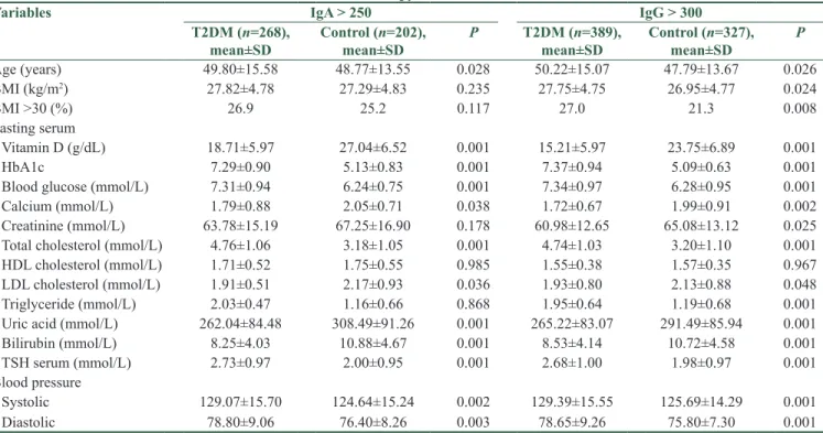
Table 3 presents the clinical and biochemistry characteristics of T2DM and controls with H. pylori infection. As shown in Table 3, the mean values were significantly higher in T2DM with H. pylori infection for IgG > 300 titer, regarding Vitamin D (P < 0.001), HbA1C (P < 0.001), FBG (P < 0.001), calcium (P = 0.002), creatinine (P = 0.025), total cholesterol (P < 0.001), LHDL (P = 0.048), triglyceride levels (P < 0.001), uric acid (P < 0.001), bilirubin (P = 0.015), TSH (P < 0.001), systolic blood pressure (P < 0.001), and diastolic (P < 0.001) blood pressure. Similarly, a positive antibody titer for H. pylori infection (IgA > 250) was found significantly higher in diabetic patients compared to controls.
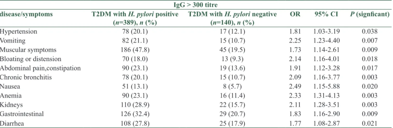
Table 4 shows symptoms and diseases among T2DM patients with H. pylori infection. The T2DM patients infected with H. pylori positive reported statistically significant higher prevalence rate of symptoms than H. pylori negative: hypertension (20.1% vs. 12.1%), vomiting (21.1% vs. 10.7%), muscular symptoms (47.8% vs. 19.5%), bloating/distension (18.0% vs. 9.3%), abdominal pain (23.1% vs. 13.6), chronic bronchitis (20.1% vs. 10.7%), nausea (13.1% vs. 5.7%), anemia (23.1% vs. 11.4%), kidneys (28.9% vs. 15.7%), gastrointestinal (GI) (32.4% vs. 20.7%), and diarrhea (27.8% vs. 17.9%).
Discussion
Our objective was to explore the association between H. pylori infection and T2DM. The current study revealed that H. pylori prevalence was significantly higher in T2DM patients than in controls (50.7% vs. 38.2% for IgA > 250 titer and 73.5% vs. 61.8% for IgG > 300 titer). This is consistent with the previously reported studies.[4‑10,16‑21] Most recently, a meta‑analysis
suggested a trend toward more frequent H. pylori infections in T2DM patients.[3] Meanwhile, the impact of H. pylori
and T2DM was explored in Bener et al.’s study[5] which
stated that the prevalence of H. pylori infection rate in T2DM patients revealed higher than in controls. In a study conducted in Italy, H. pylori infection was detected in 69% of the patients with diabetes.[22] This is confirmative with
Table 1: Sociodemographic and clinical characteristics of studied type 2 diabetes mellitus patients and controls (n=1058)
Variables T2DM patients (n=529), n (%) Controls (n=529), n (%) P
Age groups (years)
<40 134 (25.3) 124 (23.4) 0.664 40‑49 123 (23.3) 140 (26.5) 50‑59 145 (27.4) 142 (26.8) >60 and above 127 (24.0) 123 (23.3) Gender Male 261 (49.3) 267 (50.5) 0.712 Female 268 (50.7) 262 (49.5) BMI (kg/m2) 0.008 Normal (<25) 140 (26.5) 182 (34.5) Overweight (29‑30) 246 (46.5) 233 (44.2) Obese (>30) 143 (27.0) 112 (23.3)
Physical activity 30 min/day
Yes 141 (26.7) 159 (30.1) 0.220 No 388 (73.3) 370 (69.9) Household income Low 168 (31.8) 140 (26.4) 0.014 Medium 264 (49.9) 257 (48.7) High 97 (18.3) 132 (24.9) Sheesha smoking Yes 96 (18.7) 75 (14.2) 0.047 No 430 (81.3) 454 (85.8) Cigarette smoking Never 419 (79.2) 401 (75.8) 0.013 Current smoker 80 (15.1) 72 (13.6) Past smoker 30 (5.7) 56 (10.6)
Family history of hypertension
Yes 119 (22.5) 84 (15.9) 0.006
No 410 (77.5) 445 (84.1)
Metabolic syndrome ATP ‑ III
Yes 138 (26.1) 65 (12.3) 0.001
No 391 (73.9) 464 (87.7)
CHD
Yes 82 (15.5) 53 (10.0) 0.008
No 447 (84.5) 476 (90.0)
Family history of nephropathy
Yes 80 (15.1) 22 (4.2) 0.001
No 449 (84.9) 507 (95.8)
CHD: Coronary heart disease, BMI: Body mass index, T2DM: Type 2 diabetes mellitus, ATP: Acute thrombocytopenic purpura The association of H. pylori infection is made to a number
of gastrointestinal and extra‑GI diseases which has changed the approach for diagnosis among the various medical fields, and in most studies, H. pylori infection has been linked with T2DM.[1‑10,16] According to a most recent study
in Pakistan, it was observed that H. pylori infection was commonly seen among type 2 diabetic group (79%) in comparison to nondiabetic group (21%). This is consistent with the present study. A significant association implied that there stands an association between H. pylori infection and diabetes. The results are in line with another study conducted in Pakistan where hyperglycemia due to diabetes was regarded as a predisposing factor H. pylori
colonization and reported that 73% of the patients having H. pylori infection were diabetic and 51% were nondiabetic.[1,16] Furthermore, another study performed in
Africa reported that 88% of the diabetic and 67% of the nondiabetic patients were found to have a positive status for anti‑H. pylori antibodies.[18] This is again confirmative
with the current study.
In fact, worldwide, about over 4 million death patients had T2DM as well as many previous ones showed a high correlation between H. pylori infection and T2DM.[2‑10,18]
Many important factors are considered in the development of H. pylori‑associated gastroduodenal diseases, including
some risk factors such as environmental‑related factors, hygiene, age, gender, and genetic susceptibility.[5]
H. pylori infection plays an important role in the development of GI complications and has a significant role in systemic inflammation. It has some extraGI manifestations like endocrine diseases. In a study conducted in Iran,[8] the prevalence of H. pylori seropositive
was 65.9% in diabetic versus 50.5% in controls, and the difference was statistically significant. Similarly, in another study in Iran, the rate of H. pylori was significantly higher in diabetic patients compared to controls, 55.8% versus 44.2%, respectively.[2] The prevalence of H. pylori
infection in T2DM patients with obesity has been higher than the control group in Qatar (24% versus 27.5%). This is consistent with the current study. Among the patients referring to diabetes clinics, as many as 75% of them report significant GI symptoms.[18‑20] More recently, a
study conducted on diabetic patients in Iran[2] showed that
H. pylori infection increases the prevalence of metabolic syndrome through an increase in insulin resistance, this consistent with the current study.
A study, which has been performed in India,[4] reported
that CHD was more prevalent in people with diabetes with H. pylori infection than those without H. pylori (57%).
Table 2: Prevalence of Helicobacter pylori infection in the studied type 2 diabetes mellitus and helthy controls
Charecteristics T2DM patients (n=529), n (%) Control subject (n=529), n (%) P
IgA > 250 titre
H. pylori positive 268 (50.7) 202 (38.2) <0.001
H. pylori negative 261 (49.3) 361 (61.8)
H. pylori positive by gender
Male 173 (64.5) 91 (45.0) 0.001
Female 95 (35.5) 111 (55.0) 0.001
IgG > 300 titre
H. pylori positive 389 (73.5) 327 (61.8) 0.001
H. pylori negative 140 (26.5) 202 (38.2)
H. pylori positive by gender
Male 216 (55.5) 166 (50.8) 0.209
Female 173 (44.4) 161 (49.2) 0.200
H. pylori: Helicobacter pylori, T2DM: Type 2 diabetes mellitus
Table 3: The baseline clinical and biochemistry charecteristics of type 2 diabetes mellitus and controls with Helicobacter pylori infection
Variables IgA > 250 IgG > 300
T2DM (n=268),
mean±SD Control (n=202), mean±SD P T2DM (n=389), mean±SD Control (n=327), mean±SD P
Age (years) 49.80±15.58 48.77±13.55 0.028 50.22±15.07 47.79±13.67 0.026 BMI (kg/m2) 27.82±4.78 27.29±4.83 0.235 27.75±4.75 26.95±4.77 0.024 BMI >30 (%) 26.9 25.2 0.117 27.0 21.3 0.008 Fasting serum Vitamin D (g/dL) 18.71±5.97 27.04±6.52 0.001 15.21±5.97 23.75±6.89 0.001 HbA1c 7.29±0.90 5.13±0.83 0.001 7.37±0.94 5.09±0.63 0.001
Blood glucose (mmol/L) 7.31±0.94 6.24±0.75 0.001 7.34±0.97 6.28±0.95 0.001
Calcium (mmol/L) 1.79±0.88 2.05±0.71 0.038 1.72±0.67 1.99±0.91 0.002
Creatinine (mmol/L) 63.78±15.19 67.25±16.90 0.178 60.98±12.65 65.08±13.12 0.025 Total cholesterol (mmol/L) 4.76±1.06 3.18±1.05 0.001 4.74±1.03 3.20±1.10 0.001
HDL cholesterol (mmol/L) 1.71±0.52 1.75±0.55 0.985 1.55±0.38 1.57±0.35 0.967
LDL cholesterol (mmol/L) 1.91±0.51 2.17±0.93 0.036 1.93±0.80 2.13±0.88 0.048
Triglyceride (mmol/L) 2.03±0.47 1.16±0.66 0.868 1.95±0.64 1.19±0.68 0.001
Uric acid (mmol/L) 262.04±84.48 308.49±91.26 0.001 265.22±83.07 291.49±85.94 0.001
Bilirubin (mmol/L) 8.25±4.03 10.88±4.67 0.001 8.53±4.14 10.72±4.58 0.001
TSH serum (mmol/L) 2.73±0.97 2.00±0.95 0.001 2.68±1.00 1.98±0.97 0.001
Blood pressure
Systolic 129.07±15.70 124.64±15.24 0.002 129.39±15.55 125.69±14.29 0.001
Diastolic 78.80±9.06 76.40±8.26 0.003 78.65±9.26 75.80±7.30 0.001
BMI: Body mass index, HbA1c: Glycosylated hemoglobin, HDL: High‑density lipoprotein, LDL: Low‑density lipoprotein, TSH: Thyroid‑ stimulating hormone, T2DM: Type 2 diabetes mellitus, SD: Standard deviation
Table 4: The comparison reported symptoms and diseases among between the parameters of two groups of seropositive diabetic patients with seronegative diabetic patients (n=529)
IgG > 300 titre
disease/symptoms T2DM with H. pylori positive
(n=389), n (%) T2DM with H. pylori negative (n=140), n (%) OR 95% CI P (signficant)
Hypertension 78 (20.1) 17 (12.1) 1.81 1.03‑3.19 0.038 Vomiting 82 (21.1) 15 (10.7) 2.25 1.23‑4.40 0.007 Muscular symptoms 186 (47.8) 45 (19.5) 1.73 1.14‑2.61 0.009 Bloating or distension 70 (18.0) 13 (9.3) 2.14 1.16‑4.01 0.018 Abdominal pain,constipation 90 (23.1) 19 (13.6) 1.91 1.12‑3.28 0.017 Chronic bronchitis 78 (20.1) 15 (10.7) 2.09 1.16‑3.77 0.003 Nausea 51 (13.1) 8 (5.7) 2.49 1.15‑5.88 0.020 Anemia 90 (23.1) 16 (11.4) 2.33 1.31‑4.13 0.003 Kidneys 110 (28.9) 22 (15.7) 2.11 1.28‑3.51 0.003 Gastrointestinal 126 (32.4) 29 (20.7) 1.83 1.16‑2.90 0.009 Diarrhea 108 (27.8) 25 (17.9) 1.77 1.08‑2.87 0.021
H. pylori: Helicobacter pylori, T2DM: Type 2 diabetes mellitus, OR: Odds ratio, CI: Confidence interval Furthermore studies in Pakistan,[1,16] in Qatar,[5] in Iran,[2,6,8,10]
in Italy[21] and in China,[19,22] revealed significantly high risk
H. Pylori of infection among Diabetes than in control. This is consistent with the current study, in which H. pylori infection was found a statistically significantly high prevalence rate of T2DM compared to the control group.
Conclusions
The current study suggests that H. pylori infection is one of the risk factors that may be considered as a marker in the evaluation of diabetic patients. This study revealed that H. pylori infections were higher in diabetic patients compared to controls. The T2DM patients infected with H. pylori positive reported a higher prevalence rate of symptoms than H. pylori negative.
Acknowledgment
This work was generously supported and funded by the Qatar Diabetes Association, Qatar Foundation. The authors would like to thank the Istanbul Medipol University for their support and ethical approval (Research Protocol and IRB# 10840098‑604.01.01‑E.8421 and Research Protool and IRB# 10840098‑604.01.01‑E.3193).
Financial support and sponsorship
Nil.
Conflicts of interest
There are no conflicts of interest.
References
1. Devrajani BR, Shah SZ, Soomro AA, Devrajani T. Type 2 diabetes mellitus: A risk factor for Helicobacter pylori infection: a hospital based case‑control study. Int J Diabetes Dev Ctries 2010;30:22‑6.
2. Vafaeimanesh J, Parham M, Seyyedmajidi M, Bagherzadeh M. Helicobacter pylori Infection and Insulin Resistance in Diabetic and Nondiabetic Population. ScientificWorldJournal. 2014;2014:391250.
3. Zhou X, Zhang C, Wu J, Zhang G. Association between Helicobacter pylori infection and diabetes mellitus: A meta‑analysis of observational studies. Diabetes res Clin Pract 2013;99:200‑8.
4. Agrawal RP, Sharma R, Garg D, Pokharna R, Kochar DK, Kothari RP. role of Helicobacter pylori in causation of diabetic gastropathies and non‑gastrointestinal complications in type 2 diabetes. J Indian Med Assoc 2010;108:140‑3.
5. Bener A, Micallef R, Afifi M, Derbala M, Al‑Mulla HM, Usmani MA. Association between type 2 diabetes mellitus and Helicobacter pylori infection. Turk J Gastroenterol 2007;18:225‑9.
6. Jafarzadeh A, Rezayati MT, Nemati M. Helicobacter pylori seropositivity in patients with type 2 diabetes mellitus in south‑east of Iran. Acta Med Iran 2013;51:892‑6.
7. Marrollo M, Latella G, Melideo D, Storelli E, Iannarelli R, Stornelli P, et al. Increased prevalence of Helicobacter pylori in patients with diabetes mellitus. Dig Liver Dis 2001;33:21‑9. 8. Vafaeimanesh J, Parham M, Bagherzadeh M. Helicobacter
pylori infection prevalence: Is it different in diabetics and non‑diabetics? Indian J Endocrinol Metab 2015;19:364‑8. 9. Jeon CY, Haan MN, Cheng C, Clayton ER, Mayeda ER,
Miller JW, et al. Helicobacter pylori infection is associated with an increased rate of diabetes. Diabetes Care 2012;35:520‑5. 10. Hosseininasab Nodoushan SA, Nabavi A. The interaction of
Helicobacter pylori infection and type 2 diabetes mellitus. Adv Biomed Res 2019;8:15.
11. Fernandini‑Paredes GG, Mezones‑Holguin E, Vargas‑Gonzales R, Pozo‑Briceño E, Rodriguez‑Morales AJ. In patients with type 2 diabetes mellitus, are glycosylated hemoglobin levels higher for those with Helicobacter pylori infection than those without infection? Clin Infect Dis 2008;47:144‑6.
12. Alberti KG, Eckel RH, Grundy SM, Zimmet PZ, Cleeman JI, Donato KA, et al. Harmonizing the metabolic syndrome: A joint interim statement of the international diabetes federation task force on epidemiology and prevention; national heart, lung, and blood institute; american heart association; world heart federation; international atherosclerosis society; and international association for the study of obesity. Circulation 2009;120:1640‑5. 13. Alberti KG, Zimmet P, Shaw J. Metabolic syndrome‑a new
world‑wide definition. A consensus statement from the international diabetes federation. Diabet Med 2006;23:469‑80. 14. Andersen LP, Espersen F, Souckova A, Sedlackova M, Soucek A.
isolation and preliminary evaluation of a low‑molecular‑mass antigen preparation for improved detection of Helicobacter pylori immunoglobulin g antibodies. clin diagn lab immunol 1995;2:156‑9.
15. Hafiz QM, Ikram O, Zia MT, Theba FK, Ikram N, Tariq A. Helicobacter pylori infection among type 2 diabetics: A case control study. Int J Res Med Sci 2020;8:1047‑50.
16. Ko GT, Chan FK, Chan WB, Sung JJ, Tsoi CL, To KF, et al. Helicobacter pylori infection in chinese subjects with type 2 diabetes. Endocr Res 2001;27:171‑7.
17. Ebule IA, Djune FA, Njeambosay BA, Doh GN, Metaghue G. Association of Helicobacter pylori infection and diabetes mellitus type 2 subjects in yaounde cameroon using a panel of serum biomarkers (PGII, HPIGG): A case control study.J Clin Gastroenterol Treat 2017;3:53‑8.
18. Wan Z, Song l, Hu L, Hu M, Lei X, Huang Y, et al. Helicobacter pylori infection is associated with diabetes among chinese adults.
J Diabetes Investig 2020;11:199‑205.
19. Cheng KP, Yang YJ, Hung HC, Lin CH, Wu CT, Hung MH, et al. Helicobacter pylori eradication improves glycemic control in type 2 diabetes patients with asymptomatic active Helicobacter pylori infection. J Diabetes Investig 2019;10:1092‑101.
20. Senturk O, Canturk Z, Cetinarslan B, Ercin C, Hulagu S, Canturk NZ. Prevalence and comparisons of five different diagnostic methods for Helicobacter pylori in diabetic patients. Endocr Res 2001;27:179‑89.
21. Quatrini M, Boarino V, Ghidoni A, Baldassarri AR, Bianchi PA, Bardella MT. Helicobacter pylori prevalence in patients with diabetes and its relationship to dyspeptic symptoms. J Clin Gastroenterol 2001;32:215‑7.
22. So WY, Tong PC, Ko GT, Ma RC, Ozaki R, Kong AP, et al. Low plasma adiponectin level, white blood cell count and Helicobacter pylori titre independently predict abnormal pancreatic beta‑cell function. Diabetes Res Clin Pract 2009;86:89‑95.
Submission of Manuscript for publication
Dear Sir,
We intend to publish an article entitled
__________________________________________________________________________________
in your journal.
On behalf of all the contributors I will act and guarantor and will correspond with the journal from this
point onward.
Prior presentation of the data reported in this manuscript:
Organisation
Place
Date
We have done sufficient work in the field to justify authorship for this manuscript.
We hereby transfer, assign, or otherwise convey all copyright ownership, including any and all rights
incidental thereto, exclusively to the journal, in the event that such work is published by the journal.
Thank you,
Yours’ sincerely,
Name of corresponding contributor
Signature
Type of manuscript:
Running title:
Contributors:
First name Middle name initial Last name Highest academic degreeNames of departments and institutions (including city and state)
Email addresses
1
2
3
4
5
6
Address:
Phone numbers:
Facsimile numbers:
E-mail address:
Total number of pages:
Total number of tables:
Total number of figures:
Total number of supplementary files:
Word counts: For abstract:
For the text:
Acknowledgement:
Conflict of interest:
Contribution details (to be ticked marked as applicable):
Contributor 1 Contributor 2 Contributor 3 Contributor 4 Contributor 5 Contributor 6