Clinical Investigation
Native Electrocardiographic QRS Duration after Cardiac
Resynchronization Therapy: The Impact on Clinical
Outcomes and Prognosis
OGUZ KARACA, MD,1BEYTULLAH CAKAL, MD,1MEHMET ONUR OMAYGENC, MD,1HACI MURAT GUNES, MD,1 SINEM DENIZ CAKAL, MD,1FILIZ KIZILIRMAK, MD,1TAYYAR GOKDENIZ, MD,1IRFAN BARUTCU, MD,1
BILAL BOZTOSUN, MD,1AND FETHI KILICASLAN, MD2 Istanbul, Turkey
ABSTRACT
Background: We investigated whether reversed electrical remodeling (RER), defined as narrowing of the native electrocardiographic QRS duration after cardiac resynchronization therapy (CRT), might predict prog-nosis and improvement in echocardiographic outcomes.
Methods and Results: A total of 110 CRT recipients were retrospectively analyzed for the end points of death and hospitalization during 18± 3 months. Native QRS durations were recorded at baseline and 6 months after CRT (when pacing was switched off to obtain an electrocardiogram) to determine RER. CRT re-sponse and mitral regurgitation (MR) improvement were defined as≥15% reduction in left ventricular end-systolic volume and absolute reduction in regurgitant volume (RegV) at 6 months, respectively. Overall, 48 patients (44%) had RER, which was associated with functional improvement (77% vs 34%; P< .001) and CRT response (81% vs 52%; P< .001) compared with those without RER. The change in the intrinsic QRS duration correlated with the reduction in RegV (r= 0.51; P < .001) and in tenting area (r = 0.34; P < .001). RER was a predictor of MR improvement (P= .023), survival (P = .043), and event-free survival (P = .028) according to multivariate analyses.
Conclusions: Narrowing of the intrinsic QRS duration is associated with functional and echocardiographic CRT response, reduction in MR, and favorable prognosis after CRT. (J Cardiac Fail 2016;22:772–780) Key Words: Native QRS duration, reversed electrical remodeling, cardiac resynchronization therapy response, mitral regurgitation, prognosis.
Cardiac resynchronization therapy (CRT) is an estab-lished therapeutic option for systolic heart failure concomitant with electrical dyssynchrony.1–4The extent of baseline
elec-trical dyssynchrony is currently determined by prolongation of the QRS duration with a left bundle branch block (LBBB) morphology, which is a well known predictor of CRT
response.3,4 Restoration of the dyssynchronous conduction
within the His-Purkinje system by means of biventricular pacing has been proposed as the mechanism of action that correlates with the clinical and echocardiographic improve-ments after CRT.4,5Narrowing of the native QRS duration as
a marker of reversed electrical remodeling (RER) after CRT has been shown in previous studies to correlate with favor-able structural changes and CRT response.5,6
Mitral regurgitation (MR), a significant prognostic marker in patients with heart failure, has been shown to be reduced by CRT, although the precise mechanisms are not fully understood.7–9
The impact of native QRS narrowing on the degree of MR and on the geometric changes of the mitral valve following CRT have not been addressed previously. The objectives of the present study were: 1) to evaluate the rela-tionship of CRT-induced RER to the improvement in functional status, CRT response, MR, and mitral geometry; and 2) to test the hypothesis that narrowing of the native QRS
From the1Department of Cardiology, Medipol University Faculty of Med-icine, Istanbul, Turkey and2Department of Cardiac Electrophysiology, Medipol University Faculty of Medicine, Istanbul, Turkey.
Manuscript received September 9, 2015; revised manuscript received March 9, 2016; revised manuscript accepted April 1, 2016.
Reprint requests: Oguz Karaca, MD, Cardiology Department, Medipol University Faculty of Medicine, TEM Otoyolu Goztepe Cikisi No 1, 34214 Bagcilar, Istanbul, Turkey. Tel:+90 505 355 76 00; Fax: +90 212 460 70 70. E-mail:oguzkaraca@hotmail.com.
Funding: None declared.
See page 779 for disclosure information. 1071-9164/$ - see front matter © 2016 Elsevier Inc. All rights reserved.
http://dx.doi.org/10.1016/j.cardfail.2016.04.001
duration might predict prognostic outcomes such as reduc-tion in MR severity and decrease in death and hospitalizareduc-tion during follow-up.
Methods Patient Selection and Study Design
A total of 172 patients receiving a biventricular device with or without a defibrillator (CRT-D or CRT-P) were retrospec-tively analyzed in the study. Compatibly with current guidelines,10
all patients had a left ventricular ejection frac-tion (LVEF)≤35%, an increased QRS duration (≥120 ms), and symptomatic heart failure (New York Heart Association [NYHA] functional class II–IV) despite optimal medical therapy. Patients with right ventricular pacing upgraded to CRT and those with pacemaker dependency were excluded. Pa-tients with organic MR, defined as moderate to severe annular calcifications, rheumatic valve disease, mitral valve pro-lapse with or without flail leaflet/chordae, or a history of mitral valve repair or replacement, were excluded.11Patients with
inadequate image quality to calculate left ventricular (LV) volumes (n= 12) and those with trivial MR that precluded the quantification of MR (n= 44) were also excluded from the analyses. Patients who failed to continue follow-up visits (n= 6) were excluded, leading to a final study population of 110 CRT recipients with MR. The study protocol was ap-proved by the local Ethics Committee, and written informed consent was obtained from every participant.
Electrocardiographic Recordings
Baseline, post-implantation (paced), and follow-up intrinsic (when the CRT device was switched off) 12-lead elec-trocardiographic (ECG) tracings were recorded on a chart paper at a speed of 50 mm/s with a gain setting of 10 mm/mV. QRS duration was defined as the widest interval in any of the 12 leads. QRS duration was manually measured and double-checked with the computer output. Baseline QRS morphologies were recorded as either LBBB or non-LBBB based on well defined criteria.12
Paced QRS intervals were calculated after echocardiographic device optimization, which was per-formed during the hospital stay. At 6 months of follow-up, CRT devices were switched off for 10 seconds to assess the native conduction. The intrinsic QRS duration on the follow-up ECG was included in the study data to determine RER, which was defined as narrowing of the native QRS duration after CRT.
Device Implantation and Programming
Transvenous approach via the left subclavian vein was used to implant CRT-D or CRT-P devices under local anesthesia in the vast majority of patients. The right atrial lead was located in the right atrial free wall (in patients with sinus rhythm) and the right ventricular lead was located in the right ven-tricular apex or the outflow tract according to the operator’s choice. Following coronary sinus angiography, the LV lead
was inserted preferably in the posterolateral vein as recommended.13In case of anatomic difficulties in
implant-ing the LV lead in the optimal position (in 12 patients), minimal invasive surgical approach was preferred to implant an epi-cardial LV lead. Use of a defibrillator lead depended on the clinical indication or the operator’s choice. Initial device pro-gramming was performed by setting default values of 100 ms for the sensed AV interval, 130 ms for the paced AV inter-val, and 20 ms for the VV interval (LV preceding RV). Both AV and VV intervals were checked before patient discharge and optimized with a previously described echocardiographic method in case of necessity.14The ECG obtained after
opti-mization was used to measure the paced QRS duration to include in the study data.
Echocardiographic Analyses
Baseline and 6-month follow-up echocardiography were performed with the use of a commercially available system (Vivid E9; General Electric, Milwaukee, Wisconsin) using a 3.5-MHz transducer. The cine-loops for measurements of LV volumes, mitral deformation indices, and MR quantifica-tion were recorded in the left lateral decubitus posiquantifica-tion. Offline analysis was performed by 2 expert sonographers who were blinded to the study data.
LV Measures
Apical 2- and 4-chamber views were used to calculate LV volumes in both end-systole (LVESV) and end-diastole (LVEDV); once obtained, the results were used to calculate the left ventricular ejection fraction (LVEF) by means of the biplane Simpson method.15
Mitral Valve Deformation and Quantification of MR
Mitral geometry was assessed in the parasternal long-axis view at mid-systole. Tenting area (TA) was measured as the area enclosed between the annular plane and the coap-tation point of the mitral leaflets.16
Tenting distance (TD) was measured as the perpendicular line between the annular plane and the coaptation point of the mitral valve leaflets.16The
change in TA at 6 months compared with baseline was defined asΔTA.
The proximal isovelocity surface area (PISA) method was used to determine the severity of MR, as described previously.17
MR was quantified as the effective regurgitant orifice area (EROA) and the regurgitant volume (RegV). Depending on the change in RegV at 6 months, patients were defined as either MR responders (RegV reduced at follow-up) or MR nonresponders (no change or an increase in RegV) at 6 months. The change in RegV at 6 months compared with baseline was defined asΔRegV.
Reproducibility
Twenty randomly selected patients were retrospectively reevaluated to test the reproducibility of the main
echocardiographic parameters in the study. Intraobserver vari-ability was found to be 8% and interobserver varivari-ability as 9% for measuring RegV, and these values were found to be 5% and 8%, respectively, for measuring LVESV, all of which were within a clinically acceptable range.
Follow-Up and Definition of Outcomes
The end points of the study were designated as all-cause death and heart failure–related hospitalization. Patients were followed for a mean period of 18± 3 months (range 1–24 mo) beginning from the day of CRT implantation. Regular clin-ical visits were scheduled for intervals of 3 months. The follow-up data were obtained through regular visits, by telephone calls, and from hospital records. Only 1 event was counted for each patient during the follow-up.
Improvement in functional status was defined as≥1 grade decrease in NYHA functional class at 6 months of follow-up. Response to CRT was defined as a≥15% reduction in LVESV at 6 months. Improvement in MR (MR response) was defined as absolute reduction in RegV at 6 months com-pared with baseline. The study population (n= 110) was divided into 2 groups based on narrowing of the intrinsic QRS interval (RER) assessed at 6 months: patients without RER (n= 62) and patients with RER (n = 48). Both groups were compared regarding the baseline clinical, ECG, and echocardiographic features, as well as the outcome vari-ables defined as functional improvement, CRT response, MR response, death, and hospitalization.
Statistical Analyses
Statistical analyses were conducted with the use of SPSS (version 17.0; SPSS, Chicago, Illinois). Data were ex-pressed as mean± SD for continuous variables and percentage for categoric variables. Shapiro-Wilk test was used to test for normal distribution. Continuous variables were compared with the use of Student t test for independent samples that showed normal distribution, and the Mann-Whitney U test was used for nonnormally distributed samples based on RER. Asso-ciations of the categoric variables between groups were tested with the use of chi-square test. Comparison of the baseline and the follow-up values for NYHA functional class, intrin-sic QRS duration, and several echocardiographic parameters according to the status of RER was performed by means of paired-sample t test and the results were presented in a table. Statistical significance was defined as a P value<.05 for all comparisons. Pearson correlation analysis was used to test the relationship between (1) the change in intrinsic QRS du-ration and the change in RegV at 6 months and (2) the change in intrinsic QRS duration and the change in TA. The results of the correlations were shown on separate scatter-dot graphs with the corresponding r and P values.
Cox proportional hazard models were separately created to determine the predictors of all-cause death and combined death/hospitalization after CRT. In univariate analysis, vari-ables showing significant differences with outcome (P< .05)
were included in the multivariate analysis. Baseline QRS du-ration, LBBB morphology, paced QRS dudu-ration, follow-up intrinsic QRS duration, and RER (positiveΔQRS) were the independent variables to predict outcome. Time variables were defined as time to death and time to death or hospitalization in months. The results of the Cox regression were reported as hazard ratios (HRs), 95% confidence intervals (CIs), and
P values in a table.
Univariate and multivariate logistic regression analyses were performed to define the independent predictors of MR im-provement after CRT. Statistically significant (P< .05) variables in the univariate analysis were tested in the multivariate model. Baseline QRS duration, paced QRS duration, follow-up in-trinsic QRS duration, RER, baseline LVEF, baseline LVESV, and TA were the independent variables, whereas the MR re-sponse was the dependent variable of the model. Results of the regression analyses were expressed as the P value and odds ratio (OR) with 95% CI and presented in a table.
Kaplan-Meier curves were generated to analyze the effect of RER on survival and event-free survival during the time course of the study. Each analysis was demonstrated with a separate graphic, and the results were expressed as log rank and P values.
Results
The study population had a mean age of 64± 11 years (36% female), mean LVEF of 26.5± 5.7% ,and mean baseline QRS duration of 160.5± 20.9 ms. Overall, 48 patients (44%) had RER, defined as narrowing of the intrinsic QRS duration as-sessed while CRT was switched off at 6 months. Comparison of the baseline features of the study population based on RER is presented inTable 1. In general, the 2 groups were similar except for the etiology of heart failure and the paced QRS durations after CRT. Patients with RER were more likely to have nonischemic cardiomyopathy (60% vs 55%; P= .032) and had shorter paced QRS durations (149.3± 20.1 ms vs 160.6± 26.5 ms; P = .012) than patients without RER.
Overall, the end point of all-cause death was reached in 17 patients (4 patients with RER vs 13 patients without RER), and combined death/hospitalization was observed in 43 pa-tients (12 papa-tients with RER vs 31 papa-tients without RER) during the mean follow-up of 18± 3 months (range 1–24 mo). A total of 71 patients (65%) responded to CRT, and MR im-provement was observed in 58 patients (53%) at 6 months.
Table 2presents the baseline and CRT-induced changes in echocardiographic parameters (including LVESV, LVEDV, LVEF, EROA, RegV, TA, and TD), as well as the change in severe func-tional status (NYHA III/IV) and intrinsic QRS duration in the overall population and in groups based on RER. Reduction in NYHA III/IV status and improvements in LV volumes/function were observed in the overall population and in both RER groups. However, parameters of MR quantification (EROA and RegV) and mitral geometry (TA and TD) seemed to improve only in patients with RER and to worsen in patients without RER (all
in MR and slight improvement in mitral geometry which did not reach statistical significance.
Functional Improvement in RER Groups
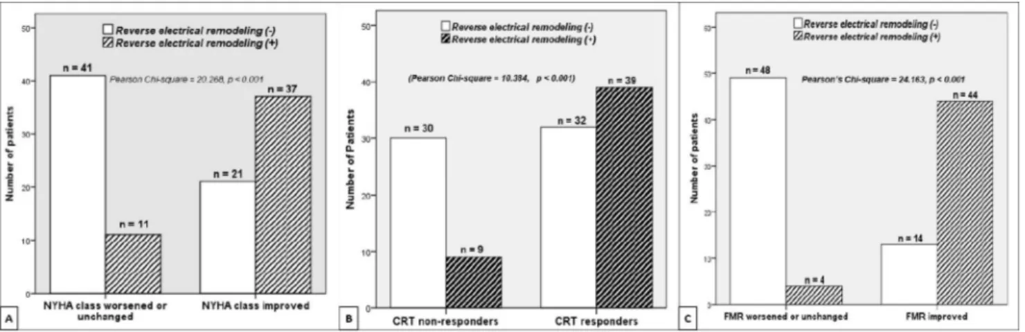
The association of the improvement in NYHA functional class based on RER is demonstrated inFig. 1A. Functional improvement was observed more frequently in patients with RER (n= 37 vs n = 21), whereas patients without RER were more likely to have NYHA functional class unchanged or worsened (n= 41 vs n = 11) after CRT (Pearson chi-square= 20.268; P < .001). In addition, although the baseline
mean NYHA functional classes were similar (2.9± 0.6 vs 2.8± 0.7; P = .683), patients with RER showed a signifi-cant functional improvement (follow-up NYHA functional classes 1.9± 0.7 vs 2.5 ± 0.9; P = .002) compared with pa-tients without RER at 6 months.
Association of RER With LV Reverse Remodeling
Comparison of the LV remodeling parameters in RER groups revealed that although the mean baseline LVESV values were similar (157.7± 49.8 mL vs 156.4 ± 51.4 mL; P = .865), follow-up mean LVESV values were significantly lower
Table 1. Comparison of the Baseline Demographic, Clinical, and Electrocardiographic Features of the Study Population According to the Narrowing of the Intrinsic QRS Duration Following CRT, Represented as Reversed Electrical Remodeling (RER)− or RER+
Overal (n= 110) RER– (n= 62) RER+ (n= 48) P Value Age, y* 66 (61–75) 64 (58–71) 67 (62–73) .096 Female sex, % (n) 36% (40) 35% (22) 38% (18) .829 BSA, m2* 1.86 (1.73–1.99) 1.89 (1.73–1.99) 1.83 (1.72–2.02) .795 BMI, kg/m2 27.5± 4.7 27.3± 4.6 27.7± 5.0 .699 Nonischemic etiology, % (n) 57% (63) 55% (34) 60% (29) .032 Hypertension % (n) 73% (80) 68% (42) 75% (36) .124 Diabetes, % (n) 33% (36) 37% (23) 27% (13) .271 Hyperlipidemia, % (n) 46% (51) 51% (32) 39% (19) .074 Atrial fibrillation,†% (n) 27% (30) 29% (18) 25% (12) .641 NYHA III/IV, % (n) 64% (71) 60% (37) 71% (34) .229 CRT-D, % (n) 89% (98) 87% (54) 92% (44) .162 LBBB morphology 85% (94) 83% (51) 89% (43) .120 Baseline QRS duration, ms 160.5± 20.9 158.8± 21.8 162.7± 19.6 .326 QRS≥150 ms, % (n) 65% (71) 60% (37) 71% (34) .225 Paced QRS duration (ms) 155.9± 25.0 160.6± 26.5 149.3± 20.1 .012 Hb, g/dL 12.4± 1.6 12.3± 1.8 12.7± 1.4 .232 Cr, mg/dL* 1.0 (0.9–1.3) 0.9 (0.8–1.4) 1.0 (0.9–1.3) .429 Medication, % (n) Beta-blocker 95% (104) 94% (58) 96% (46) .605 ACE-I/ARB 96% (105) 95% (59) 96% (46) .868 Spironolactone 76% (84) 74% (46) 79% (38) .241 Furosemide 82% (90) 82% (50) 81% (40) .442 Digoxine 40% (44) 42% (26) 37% (18) .136 Nitrate 28% (31) 31% (19) 25% (12) .347 Amiodarone 36% (40) 35% (22) 38% (18) .229 Oral anticoagulation‡ 25% (28) 27% (17) 23% (11) .162
BSA, body surface area; BMI, body mass index; NYHA, New York Heart Association functional class; CRT-D, biventricular device combined with a de-fibrillator; LBBB, left bundle branch block; Hb, hemoglobin; Cr, creatinine; ACE-I, angiotensin-converting enzyme inhibitor; ARB, angiotensin receptor blocker.
*Median (interquartile range).
†Chronic or paroxysmal atrial fibrillation.
‡Warfarin or novel agents (dabigatran, apixaban, or rivaroxaban).
Table 2. Change in Functional Status, Electrocardiographic, and Echocardiographic Parameters in Overall Population and in Groups Based on Reversed Electrical Remodeling (RER)
Overall (n= 110) RER– (n= 62) RER+ (n = 48)
Baseline Follow-Up P Value Baseline Follow-Up P Value Baseline Follow-Up P Value
NYHA III/IV, % (n) 64% (71) 35% (38) .014 60% (37) 45% (28) .031 71% (34) 21% (10) .002 Intrinsic QRS, ms 160.5± 20.9 161.6± 21.3 .242 158.8± 21.8 166.6± 21.3 <.001 162.7± 19.6 155.0± 19.7 <.001 LVESV, mL 157.0± 50.5 138.9± 51.1 <.001 156.4± 51.4 143.8± 53.6 <.001 157.7± 49.8 132.6± 47.5 <.001 LVEDV, mL 212.6± 57.9 197.4± 56.9 <.001 209.1± 57.5 198.8± 57.7 .001 217.1± 58.7 195.6± 56.4 <.001 LVEF, % 26.5± 5.7 29.8± 7.4 <.001 26.0± 5.8 28.1± 7.4 .003 27.2± 5.4 32.0± 6.9 <.001 EROA, cm2 0.24± 0.14 0.23± 0.11 .162 0.22± 0.11 0.29± 0.15 .001 0.21± 0.11 0.16± 0.10 .001 RegV, mL 30.7± 17.3 29.1± 14.8 .276 28.9± 15.0 36.5± 18.5 <.001 29.5± 14.6 22.2± 12.1 <.001 TA, cm2 3.98± 0.85 3.95± 0.86 .709 3.7± 0.7 3.8± 0.8 .029 4.3± 0.8 3.9± 0.9 .004 TD, cm 2.0± 0.4 1.9± 0.4 .788 1.9± 0.4 2.0± 0.4 .032 2.0± 0.5 1.9± 0.4 .010
EROA, effective regurgitant orifice area; LVEDV, left ventricular end-diastolic volume; LVEF, left ventricular ejection fraction; LVESV, left ventricular end-systolic volume; NYHA, New York Heart Association functional class; RegV, regurgitant volume; A, tenting area; TD, tenting distance.
(132.6± 47.5 mL vs 143.8 ± 53.6 mL, P = .025) and the mean changes in LVESV were significantly higher (25.0± 15.4 mL vs 12.5± 23.1 mL; P = .002) in patients with RER (Supplemental Fig. 1A). Similarly, mean baseline LVEF values were similar (26.0± 5.8% vs 27.2 ± 5.4%; P = .280), but mean follow-up LVEF was significantly higher (32.0± 6.9% vs 28.1± 7.4%; P = .007) and the mean change in LVEF higher (4.7± 3.8% vs 2.1 ± 5.4%; P = .005) in patients with RER compared with patients without RER (Supplemental Fig. 1B). The relationship between CRT response and RER is dem-onstrated with a clustered bar chart inFig. 1B, revealing that presence of RER was associated with CRT response, whereas the absence of RER was associated with CRT nonresponse (Pearson chi-square= 10.384; P < .001).
Impact of RER on MR Severity and Mitral Geometry
A total of 58 patients (44 patients with RER vs 14 pa-tients without RER) were found to have reduction in RegV at 6 months, whereas 52 patients (4 patients with RER vs 48 patients without RER) had no change or increase in MR after CRT (Pearson chi-square= 24.163; P < .001), as shown in Fig. 1C. The extent of change in the native QRS duration compared with the baseline QRS duration (ie, ΔQRS) showed a significant linear correlation with ΔRegV (r= 0.513; P < .001) and ΔTA (r = 0.349; P < .001) after CRT (Supplemental Fig. 2).
Predictors of Prognosis and MR Improvement
Among the several electrocardiographic parameters tested in the univariate Cox proportional hazard model for predic-tion of survival; statistically significant variables (P values <.05; paced QRS duration and RER) were entered into the multivariate analysis to determine the independent predic-tors. RER (HR 1.026, 95% CI 1.014–1.039; P= .043) was found to independently predict all-cause death. To define the
independent predictors of combined death/hospitalization, sta-tistically significant variables in the univariate model (baseline QRS duration, paced QRS duration, and RER) were tested by means of multivariate Cox regression. Paced QRS dura-tion (HR 1.012, 95% CI 1.005–1.019; P= .048) and RER (HR 2.071, 95% CI 1.208–4.910; P= .028) independently pre-dicted event-free survival during the overall follow-up (Table 3).
Table 4presents the logistical regression analyses for pre-diction of reduction in RegV (MR response) after CRT. Significant variables (P< .05) in the univariate model (paced QRS duration, RER, baseline LVESV, and baseline TA) were entered in the multivariate analysis. RER (HR 1.103, 95% CI 1.007–1.208; P= .023) and baseline TA (HR 2.011, 95% CI 1.268–2.754; P= .014) were the independent predictors of MR improvement after CRT.
Estimation of survival and event-free survival based on RER were separately analyzed by means of Kaplan-Meier method, as shown inFig. 2. Presence of RER showed statistical sig-nificance for prediction of survival (free from all-cause death: log rank 3.938; P= .047) and event-free survival (free from combined death/hospitalization: log rank 6.066; P= .014) during the mean follow-up of 18± 3 months after CRT implantation.
Discussion
In the present study, we evaluated the clinical impact of narrowing of the intrinsic QRS duration after CRT on the echocardiographic and clinical outcomes. According to the main findings of the study, a narrowed intrinsic QRS dura-tion compared with the baseline was strongly associated with higher incidence of functional improvement, greater CRT re-sponse, and reduced MR severity. Moreover, narrowing of the native QRS duration was a predictor of MR response, hos-pitalization, and death during the follow-up.
Fig. 1. Bar charts demonstrating the association between reversed electrical remodeling (RER) and functional, echocardiographic, and mitral regurgitation (MR) response at follow-up. (A) Functional response (≥1-degree improvement in New York Heart Association [NYHA] func-tional class) was observed more frequently in patients with RER, whereas patients without RER seemed to have a worsened or unchanged NYHA functional class (P< .001). (B) Cardiac resynchronization therapy (CRT) response was more frequently observed in patients with RER, whereas CRT nonresponse was more pronounced in patients without RER (P< .001). (C) MR response (reduction in RegV) was more frequently observed in patients with RER, whereas patients without RER seemed to have worsened or unchanged FMR at 6 months (P< .001).
CRT has been primarily accepted as an electrical therapy that aims to restore the synchronicity of the conduction system in the failing heart.18The evidence arising from RV-pacing
studies19supports that deterioration of intraventricular
con-duction by inducing iatrogenic LBBB further affects LV systolic dysfunction and can be reversed with the use of biventricular pacing.20In addition, complete restoration of the
electrical remodeling in a patient with LBBB resulted in superresponse to CRT, as shown by Dizon et al,21
suggest-ing that reversal of electrical remodelsuggest-ing is the main target of biventricular pacing in heart failure.
CRT-induced RER has been addressed in several previ-ous studies. Narrowing of the intrinsic QRS duration by CRT was mostly evaluated for favorable structural changes and pre-diction of CRT response. Although Stockburger et al did not find any association between structural and electrical remodeling,22
a study from Boriani et al suggested that the extent of QRS shortening by CRT was a determinant of re-duction in LVESV.6
Similarly, Yang et al found that narrowing of the intrinsic QRS complex correlated with CRT response.5 Table 3. Cox Proportional Hazard Model for Prediction of All-Cause Mortality and Combined Death and Hospitalization According to
Univariate and Multivariate Analyses
Univariate Analysis Multivariate Analysis
HR 95% CI P Value HR 95% CI P Value
Death from all causes
Baseline QRS duration 0.987 0.963–1.011 .272
LBBB morphology 0.658 0.189–2.293 .511
Paced QRS duration 1.033 1.012–1.054 .036 0.456 0.085–2.440 .359
Follow-up intrinsic QRS duration 1.000 0.979–1.021 .641
Reverse electrical remodeling 2.180 1.356–3.504 .026 1.026 1.014–1.039 .043
Combined death and hospitalization
Baseline QRS duration 1.125 1.082–1.168 .012 0.842 0.416–1.706 .112
LBBB morphology 1.430 0.755–2.709 .272
Paced QRS duration 1.026 1.014–1.039 .022 1.012 1.005–1.019 .048
Follow-up intrinsic QRS duration 0.996 0.983–1.010 .587
Reverse electrical remodeling 2.245 1.150–4.382 .018 2.071 1.208–4.910 .028
HR, hazard ratio; CI, confidence interval; LBBB, left bundle branch block.
Table 4. Univariate and Multivariate Logistic Regression Analyses to Determine Independent Predictors of Improvement in
Mitral Regurgitation (MR Response)
OR 95% CI P Value
Univariate analysis
Baseline QRS duration 1.009 0.991–1.028 .318
Paced QRS duration 1.040 1.023–1.057 .032
Follow-up intrinsic QRS duration 1.044 0.977–1.117 .201
RER 2.651 1.112–6.309 .032 Baseline LVESV 1.012 1.005–1.019 .028 Baseline LVEF 0.767 0.327–1.798 .541 TA 1.916 1.228–2.290 .012 Multivariate analysis Paced QRS duration 0.996 0.985–1.007 .415 RER 1.103 1.007–1.208 .023 Baseline LVESV 1.966 0.410–9.428 .398 TA 2.011 1.268–2.754 .014
OR, odds ratio; other abbreviations as inTables 1 and 2.
Fig. 2. Kaplan-Meier curves generated for estimation of the study end-points during a mean follow-up of 18± 3 months. (A) Estimation of survival (free from all-cause death: log rank= 3.938; P = .047). (B) Estimation of event-free survival (free from combined death/ hospitalization: log rank= 6.066; P = .014).
However, there is no uniform definition of RER in the liter-ature. Tereshenko et al accepted≥10 ms of QRS narrowing as an index of RER and found an association with de-creased mortality.23A study by Yang et al suggested that
regression of the fragmented QRS complex interval was as-sociated with improved electrical conduction that resulted in beneficial outcomes after CRT.24We observed that even minor
alterations in the native QRS duration correlated with sig-nificant echocardiographic changes after CRT. Therefore, in the present study we used the absolute QRS narrowing as an index of RER to correlate with clinical outcomes.
Consistently with the recent studies, our study once again confirmed that the presence of RER is strongly associated with increased functional and echocardiographic response after CRT. However, we investigated the effect of RER beyond the extent of LV reverse remodeling. We also correlated RER with important prognostic variables such as improvement in MR and reduction in death and hospitalization. Reversal of the intrinsic electrical dyssynchrony induced by CRT is the suggested mechanism of action that triggers the reversal of both mechanical dyssynchrony and LV remodeling, leading to favorable clinical and echocardiographic outcomes after CRT.
Comparison of the baseline characteristics in our study pop-ulation revealed that traditional CRT response predictors,25
such as female sex, baseline QRS duration, and LBBB mor-phology, did not differ in RER groups. However, patients with RER had nonischemic cardiomyopathy more frequently and tended to have narrower paced QRS durations, which may have influenced the results of the study. According to a report by Hsing et al,26paced QRS and the CRT-induced QRS change
predicted favorable clinical outcomes and LV reverse remod-eling. Similarly, QRS narrowing as indexed to baseline was significantly associated with reversed LV remodeling after CRT, as reported by Rickard et al.20Lecoq et al evaluated CRT
response in 139 patients and noted that QRS narrowing was a determinant of CRT response.3We achieved similar results
in our study regarding the importance of paced QRS dura-tion after CRT. Based on the regression analyses, paced QRS duration was found to be a univariate predictor of both death/ hospitalization and MR response, which necessitates further studies to confirm these findings.
In line with the major CRT trials, we had an overall CRT response rate of 65%. Both the overall population and the RER groups showed marked reduction in mean NYHA function-al class and marked improvements in LV remodeling parameters (LVESV, LVEDV, and LVEF). In contrast, re-duction in MR severity and improvement in mitral valve geometry were observed only in the group of patients with RER. The MR parameters assessed in the study (RegV, EROA, TA, and TD) significantly worsened in patients without RER, although they mildly (statistically insignificantly) improved in the overall population. Overall, 58 patients (53%) were MR responders, which consisted of 44 patients with RER and 14 patients without RER. Corresponding MR response rates were 92% vs 23% in the RER groups, which clarified the role of intrinsic QRS narrowing on MR severity after CRT.
Predictors of MR improvement after CRT have been widely investigated recently. It is important to differentiate func-tional MR from organic or ischemic MR both of which involve distinctive mechanisms. CRT has been shown to positively interact with and ameliorate the imbalance between the teth-ering forces (due to papillary muscle dyssynchrony and/or annular dilatation) and the impaired closing forces (due to reduced LV contractility and/or LV dyssynchrony) that were suggested as the underlying mechanisms of MR in patients with heart failure. Ypenburg et al reported that baseline LV mechanical dyssynchrony was associated with improve-ment in MR after CRT.9Sitges et al suggested that excessive
remodeling of the LV and mitral valve predicted an unfavor-able MR response after CRT.8Likewise, we found that baseline
LVESV was a univariate predictor of MR response, whereas baseline TA was an independent predictor of MR response according to multivariate analysis. Because previous studies did not provide sufficient data regarding the influence of in-trinsic QRS narrowing on MR improvement, the present study is among the first to focus on the correlation ofΔQRS with the reduction in RegV and in TA. Moreover, CRT-induced RER has never previously been suggested as a predictor of MR response.
According to the results of the present study, favorable effects of CRT on MR improvement in patients with RER might be explained by several mechanisms. First, ameliora-tion of the native conducameliora-tion after CRT results in improvement in LV remodeling parameters that subsequently leads to a de-crease in MR severity as a result of smaller LV volumes. Second, improvements in global LV remodeling in patients with RER might favorably interact with the local remodel-ing parameters actremodel-ing on the mitral apparatus, such as reduction in the mitral annular area, TA, and TD. Third, reversal of the mechanical dyssynchrony, especially at the papillary muscle level, is a mechanism for MR improvement after CRT induced by the reversal of the intrinsic electrical dyssynchrony.
Among the predictors of death and hospitalization follow-ing CRT, we achieved some “classic” and “novel” results in the present study. According to univariate analysis, tradition-al variables,25,27,28including the baseline and the paced QRS
durations, tended to affect prognosis after CRT, as ex-pected. These findings were in line with previous meta-analyses investigating the predictors of mortality and morbidity after CRT.29,30
A unique feature of the present study is that narrowing of the intrinsic QRS duration after CRT is pro-posed as an independent predictor of both survival and event-free survival by multivariate analysis.
Study Limitations
The main limitation of the study is that the study popula-tion consists of patients treated in a single center with a relatively small sample size. The overall follow-up period was 18± 3 months with a wide variation (range 1–24 months) to reach the study end points. Therefore, assessment of func-tional improvement, CRT response, and MR reduction was
done at 6 months, which is a generally accepted time inter-val to assess clinical and echocardiographic response to CRT. Patients who died within the 1st 6 months (n= 3; 1 with RER and 2 without RER) were included in the analyses with their baseline values. In addition, the absence of long-term echocardiographic data and of the association with progno-sis during the overall follow-up might also be an area of criticism.
Any method for quantification of MR involves some draw-backs. Although the addition of vena contracta and jet area assessment would have increased accuracy, the PISA method that we used in the present study to assess RegV is the cri-terion standard in current guidelines. In addition, 3-dimensional assessment by tenting volume analysis would be more precise, because the evaluation of the mitral valve remodeling is ana-tomically challenging. Another possible limitation is that there was a significant overlap between the patients with reduc-tion in the paced and in the native QRS durareduc-tions. The differentiation of a coexistence or a cause-effect relation-ship must be established in further studies. Finally, long-term clinical data on ventricular arrhythmias or need for transplant/assist device would be more comprehensive, because we focused only on major clinical end points, such as im-provement in NYHA functional class, hospitalization, and death.
Conclusion
Reversed electrical remodeling by means of narrowing of the intrinsic electrocardiographic QRS duration after CRT has clinical and prognostic implications. A narrowed intrinsic QRS interval than the baseline is associated with improved func-tional status, higher CRT response, reduced MR, and favorable prognosis after CRT. The results of the present study suggest that the assessment of the native conduction should be a part of routine clinical follow-up of patients after CRT. Further randomized studies are needed for translation of our find-ings into clinical practice with algorithms including RER assessment for early prediction of prognosis after CRT.
Disclosures
None.
Supplementary Data
Supplementary data related to this article can be found at
doi:10.1016/j.cardfail.2016.04.001.
References
1. Linde C, Ståhlberg M, Benson L, Braunschweig F, Edner M, Dahlström U, et al. Gender, underutilization of cardiac resynchronization therapy, and prognostic impact of QRS prolongation and left bundle branch block in heart failure. Europace 2015;17:424–31.
2. Gasparini M, Leclercq C, Yu CM, Auricchio A, Steinberg JS, Lamp B, et al. Absolute survival after cardiac resynchronization therapy according
to baseline QRS duration: a multinational 10-year experience: data from the Multicenter International CRT Study. Am Heart J 2014;167:203–9, e1.
3. Lecoq G, Leclercq C, Leray E, Crocq C, Alonso C, de Place C, et al. Clinical and electrocardiographic predictors of a positive response to cardiac resynchronization therapy in advanced heart failure. Eur Heart J 2005;26:1094–100.
4. Sassone B, Gambetti S, Bertini M, Beltrami M, Mascioli G, Bressan S, et al. Relation of QRS duration to response to cardiac resynchronization therapy. Am J Cardiol 2015;115:214–9.
5. Yang XW, Hua W, Wang J, Liu ZM, Ding LG, Chen KP, et al. Native QRS narrowing reflects electrical reversal and associates with anatomical reversal in cardiac resynchronization therapy. J Interv Card Electrophysiol 2014;41:161–8.
6. Boriani G, Biffi M, Martignani C, Ziacchi M, Saporito D, Grigioni F, et al. Electrocardiographic remodeling during cardiac resynchronization therapy. Int J Cardiol 2006;108:165–70.
7. Onishi T, Onishi T, Marek JJ, Ahmed M, Haberman SC,
Oyenuga O, et al. Mechanistic features associated with improvement in mitral regurgitation after cardiac resynchronization therapy and their relation to long-term patient outcome. Circ Heart Fail 2013;6:685– 93.
8. Sitges M, Vidal B, Delgado V, Mont L, Garcia-Alvarez A, Tolosana JM, et al. Long-term effect of cardiac resynchronization therapy on functional mitral valve regurgitation. Am J Cardiol 2009;104: 383–8.
9. Ypenburg C, Lancellotti P, Tops LF, Bleeker GB, Holman ER, Piérard LA, et al. Acute effects of initiation and withdrawal of cardiac resynchronization therapy on papillary muscle dyssynchrony and mitral regurgitation. J Am Coll Cardiol 2007;50:2071–7.
10. Brignole M, Auricchio A, Baron-Esquivias G, Bordachar P, Boriani G, Breithardt OA, et al. 2013 ESC guidelines on cardiac pacing and cardiac resynchronization therapy: the Task Force on Cardiac Pacing and Resynchronization Therapy of the European Society of Cardiology (ESC). Europace 2013;15:1070–118.
11.Pino PG, Galati A, Terranova A. Functional mitral regurgitation in heart failure. J Cardiovasc Med (Hagerstown) 2006;7:514–23.
12. Strauss DG, Selvester RH, Wagner GS. Defining left bundle branch block in the era of cardiac resynchronization therapy. Am J Cardiol 2011;107:927–34.
13. Dong YX, Powell BD, Asirvatham SJ, Friedman PA, Rea RF, Webster TL, et al. Left ventricular lead position for cardiac resynchronization: a comprehensive cinegraphic, echocardiographic, clinical, and survival analysis. Europace 2012;14:1139–47.
14. Barold SS, Ilercil A, Herweg B. Echocardiographic optimization of the atrioventricular and interventricular intervals during cardiac resynchronization. Europace 2008;10(Suppl 3):iii88–95.
15. Lang RM, Badano LP, Mor-Avi V, Afilalo J, Armstrong A, Ernande L, et al. Recommendations for cardiac chamber quantification by echocardiography in adults: an update from the American Society of Echocardiography and the European Association of Cardiovascular Imaging. Eur Heart J Cardiovasc Imaging 2015;16:233–70.
16. Karaca O, Avci A, Guler GB, Alizade E, Guler E, Gecmen C, et al. Tenting area reflects disease severity and prognosis in patients with non-ischaemic dilated cardiomyopathy and functional mitral regurgitation. Eur J Heart Fail 2011;13:284–91.
17. Enriquez-Sarano M, Miller FA Jr, Hayes SN, Bailey KR, Tajik AJ, Seward JB. Effective mitral regurgitant orifice area: clinical use and pitfalls of the proximal isovelocity surface area method. J Am Coll Cardiol 1995;25:703–9.
18. Floré V, Bartunek J, Goethals M, Verstreken S, Timmermans W, De Pauw F, et al. Electrical remodeling reflected by QRS and T vector changes following cardiac resynchronization therapy is related to survival in heart failure patients with left bundle branch block. J Electrocardiol 2015;48:578–85.
19. Vogt J, Krahnefeld O, Lamp B, Hansky B, Kirkels H, Minami K, et al. Electrocardiographic remodeling in patients paced for heart failure. Am J Cardiol 2000;86(9A):152K-6K.
20. Rickard J, Popovic Z, Verhaert D, Sraow D, Baranowski B, Martin DO, et al. The QRS narrowing index predicts reverse left ventricular remodeling following cardiac resynchronization therapy. Pacing Clin Electrophysiol 2011;34:604–11.
21. Dizon J, Horn E, Neglia J, Medina N, Garan H. Loss of left bundle branch block following biventricular pacing therapy for heart failure: evidence for electrical remodeling? J Interv Card Electrophysiol 2004;10:47–50.
22. Stockburger M, Nitardy A, Fateh-Moghadam S, Krebs A, Celebi O, Karhausen T, et al. Electrical remodeling and cardiac dimensions in patients treated by cardiac resynchronization and heart failure controls. Pacing Clin Electrophysiol 2008;31:70–7.
23. Tereshchenko LG, Henrikson CA, Stempniewicz P, Han L, Berger RD. Antiarrhythmic effect of reverse electrical remodeling associated with cardiac resynchronization therapy. Pacing Clin Electrophysiol 2011;34:357–64.
24. Yang XW, Hua W, Wang J, Liu ZM, Ding LG, Chen KP, et al. Regression of fragmented QRS complex: a marker of electrical reverse remodeling in cardiac resynchronization therapy. Ann Noninvasive Electrocardiol 2015;20:18–27.
25. Hsu JC, Solomon SD, Bourgoun M, McNitt S, Goldenberg I, Klein H, et al. Predictors of super-response to cardiac resynchronization therapy and associated improvement in clinical outcome: the MADIT-CRT
(Multicenter Automatic Defibrillator Implantation Trial With Cardiac Resynchronization Therapy) study. J Am Coll Cardiol 2012;59:2366–73.
26. Hsing JM, Selzman KA, Leclercq C, Pires LA, McLaughlin MG, McRae SE, et al. Paced left ventricular QRS width and ECG parameters predict outcomes after cardiac resynchronization therapy: PROSPECT-ECG substudy. Circ Arrhythm Electrophysiol 2011;4:851–7.
27. Varma N, Manne M, Nguyen D, He J, Niebauer M, Tchou P. Probability and magnitude of response to cardiac resynchronization therapy according to QRS duration and gender in nonischemic cardiomyopathy and LBBB. Heart Rhythm 2014;11:1139–47.
28. Upadhyay GA, Chatterjee NA, Kandala J, Friedman DJ, Park MY, Tabtabai SR, et al. Assessing mitral regurgitation in the prediction of clinical outcome after cardiac resynchronization therapy. Heart Rhythm 2015;12:1201–8.
29. Sipahi I, Chou JC, Hyden M, Rowland DY, Simon DI, Fang JC. Effect of QRS morphology on clinical event reduction with cardiac resynchronization therapy: meta-analysis of randomized controlled trials. Am Heart J 2012;163:260–7, e3.
30. Cleland JG, Abraham WT, Linde C, Gold MR, Young JB,
Claude Daubert J, et al. An individual patient meta-analysis of five randomized trials assessing the effects of cardiac resynchronization therapy on morbidity and mortality in patients with symptomatic heart failure. Eur Heart J 2013;34:3547–56.