Tropical Journal of Pharmaceutical Research April 2012; 11 (2): 307-318 © Pharmacotherapy Group, Faculty of Pharmacy, University of Benin, Benin City, 300001 Nigeria. All rights reserved
.
Available online at http://www.tjpr.orghttp://dx.doi.org/10.4314/tjpr.v11i2.19
Review Article
Injectable In Situ Forming Microparticles: A Novel
Drug Delivery System
Evren Algın Yapar
1*, Özge
İ
nal
2, Yalçın Özkan
3and Tamer Baykara
41Ministry of Health of Turkey, General Directorate of Pharmaceuticals and Pharmacy, Sö
ğütözü Mah. 2176. Sok. No:5 Kat:6, 06520 Çankaya-Ankara, 2Department of Pharmaceutical Technology, Faculty of Pharmacy, University of Ankara, 06100 Tandoğan-Ankara, 3Department of Pharmaceutical Technology, Gülhane Military Medical Academy, 06018 Etlik-Ankara, 4UNAM (Institute of Materials Science and Nanotechnology), Bilkent University, 06800 Bilkent-Ankara, Turkey
Abstract
Pharmaceutical formulation research has recently been focusing on delivery systems which provide long therapeutic effects and reduced side effects, and involving simplified production stages and facilitated application process. In situ forming microparticle (ISM) systems, one of the latest approach in this field, offer a new encapsulation technique and meet the objectives stated above. Factors such as the carrier used to form the multiparticles, amount and type of drug and the vehicle type can be taken as the main performance criteria for these systems. Ongoing studies have shown that this new multiparticulate drug delivery system is suitable for achieving new implant delivery system with low risk of dose-dumping, capable of being modulated to exhibit varying release patterns, reproducible, easily applicable and well-tolerated compared with classically surgical implants.
Keywords: Injectable, In situ forming, Microparticle system, Controlled drug delivery.
Received: 5 August 2011 Revised accepted: 20 February 2012
INTRODUCTION
Various intramuscular or subcutaneous controlled drug delivery systems in the form of implants or microparticles have been developed based on biodegradable polymers. There are different techniques available to form drug–polymer composites as implants or microparticles [1-2]. Implants are generally formed by blending drug, polymer and additives followed by melt extrusion, melt compression or injection molding to obtain the desired implant size or shape [2]. Problems have occurred with these processes include elevated process temperature, poor content uniformity (especially with low-dose drugs) and often the requirement of surgical administration [1]. Additionally preparation of biodegradable implants and especially of microparticles is complicated and involves multiple-step processes and formulation parameters to be controlled, and this has also affect scale-up and cost [3].
In order to avoid inconvenient surgical insertion of large implants, injectable biodegradable and biocompatible polymeric particles (microspheres, microcapsules, nanocapsules, nanospheres etc.) could be employed for controlled drug delivery [3]. Microparticles of size < 250 µm, ideally < 125
µm, are suitable for this purpose [4]. Biodegradable polymers used for these systems are natural or synthetic in origin and are degraded in vivo by suitable biodegradation kinetics, either enzymatically or non-enzymatically or both, to produce biocompatible and toxicologically safe by-products which are further eliminated by normal metabolic pathways. The polymers selected for these systems must also meet some other requirements such as drug compatibility, suitable mechanical properties and ease of processing [5-7].
Approaches to prolonged therapeutic injectables were initially studied as formulations of viscous oils, which could
decrease drug diffusion rate [8-10]. Latest studies in this field are focused on the development of liquid drug-polymer formulations, which form implants in situ upon injection and contact with body fluids as an alternative to solid implants [11-13]. These liquid drug-polymer formulations are prepared by dissolving thermoplastic aliphatic poly(esters) such as poly(lactide) (PLA) and especially poly(lactide-co-glycolide) (PLGA) in water-miscible solvents such as N-methyl-2-pyrrolidone (NMP), dimethyl sulfoxide (DMSO), etc. Upon injection of drug-containing polymer solution, the polymer solidifies at the site of injection and forms an implant. This technology has been investigated for the delivery of model proteins, LHRH-antagonists, narcotic antagonists, growth factors, anti-inflammatory agents, antibiotic, antiemetic, antiasthmatic, antipsychotic and anticancer drugs [12,14-21].
Drug release from these systems follows Higuchi square root of time relationship [22] with a high burst release in general [23-27]. Potential solvent toxicity and high viscosity of polymer solutions causing injection problems can possibly be occurred to these systems [11-17,25-27]. To overcome these problems a novel in situ forming microparticle system has been developed [28-29]. ISM systems are based on an emulsion of an internal drug-containing polymer solution and a continuous oil or aqueous phase. After injection, the inner polymeric phase hardens upon contact with body fluids and thus forms in situ microparticles. ISM systems have significantly reduced initial burst release and viscosity (which is primarily controlled by the external phase). Thus, easier injectability and reduced pain has been achieved, compared to use of the polymer solutions (in situ implant systems). Additionally ISMs are multiparticulates, and thus minimize variations in implant morphology (after solidification) and provide more consistent and reproducible drug release profile [28-34].
METHODS OF PREPARATION OF
ISM SYSTEMS
ISM systems consist of an internal drug-containing polymer solvent phase (polymer phase) that is dispersed into an external oil or water phase. Following injection of this dispersion, the inner polymer phase hardens upon contact with body fluids and forms in situ microparticles. The preparation process for ISM is simple, when compared with classical techniques for the preparation of various other microparticles such as solvent evaporation or phase separation methods [28-29,34].
Preparation of oil-in-oil (O/O)–ISM system
The O/O–ISM system comprises of polymer solution phase and external oil phase. Polymer solution phase can be prepared by dissolving a biodegradable polymer such as PLA or PLGA in a water-miscible, biocompatible solvent (this may also act as a plasticizer for the polymer) such as NMP, 2-pyrrolidone, dimethyl sulphoxide (DMSO), triacetin and/or low molecular weight grade of polyethylene glycol {such as polyethylene glycol (PEG) 200 or 400}, which are able to form highly concentrated polymer solutions in combination with surfactants such as polyethylene glycol sorbitan monooleate (Tween 80) or polyoxyethylene–polyoxy-propylene copolymer (Pluronic F 68). Peanut oil and sesame oil (oil for injection) can be used as a biocompatible external oil phase with surfactants such as sorbitan monooleate (Span 80) or triglyceride (Miglyol 812) with/without aluminum stearate or aluminum-monostearate. Subsequently, accurately weighed internal and external phases (in different ratios) are loaded preferably into polypropylene syringes A and B, respectively. The two syringes are coupled with a connector and emulsification can be achieved by pushing the content of syringe A into syringe B to and fro for 50 mixing cycles. At the end of this, the contents are pushed into one syringe, the connector is removed and a
needle is attached, and is ready for use [19-20, 31-38].
Preparation of oil in-water (O/W)–ISM system
The O/W–ISM system is able to prepare similar with O/O–ISM system, except some differences in the following aspects. This system consists of an internal polymer solution phase in a hydrophobic solvent such as ethyl acetate and an external aqueous phase containing aqueous surfactant like Poloxamer 188 (Lutrol F 68). Emulsification of O/W–ISM can be achieved by 25 mixing cycles in a two-syringe system as described above for O/O-ISM system [30, 39].
PREVIOUS
STUDIES
ON
ISM
SYSTEMS
In vitro studiesThe ISM system described by Jain et al is a solution of PLGA, triacetin, a model protein cytochrome c, PEG 400 and Tween 80 (Oil Phase 1) which is added dropwise with continuous homogenization to Miglyol 812-Span 80 solution (Oil Phase 2), thereby inducing phase separation (coacervation) of PLGA and forming drug containing PLGA microglobules dispersed in the continuous phase. This dispersion has a viscous consistency, but is sufficiently syringeable. When injected, it comes in contact with water from aqueous buffer or physiological fluid, thus microglobules harden to form cytochrome c entrapped solid matrix type microspheres which has released the drug in a controlled fashion [35, 40]. Figure 1 shows schematic representation of this microsphere formation process [35].
Jain et al also investigated the effects of the following formulation variables on the release characteristics of ISMs: (i) concentrations of hydrophilic solvent (PEG 400), encapsulated drug and hydrophilic excipient (mannitol); and (ii) types of encapsulated drug, red dye (micromolecules), Cytochrome c and
myoglobin (macromolecules)] and vehicles. Moreover, effects of formulation properties, process, and storage conditions (15 days/48
o
C) on physical stability of encapsulated protein were evaluated. It was observed that in vitro drug release increased with decrease in PEG 400 concentration, and increase in drug and mannitol concentrations.
Figure 1: Schematic representation of in situ
PLGA microsphere formation process [35]. Drug release decreased with increase in molecular weight of the encapsulated drug. Substitution of triacetin with triethyl citrate and Miglyol 812 with soybean oil resulted in variation in release pattern of drug. Soybean oil (mixed triglyceride of unsaturated and saturated fatty acids) which is relatively more hydrophobic than Miglyol 812 (mixed C8-C10
triglyceride of saturated fatty acids) produced relatively more hydrophobic PLGA microglobule dispersion, and thereby delayed the dissolution of released myoglobin [35].
The above novel microencapsulation process overcomes some of the disadvantages associated with other methods by (a) excluding the use of unacceptable organic solvents like dichloromethane or NMP and using acceptable vehicle mixture instead to prepare biodegradable PLGA microspheres, (b) forming drug containing PLGA microglobules (pre-microspheres or embryonic microspheres) which can be considered as precursors of the final microsphere product; these on coming into contact with water harden to form discreet PLGA microspheres which subsequently exhibit non-variable, predictable, and controlled drug release profile, and (c) precluding the need for reconstitution of PLGA microspheres before their administration [41-42]. Thus, this novel microencapsulation method can be viewed as a modified coacervation process, wherein drug containing microglobules are converted to microspheres in situ [40-43].
Luan and Bodmeier investigated the influence of PLGA type (molecular weight and end-group functionality) on leuprolide release from ISM systems which were based on an emulsion of PLGA solution in NMP dispersed in an oil phase (sesame oil containing 2 % Span 80 and 2.5 % aluminum stearate). The results of in vitro drug release were in contrast to those for microparticles prepared by classical solvent evaporation method. The use of lower molecular weight PLGA resulted in lower initial release from ISMs than when higher molecular weight PLGA was used. ISMs prepared with PLGA combinations showed a decrease in initial release when low-molecular-weight PLGA content was increased. Slow solvent diffusion from low-molecular-weight PLGA droplets into release medium led to a less porous structure of solidified microparticles, which explains the lower initial release. Moreover PLGA with free carboxylic acid end groups led to a lower drug release compared to PLGA with esterified end groups [32].
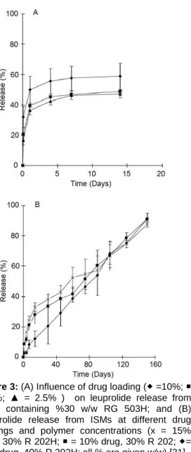
The influence of various formulation and processing parameters on drug release from ISM systems has been investigated in another study by Luan and Bodmeier. The systems consisted of leuprolide acetate-containing internal polymer phase {PLGA (RG 503H)/PLA (R 202H) and NMP} and external oil phase (peanut/sesame oil, Span 80 and aluminum monostearate). In vitro solidification and dissolution studies releaved that in situ forming microparticles prepared by using PLGA were spherical and had a smooth surface but a porous inner structure as seen in Figure 2A. Drug release from these ISMs [31] was occurred in two phases with a high initial release followed by an almost constant and slower release, as shown in Figure 3A. Drug release profiles obtained from ISMs prepared by using PLA were almost monophasic (Figure 3B) compared to ISMs that contained PLGA (Figure 3A). In particular, ISMs with 10 % drug loading and 40 % polymer concentration showed lower initial and almost linear drug release from days 2 to 150, and also exhibited a smooth surface with a less porous inner structure, which explains the slower drug release compared to others, as shown in Figures 2B and 3B. In contrast to RG 503H, R 202H led to a much lower initial leuprolide release {Figs 3A and 3B); this could possibly be explained by the higher carboxylic acid content of lower molecular weight PLA (R 202H acidic number is 10 mg KOH/g versus 4 mg KOH/g for RG 503H)}. In both systems, leuprolide acetate and PLGA or PLA were dissolved in polar solvent NMP. Therefore existence of ionic interactions between polymer and drug was also possible and a stronger interaction in the presence of R 202H could impede drug loss from polymer solution and thus lead to a lower initial release [31].
Kranz and Bodmeier investigated in vitro drug (diltiazem hydrochloride and buserelin acetate) release from ISM systems (PLA or PLGA in 2-pyrrolidone, NMP or DMSO dispersed into peanut oil phase) as a function of type of solvent and polymer, concentration
of polymer and ratio of internal polymer phase:external oil phase.
Figure 2: Scanning electron micrographs of ISMs
(A) prepared with 30 % w/w RG 503H and 5 % w/w drug loading and (B) prepared with 40 % w/w R 202H and 10 % w/w drug loading (2B) [31]. According to the data obtained, initial drug release from ISM systems decreased with increasing polymer concentration and decreasing polymer phase:external oil phase ratio. Decrease in the release of the drug was in the rank order of DMSO > NMP > 2-pyrrolidone, for the biocompatible solvent. In contrast to the release of low molecular weight diltiazem hydrochloride, peptide release (buserelin acetate) was strongly dependent on polymer degradation/erosion. The results confirmed that ISM system significantly reduced burst effect due to the presence of an external oil phase compared to in situ implant systems [33].
In another study by Kranz and Bodmeier [36], the effects of key formulation variables on the release of bupivacaine hydrochloride from ISM systems and in situ forming implants were investigated. The ISM systems were prepared as a combination of PLA–solvent phase dispersed into an external peanut oil phase. The solvent release from ISMs and in situ forming implants into phosphate buffer which influences polymer precipitation rate was investigated as a function of the type of
Figure 3: (A) Influence of drug loading ( =10%;
= 5%; ▲ = 2.5% ) on leuprolide release from ISMs containing %30 w/w RG 503H; and (B) Leuprolide release from ISMs at different drug loadings and polymer concentrations (x = 15% drug, 30% R 202H; = 10% drug, 30% R 202; = 10% drug, 40% R 202H; all % are given w/w) [31].
solvent (NMP, DMSO and 2-pyrrolidone), polymer concentration and polymer:oil phase ratio. Upon contact with the release medium, solvent release into buffer medium from ISMs was significantly slower compared to in situ forming implant solutions due to the presence of an external oil phase. The solvent release of ISM systems into phosphate buffer decreased with increasing polymer concentration and decreasing polymer:oil phase ratio. The type of solvent used
decreased solvent release rate in the rank order: DMSO > NMP >> 2-pyrrolidone. A slower solvent release into the aqueous medium resulted in less porous microparticles, thus explaining the reduced initial drug release from ISM systems compared to in situ forming implant solutions [36].
Incorporation of a model protein hen egg white lysozyme with ISM system was also investigated by Körber and Bodmeier. Ternary solvent blends of DMSO, ethyl acetate and water were used to adjust the protein solubility in order to facilitate the incorporation of either dispersed or dissolved protein into the polymer solution to achieve a good entrapment and to get a low initial release. The ISM systems consisted of lysozyme-containing polymer solutions (40 % PLGA-RG 502H) in sesame oil (external phase). The release of lysozyme from ISMs was only slightly affected by the investigated DMSO/ethyl acetate/water ratios (100/0/0, 75/25/0 and 70.5/23.5/6) and was incomplete. This result could be attributed to the protein, which is located in the polymer rich phase following solidification of ISMs and adsorption of the positively charged lysozyme to the negatively charged PLGA [44].
Stability of ISM systems
Some aspects of the stability of ISM systems have been investigated by researchers. Jain et al investigated the effect of myoglobin (macromolecules) on the release characteristics of ISMs and reported that the physical stability of myoglobin (helical structure) was unaffected by formulation, process, and storage conditions [35]. Kranz et al [37] also investigated the stability of ISM systems and showed that the stability of the ISM increased by dissolving Pluronic F68 in the polymer phase while placing aluminum monostearate in the oil phase of the system.
The stability of PLGA and leuprolide acetate in in-situ forming systems, either ISMs or polymer solutions and lyophilized sponges,
was investigated by Dong et al. It was found that degradation of PLGA increased with increasing storage temperature and water content in biocompatible solvents. Faster degradation occurred in polar protic solvents (2-pyrrolidone, PEG 400, triethyl citrate) than in polar aprotic solvents (NMP, DMSO, triacetin, ethyl acetate). The presence of leuprolide acetate significantly accelerated the PLGA degradation, especially in solution state. PLGA was stable in oily (sesame oil, soybean oil, medium chain triglyceride) suspensions at 4 °C and degraded only slightly faster than solid powder at 25 °C. No interaction between oils and PLGA was observed as indicated by an unchanged Tg of approximately 47 °C. Finally, leuprolide acetate was chemically stable in sponges, oils and polymer solutions in suspension state, but unstable (aggregation) when dissolved in the polymer solutions and stored at 25 and 40 °C [45].
The stability of Montelukast and haloperidol ISM systems were studied by Ahmed et al [19,20] while meloxicam ISM system was studied by Ibrahim et al [46]. These studies indicate that the best temperature of storage was 4 ºC for both systems which retained their stability for more than 12 months at 4 ºC.
In vitro - in vivo studies
The influence of various preparation and
formulation parameters on in vitro and in vivo release of bupivacaine hydrochloride
from ISM systems, which were prepared by emulsifying drug-containing polymer solutions (PLGA in 2-pyrrolidone) into a peanut oil phase at a polymer to oil phase ratio of 1:1, 0.5:1, 0.25:1 and 0.1:1, was investigated by Kranz et al [37]. Ready-to-inject formulations were prepared by probe sonication. Alternatively, emulsion formation prior to injection was carried out using two syringes filled with polymer phase in the first and peanut oil phase in the second container. In vitro drug release was highly influenced by the polymer to oil phase ratio and initial drug
release was reduced with decreasing polymer phase/external oil phase ratio. A hot plate model has been used in order to quantify the pharmacodynamic effect of bupivacaine release from ISM systems in male Sprague–Dawley rats. ISM with a polymer/oil phase ratio of 0.25:1 and 0.5:1 were selected for in vivo studies due to reduced initial in vitro release of drug. The in vivo drug release studies were in good agreement with in vitro drug release data. With ISM system, the analgesic effect of bupivacaine hydrochloride was prolonged when compared to the injection of a drug solution or drug-polymer solution [37].
Rungseevijitprapaa and Bodmeier [30] evaluated the forces required to inject biodegradable emulsions of the ISM systems via different sizes of syringe and needle into a newly developed chicken meat model using the compression mode of a texture analyzer (Figure 4). The injection forces obtained were finally correlated with the injectability of formulations into living rats. The results indicate that the flow of ISM formulations through the needle could be described by the well-known Poiseuille equation. The injection forces obtained were directly proportional to the form ulations’ viscosity, injection speed
Figure 4: Illustration of the setup for injection force
and the second power of the syringe plunger radius, and inversely proportional to the fourth power of the needle radius. This test method could differentiate very well the required forces from various formulations and injection conditions as well as different injection sites (muscle, subcutaneous tissues or air). In comparison to polymer solutions that form implants in situ, the ISM systems were more easily injectable with smaller needle size, and thus it was expected to be less painful and give better patient compliance [30].
Ahmed et al [19] studied the controlled release of montelukast (MK) using in situ forming implants and ISM systems. PLGA solutions in NMP, DMSO, ethyl acetate or triacetin were used as in situ forming implants while ISMs were prepared by mixing PLGA in NMP or DMSO and emulsified into peanut oil. In vitro dissolution studies showed that in comparison with implant system, the ISM system showed lower initial burst and slower release of MK. In vivo studies were performed on male Sprague Dawley rats followed by intramuscular injection of the formulations. The pharmacokinetic data obtained demonstrated that sustained levels of MK plasma concentration (range 2432 - 48 ng/mL) was achieved for > 17 days for the implant-treated group and > 28 days (concentration range: 1466 - 62 ng/mL) for ISM-treated group. Similar formulations of haloperidol with MK were also studied by Ahmed et al [20]. In vitro release profiles of haloperidol were similar for ISM systems and in situ forming implants. The pharmacokinetic data demonstrated the sustained level of haloperidol plasma concentration for > 20 days (concentration range 60 - 8 ng/mL) in animals injected with implant formulation and > 30 days (concentration range: 44 - 6 ng/mL) in ISM-treated group. The results obtained for MK and haloperidol were encouraging when used in ISM systems for controlled drug delivery [19,20].
Ibrahim et al investigated the extended release of meloxicam from ISM systems by
both in vitro and in vivo studies. ISM systems were prepared by emulsifying drug-containing polymer solutions (PLGA in NMP) into peanut oil. Three distinct phases of drug release; initial burst, constant release, and rapid release were noticed for in vitro dissolution studies. Pharmacokinetics study revealed low initial burst and sustained drug release for 21 days. In vivo drug release was faster than in vitro release. The data obtained showed that injectable ISM formulations would be a viable approach for providing extended delivery of meloxicam with low initial burst and long shelf-life [46].
Myotoxicity studies on ISM systems
The potential in vitro and in vivo myotoxicity of ISM and in situ implant systems was investigated by Kranz et al [34]. Acute myotoxicity was evaluated in vitro using isolated rodent skeletal muscle model by measuring the cumulative creatine kinase (CK) efflux. Following intramuscular injection (i.m.) to male Sprague Dawley rats, the area under plasma CK-curve was used to evaluate muscle damage for in vivo study. While PLA in 2-pyrrolidone dispersed into peanut oil phase was used as ISM system, in situ forming implants were prepared as PLA solutions in 2-pyrrolidone, NMP or DMSO. Phenytoin and normal saline served as positive and negative controls, respectively. PLA in different solvents (in situ forming implants) resulted in 14.4 – 24.3 times higher CK-values compared to normal saline, indicating a high myotoxic potential. With ISM system, the CK-release was significantly lower, decreased with lower polymer phase: oil phase ratio, and approached the values of normal saline at a ratio of 1:4. Bupivacaine HCl- and buserelin acetate- containing ISM systems gave significantly lower CK-levels compared to the corresponding drug formulation in normal saline. As a result, in vivo studies confirmed the in vitro data and showed the good muscle compatibility of the ISM systems [34].
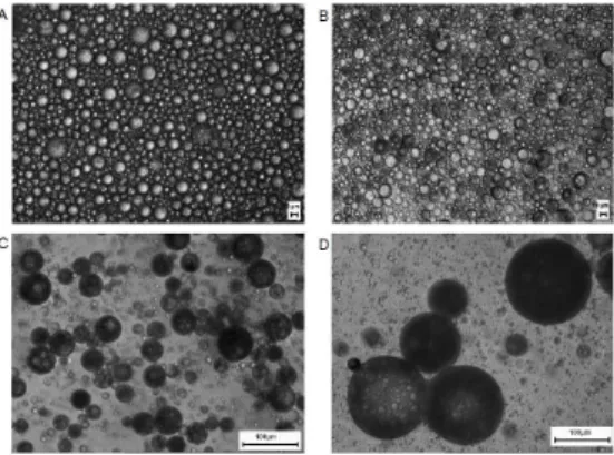
Rungseevijitprapa et al have studied the myotoxicity potential of solvents used for preparation of polymer solutions and O/W-ISM systems. The acute myotoxicity was investigated using in vitro isolated rodent skeletal muscle model by measuring the cumulative CK efflux. Phenytoin and isotonic sodium chloride solution served as positive and negative controls, respectively. The results obtained from in vitro myotoxicity studies suggested that five partially water miscible solvents caused muscle damage in the following rank order: benzyl alcohol > triethyl citrate > triacetin > propylene carbonate > ethyl acetate. Microscopic pictures of O/W-ISM formulation with varying solvent systems for the polymer are presented in Figure 5. Myotoxicity of ethyl acetate was comparable to that of isotonic sodium chloride solution. Emulsions of undiluted solvents and an aqueous 0.5 % Pluronic F 68 solution (ratio 1:4) could dramatically reduce the myotoxicities to 24 – 65 %. The myotoxicity of O/W-ISM system was less than those of the polymer solutions and undiluted solvents. The cumulative CK level obtained from muscle-treated O/W-ISM with phase ratio 1:4 was comparable to those obtained from the negative controls. The area
Figure 5: Photomicrograohs of O/W-ISM
formulations comprising internal phase (40% PLGA 502H in solvent) and external phase (0.5% Lutrol F 68 with a phase ratio of 1:1); (A) 60% ethyl acetate; (B) 40% ethyl acetate/20% PEG 400; (C) 30% ethyl acetate/30% PEG 400 and (D) 20% ethyl acetate/40% PEG 400. Magnification 20 x [39].
under CK plasma curve obtained following intramuscular injection of formulations to Sprague–Dawley rats was used to evaluate in vivo myotoxicity. In vivo results correlated well with in vitro data and confirmed the good muscle compatibility of O/W-ISM formulations [39].
Recent studies by a firm (Rovi) have enabled it to develop commercial formulations of “in situ microparticles” (ISM™) for new therapeutic applications. Extended release formulations based on ISM™ technology are currently being developed for psychiatric and oncologic drugs due to their industrial potential, as well as commercial and sanitary interest. In September 2010, the experimental stage has begun for the first Phase I trial of Risperidone-ISM™ on healthy volunteers, which is the first candidate for this drug delivery system. This first trial aims mainly to evaluate the pharmacokinetics and tolerability of a single intramuscular administration of Risperidone in an ISM™ formulation. This trial will serve not only to confirm the pharmacokinetic profile of this innovative depot formulation for the monthly administration of an approved anti-psychotic, but it will also serve as a proof of concept for validating ISM technology as a base platform for other developments. In this regard, other new formulations with ISM™, for monthly administration of another widely used anti-psychotic, and for quarterly administration of an approved aromatase inhibitor that is currently used extensively in the treatment of hormone-dependent breast cancer, are already in a pre-clinical phase [47].
CONCLUSION
In situ forming microparticle (ISM) systems offer a new encapsulation technique that provides prolonged release of drug along with much greater ease of preparation and administration than conventional micropar-ticles and surgically implanted systems. ISMs are an attractive alternative to parenteral drug delivery, especially those prepared by existing complicated microencapsulation
methods. Moreover, recent developments in this field such as the first Phase I trial of Risperidone-ISM™ suggest there is a possibility for prolonged release of bioactive molecules applied as intramuscular injection.
REFERENCES
1. Benoit JP, Marchais H, Rolland H, Vande-Velde V. Biodegradable microspheres: advances in production technology. In Benita, S, Ed. Microencapsulation methods and industrial applications. New York: Marcel Dekker; 1996; pp 35–72.
2. Rothen-Weinhold A, Besseghir K, Vuaridel E, Sublet E, Oudry N, Kubel F, Gurny R. Injection-molding versus extrusion as manufacturing technique for the preparation of biodegradable implants. Eur J Pharm Biopharm 1999; 48: 113–121.
3. Jalil R, Nixon JR. Biodegradable poly(lactic acid) and poly(lactide-co-glycolide) microcapsules: problems associated with preparative techniques and release properties. J Microencapsulation 1990; 7: 297-325. 4. Tice TR, Tabibi ES. Parenteral drug delivery:
injectables. In Kydonieus, A, editor. Treatise on controlled drug delivery: Fundamentals optimization, applications. New York: Marcel Dekker; 1991; 1991; pp 315-339.
5. Wu XS. Synthesis and properties of biodegradable lactic/glycolic acid polymers. In: Wise DL; Trantolo DJ; Altobelli DE; Yaszemski MJ; Gresser JD; Schwartz ER, editors. Encyclopedic handbook of biomaterials and bioengineering. Part A: Materials, New York: Marcel Dekker; 1995; pp 1015-1054.
6. Wu XS. Preparation, characterization, and drug delivery applications of microspheres based on biodegradable lactic/glycolic acid polymers. In: Wise DL; Trantolo DJ; Altobelli DE; Yaszemski MJ; Gresser JD; Schwartz ER, editors. Encyclopedic handbook of biomaterials and bioengineering. Part A: Materials, New York: Marcel Dekker; 1995; pp 1151-1200.
7. Lewis DH. Controlled release of bioactive agents from lactide/glycolide polymers. In Chasin M; Langer R, editors. Biodegradable polymers as drug delivery systems. New York: Marcel Dekker; 1990; pp 1-41.
8. Schultz K, Mollgaard B, Fisher AN, Illum L, Larsen C. Intramuscular rate of disappearance of oily vehicles in rabbits investigated by gamma-scintigraphy. Int J Pharm 1998; 169: 121–126. 9. Bjerregaard S, Pedersen H, Vedstesen H,
Vermehren C, Söderberg I, Frokjaer S. Parenteral water/oil emulsions containing hydrophilic compounds with enhanced in vivo retention: formulation, rheological characterization and study of in vivo fate using
whole body gamma-scintigraphy. Int J Pharm 2001; 215: 13-27.
10. Bjerregaard S, Pedersen H, Wulf-Andersen L, Stephens RW, Lund LR, Vermehren C, Söderberg I, Frokjaer S. Sustained elevated plasma aprotinin concentration in mice following intraperitoneal injections of w/o emulsions incorporating aprotinin. J Control Release 2001; 71: 87–98.
11. Dunn RL, English JP, Cowsar DR, Vanderbilt DP. Biodegradable in-situ forming implants and methods of producing the same. US Patent 4938763, 1990. http://www.google.com/pat ents/about/4938763_Biodegradable_in_situ_fo rming_ im.html?id=DRYmAAAAEBAJ Accessed 27 July, 2011. Available from: http://www.patents.com/us-4938763.html 12. Tipton AJ, Fujita SM. A biodegradable injectable
delivery system for nonsteroidal anti-inflammatory drugs. Pharm Res 1991; 9 (Suppl): S-196.
13. Shah NH, Railkar AS, Chen FC, Tarantino R, Kumar S, Murjani M, Palmer D, Infeld MH, Malick AW. A biodegradable injectable implant for delivering micro and macromolecules using poly (lactic-co-glycolic) acid (PLGA) copolymers. J Control Release 1993; 27: 139– 147.
14. Coonts BA, Lowe BK, Norton RL, Tipton AJ, Yewey GL, Dunn RL. Plasma concentrations of naltrexone following subcutaneous and intramuscular injections of Atrigel™ formulations in dogs. Pharm Res 1993; 10: S-192.
15. Duysen EG, Yewey GL, Southard JL, Dunn RL, Huffer W. Release of bioactive growth factors from the Atrigel delivery system in tibial defect and dermal wound models. Pharm Res 1993; 10: S-83.
16. Lambert WJ, Peck KD. Development of an in-situ forming biodegradable poly-lactide-co-glycolide system for the controlled release of proteins. J Control Release 1995; 33: 189– 195.
17. Radomsky ML, Brouwer G, Floy BJ, Loury DJ, Chu F, Tipton AJ, Sanders LM. The controlled release of ganirelix from the Atrigel™ injectable implant system. Proc Int Symp Control Rel Bioact Mater 1993; 20: 458–459. 18. Alğın-Yapar E, Arı N, Baykara T. Investigation of in
vitro and in vivo performance of injectable in situ implants. Turkish J Pharm Sci 2010; 7(1): 9-20.
19. Ahmed TA, Ibrahim HM, Ibrahim F, Samy AM, Kaseem A, Nutan MT, Hussain MD. Development of injectable in situ implant and microparticle formulations for controlled delivery of montelukast. AAPS Journal 2010; 12: 78 (S2). www.aapsj.org/abstracts/ AM_2010/R6278.pdf Accessed 05 August, 2011
20. Ahmed TA, Ibrahim HM, Khalifa S, Samy AM, Kaseem A, Nutan MT, Hussain MD. Controlled release of haloperidol from biodegradable injectable in situ implant and microparticle formulations. AAPS Journal 2010; 12: 77 (S2). www.aapsj.org/abstracts/AM_2010/R6277.pdf. Accessed 05 August 2011.
21. Wischke C, Zhang Y, Mittal S, Schwendeman SP. Development of PLGA-based injectable delivery systems for hydrophobic fenretinide. Pharm Res 2010; 27: 2063–2074. (DOI 10.1007/s11095-010-0202-y)
22. Higuchi T. Mechanism of sustained-action medication. J Pharm Sci 1963; 52: 1145–1149. 23. Alğın-Yapar E, Baykara T. Effects of variations in formulation components on drug release from injectable in situ implants. Med Sci Tech Res 2009; 2(2): 21-29.
24. Alğın-Yapar E, Baykara T. Effects of solvent combinations on drug release from injectable phase sensitive liquid implant systems. Turkish J Pharm Sci 2010; 7(1): 47-54.
25. Eliaz RE, Kost J. Characterization of a polymeric PLGA-injectable implant delivery system for controlled release of proteins. J Biomed Mater Res 2000; 50: 388-396.
26. McHugh AJ. The role of polymer membrane formation in sustained release drug delivery systems. J Control Release 2005; 109: 211-221.
27. Bakhshi R, Vasheghani-Farahani E, Mobedi H, Jamshidi A, Khakpour M. The effect of additives on naltrexone hydrochloride release and solvent removal rate from an injectable in situ forming PLGA implant. Polym Adv Technol 2006; 17: 354-359.
28. Bodmeier R. Verfahren zur in-situ Herstellung von Partikeln. Offenlegungsschrift, DE. Patent 1997; 197,24,784.
29. Kranz H, Bodmeier R. A biodegradable in-situ forming system for controlled drug release. Pharm Sci 1998; 1 (Suppl): 414.
30. Rungseevijitprapa W, Bodmeier R. Injectability of biodegradable in situ forming microparticle systems (ISM). Eur J Pharm Sci 2009; 36(4-5): 524–531.
31. Luan X, Bodmeier R. In situ forming microparticle system for controlled delivery of leuprolide acetate: influence of the formulation and processing parameters. Eur J Pharm Sci 2006; 27: 143-149.
32. Luan X, Bodmeier R. Influence of the poly(lactide-co-glycolide) type on the leuprolide release from in situ forming microparticle systems. J Control Release 2006; 110: 266-272.
33. Kranz H, Bodmeier R. A novel in situ forming drug delivery system for controlled parenteral drug delivery. Int J Pharm 2007; 332: 107–114. 34. Kranz H, Brazeau GA, Napaporn J, Martin RL,
Millard W, Bodmeier R. Myotoxicity studies of injectable biodegradable in-situ forming drug delivery systems. Int J Pharm 2001; 212: 11– 18.
35. Jain RA, Rhodes CT, Railkar AM, Malick AW, Shah NH. Controlled release of drugs from injectable in situ formed biodegradable PLGA microspheres: effect of various formulation variables. Eur J Pharm Biopharm 2000; 50(2): 257-262.
36. Kranz H, Bodmeier R. Structure formation and characterization of injectable drug loaded biodegradable devices: In situ implants versus in situ microparticles. Eur J Pharm Sci 2008; 34: 164–172.
37. Kranz H, Yılmaz E, Brazeau GA, Bodmeier R. In vitro and in vivo drug release from a novel in situ forming drug delivery system. Pharm Res 2008; 25(6): 1347-1354. (DOI: 10.1007/s110 95-007-9478-y.)
38. Jain RA. The manufacturing techniques of various drug-loaded biodegradable poly(lactide-co-glycolide) (PLGA) devices. Biomaterials 2000; 21: 2475-2490.
39. Rungseevijitprapa W, Brazeau GA, Simkins JW, Bodmeier R. Myotoxicity studies of O/W-in situ forming microparticle systems. Eur J Pharm Biopharm 2008; 69(1): 126–133.
40. Jain RA, Rhodes CT, Railkar AM, Malick AW, Shah NH. Comparison of various injectable protein-loaded biodegradable poly(Lactide-co-glycolide) (PLGA) devices: in-situ-formed implant versus in-situ-formed microspheres versus isolated microspheres. Pharm Dev Tech 2000; 5(2): 201-207.
41. Jain RA, Rhodes CT, Railkar AM, Malick AW, Shah NH. Controlled release of drugs from a novel injectable in situ formed biodegradable PLGA microsphere system. Pharm Sci 1998; 1(Suppl): 1298.
42. Jain RA, Rhodes CT, Railkar AM, Malick AW, Shah NH. Comparision of various protein loaded biodegradable PLGA devices. Proceedings of the International Symposium on Control Rel Bioact Mater 1999; p 26. 43. Jain R. In situ microencapsulation. [PhD
dissertation]. [Kingston, Rhode Island]: The University of Rhode Island, 1998.
44. Körber M, Bodmeier R. Development of an in situ forming PLGA drug delivery system I. Characterization of a non-aqueous protein precipitation. Eur J Pharm Sci 2008; 35: 283–292.
45. Dong WY, Körber M, Esguerra LV, Bodmeier R. Stability of poly(D,L- lactide-co-glycolide) and leuprolide acetate in in-situ forming drug delivery systems. J Control Release 2006; 115: 158–167.
46. Ibrahim HM, Ahmed TA, Ismail HR, Samy AM, Kaseem AA, Hussain MD, Nutan MT. Development of extended release biodegradable injectable in situ microparticle formulation of meloxicam. AAPS Journal 2010; 12: 89. (S2). Available from: www.aapsj.org/abstracts/AM_2010/R6289.pdf www.aapsj.org/abstracts/AM_2010/R6289.pdf. Accessed 05 August 2011.
47. Prolonged Release Injectable Formulations. Rovi Pharmaceuticals. [Homepage on the Internet] http://www.rovi.es/id_formulaciones_english.ht ml. Accessed 06 August 2011.
![Figure 1: Schematic representation of in situ PLGA microsphere formation process [35]](https://thumb-eu.123doks.com/thumbv2/9libnet/5830262.119376/4.892.146.418.315.720/figure-schematic-representation-situ-plga-microsphere-formation-process.webp)
![Figure 2: Scanning electron micrographs of ISMs (A) prepared with 30 % w/w RG 503H and 5 % w/w drug loading and (B) prepared with 40 % w/w R 202H and 10 % w/w drug loading (2B) [31]](https://thumb-eu.123doks.com/thumbv2/9libnet/5830262.119376/5.892.473.762.211.475/figure-scanning-electron-micrographs-prepared-loading-prepared-loading.webp)
![Figure 4: Illustration of the setup for injection force measurement with chicken meat model [30]](https://thumb-eu.123doks.com/thumbv2/9libnet/5830262.119376/7.892.525.716.713.948/figure-illustration-setup-injection-force-measurement-chicken-model.webp)