A Brief Reconnoitre about Effects of MMP9 on
Aortic Dissection
MMP9 Geninin Aort Diseksiyonundaki Olası Etkilerinin Araştırılması
Burcu Salman Yaylaz
1, Melda Sariman
2, Ahmet Ekmekçi
3, Emel Ergül
4,
Mahmut Uluganyan
5, Fulya Coşan
6, Özgün Melike Gedar Totuk
7, Neslihan Abaci
1 1Department of Genetics, Aziz Sancar Institute of Experimental Medicine, İstanbul University, İstanbul, Turkey2İstinye University Molecular Cancer Research Center, İstanbul, Turkey 3Department of Cardiology, Hospital of Medical Park, İstanbul, Turkey
4Department of Medical Biology and Genetics, Faculty of Medicine, Kocaeli University, Kocaeli, Turkey 5Department of Cardiology, Faculty of Medicine, Bezmialem University, İstanbul, Turkey
6Department of Rheumatology, Faculty of Medicine, Bahçeşehir University, İstanbul, Turkey 7Department of Ophthalmology, Faculty of Medicine, Bahçeşehir University, İstanbul, Turkey
ORCID ID: B.S.Y. 0000-0002-9144-3899; M.S. 0000-0003-0898-529X; A.E. 0000-0001-5424-149X; E.E. 0000-0003-0473-4020; M.U. 0000-0002-4578-4537; F.C. 0000-0002-5630-8640; Ö.M.G.T. 0000-0003-1863-6501; N.A. 0000-0002-9962-4010
Cite this article as: Salman Yaylaz B, Sariman M, Ekmekçi A, Ergul E, Uluganyan M, Coşan F, Gedar Totuk ÖM, Abacı N. A brief reconnoitre about
effects of MMP9 on aortic dissection. Experimed 2021; 11(1): 12-20.
ABSTRACT
Objective: Matrix metalloproteinases (MMPs) are the extracellular
ma-trix regulators that frequently investigate cardiovascular diseases and cancer metastasis. Our study aimed to examine specific polymorphisms in the MMP9 gene in our patients with aortic dissection and compare the effect of MMP9 on aortic dissection with expression datasets.
Materials and Methods: Q279R and P574R polymorphisms were
analyzed in 44 aortic dissection patients and 40 healthy donors via polymerase chain reaction-restriction fragment length polymorphism. (PCR-RFLP) methods. Q279R and P574R prevalence was statistically compared with the medical data of the patients. Additionally, we col-lected datasets of aortic dissection from NCBI GEO to reanalyze GEO2R and RStudio to see metalloproteinase activity on samples. Later, enrich-ment analysis was processed on widely used databases.
Results: Genotypic distribution of alleles was similar in the two study
groups. In addition to this, female CG carriers had a higher risk of de-veloping aortic dissection than those of males. As the results of the protein-protein interaction analysis of MMP9 and patients’ clinical data, hypertension was found to be the significant outcome of P574R varia-tion in the patients. In array analysis, MMP9 expression did not change critically, but TIMPs had been downregulated in many samples. Also,
MMP9 targeted miRNA expression levels were detected as low in aortic
tissue and blood.
Conclusion: Q279R and P574R are two polymorphisms that do not
di-rectly affect MMP9 protein structure. Consequently, studied polymor-phisms and performed meta-analysis show that MMP9 does not spark off the phenotype but sets the stage for aortic dissection development as seen in the statistical results. Furthermore, enrichment analysis on datasets shows MMP9 was not a primary reason for vascular remodeling.
Keywords: Aortic dissection, MMP9, Q279R, P574R, gene expression
data
ÖZ
Amaç: Kardiyovasküler hastalıklar ve kanser metastazında sıklıkla
araştı-rılan matriks metalloproteinazlar (MMPs) ekstraselüler matriks düzenle-yicileridir. Çalışmamız, aort diseksiyonu olan hastalarda MMP9 genindeki spesifik polimorfizmleri incelemeyi ve MMP9'un aort diseksiyonu üzerin-deki etkisini ekspresyon veri setleri ile karşılaştırmayı amaçlamaktadır.
Gereç ve Yöntem: Q279R ve P574R polimorfizmleri 44 aort diseksiyon
tanısı almış ve 40 sağlıklı bireyde polimeraz zincir reaksiyonu - restrik-siyon parça uzunluğu polimorfizmi (PCR-RFLP) yöntemiyle çalışıldı. Q279R ve P574R prevalansı istatistiksel olarak hastaların tıbbi verileriyle karşılaştırıldı. Buna ek olarak, NCBI GEO veri tabanından aort diseksiyon veri setleri toplandı ve MMP9 ifadesindeki farklılıkları görmek amacıyla bu veri setleri GEO2R ve RStudio ile yeniden analiz edildi. Elde edilen sonuçların informatik analizi için çevrimiçi veri tabanları kullanıldı.
Bulgular: CG alleli taşıyıcısı kadınların aort diseksiyonu geliştirme riski
erkeklerden daha yüksek bulunmasına rağmen her iki çalışma grubun-da grubun-da allellerin genotipik grubun-dağılımı benzer bulunmuştur. MMP9'un pro-tein-protein etkileşim analizinin ve hastaların tıbbi verilerinin incelen-mesinin sonucu olarak, P574R hipertansiyonu olan hastalarda önemli bir bulgu olarak değerlendirilmiştir. Array verisi analizinde ise MMP9 ifa-desinde kritik bir değişim gözlemlenmemiş olup, birçok örnekte TIMP ifade seviyelerinde azalma tespit edilmiştir. Ayrıca MMP9’u hedefleyen miRNA ekspresyon seviyelerinin aort dokusu ve kanda düşük olduğu saptanmıştır.
Sonuç: Q279R ve P574R, MMP9 protein yapısını doğrudan etkilemeyen
iki polimorfizmdir. İncelenen polimorfizmler ve gerçekleştirilen meta-a-nalizler, MMP9'un fenotipi doğrudan etkilemediği, ancak istatistiksel sonuçlarda görüldüğü gibi aort diseksiyonu gelişimi için zemin hazır-ladığını göstermektedir.
Anahtar Kelimeler: Aort diseksiyon, MMP9, Q279R, P574R, gen ifade
verisi
Corresponding Author/Sorumlu Yazar: Neslihan Abacı E-mail: [email protected] Submitted/Başvuru: 09.02.2021 Accepted/Kabul: 21.03.2021
Content of this journal is licensed under a Creative Commons Attribution-NonCommercial 4.0 International License.
13
INTRODUCTION
Aortic dissection is caused by the creation of a false lumen in the aorta. An intimomedial tear allows blood flow to enter the aortic wall by creating a false lumen or a secondary channel, called an aortic dissection, a catastrophic manifestation of the acute aortic syndrome. Aortic dissection has the highest mor-tality rate among cardiovascular diseases due to the rising of lethality rate at 1% per hour. Diagnosis of dissection is done by scanning the aorta and its branches with computed to-mography (1-3). Predisposition to acute aortic dissection has been associated with atherosclerosis vasculitis, bicuspid aortic valves, male gender, long-term arterial hypertension, and col-lagen-based disorders such as Marfan’s and Ehlers-Danlos syn-dromes (4-6).
Matrix metalloproteinases (MMPs) are extracellularly acting, zinc-dependent endopeptidases that break the peptide bonds of nonterminal amino acids. MMPs are involved in the devel-opmental stages of the extracellular matrix and have been im-plicated in several collagen-based disorders (7,8). MMPs bind to adhesion and extracellular matrix proteins and have been divided into types based on their substrates, including mem-brane, gelatinase, stromelysin, matrilysin, and collagenase (9,10). Matrix metalloproteinase 9 (MMP9), also known as the 92 kDa type IV collagenase or gelatinase B, is categorized in the gelatinase subgroup and has a significant role in extracellular matrix degradation (7,8). MMP9 in humans contains an NH2-ter-minal prodomain, a COOH-terNH2-ter-minal hemopexin-like domain, a catalytic domain, and a linker domain. The MMP9 gene is posi-tioned at the chromosome region 20q11.2-13.1.8 and has 13 exons and 12 introns. MMP9 is secreted from various types of cells, such as fibroblasts, macrophages, and neutrophils (8-11).
MMP9 is positively regulated by transcription factors (such as
NF-kappaβ and activator protein-1) and polyomavirus en-hancer A-binding protein-3. Tissue inhibitors of metallopro-teinases inhibit MMP9 by binding to the zymogen forms of the enzyme. Recent studies have revealed that MMP9 polymor-phisms are associated with cardiovascular diseases, such as hypertension, myocardial infarction, and atherosclerosis. The most thoroughly investigated polymorphisms in the literature are: C-1562T, Q279R, 836GA, R668Q, and P574R (8).
In this study, we investigated the MMP9, Q279R and P574R vari-ations in Turkish aortic dissection patients. In addition, we an-alysed our results with the published epigenome, expression, and miRNA array data.
MATERIALS AND METHODS
Our study was designed as a retrospective case-control study, and a case group was built comprising 44 patients who under-went aortic dissection surgery between 2007 and 2011. The patient’s diagnosis was confirmed with echocardiography and computed tomography imaging techniques that demonstrat-ed an intimal flap with a true and a false lumen. As hypercho-lesterolemia, diabetes mellitus, hypertension, and hypertri-glyceridemia are risk factors for aortic dissection, the patients’ blood pressure, fasting glucose, triglyceride, and low-density lipoprotein levels were recorded. Iatrogenic, syndromic, and traumatic aortic dissection patients were excluded from the study. The control group consisted of 40 individuals with nor-mal findings on their physical and echocardiographic exam-inations. This study has the ethics committee approval from Istanbul University (Istanbul Medical Faculty Clinical Research Ethics Committee; 2018/1252), and an informed consent was obtained from all selected individuals.
Genomic DNA was extracted from the peripheral blood of both the patient and control group subjects. The polymerase chain reaction (PCR) was used to amplify the targeted genomic se-quences. The primers designed for the targeted regions are shown in Table 1. Amplification was conducted using a total volume of 25 µl comprising: 100 ng genomic DNA; a reaction buffer includes 25 mM magnesium chloride and 10X potas-sium chloride; 1 U Taq polymerase; 2.5 mM deoxynucleotide; and 10 pmol of each primer. The PCR thermal cycling steps were performed at 95°C for 2 minutes (initiation), 94°C for 30 seconds (denaturation), 55°C for 45 seconds (annealing), 72°C for 30 seconds (extension), and 72°C for 5 minutes (final exten-sion). The PCR products were analyzed on a 2% agarose gel, and electrophoresis was performed at 80 V for 30 minutes. The prevalence of two single nucleotide polymorphisms of the
MMP9 gene, Q279R, and P574R, was evaluated in the patient
and control groups. The Q279R polymorphism has three gen-otypes: the common genotype is AA, and the less common genotypes are AG and GG. The other studied single nucleo-tide polymorphism, P574R, has only two genotypes: the wide-spread genotype is CC, and the less common genotype is CG. The reaction products were digested with the MspI and BsrBI restriction enzymes. For the digestion step, 20 µl of PCR prod-uct was mixed with 5 µl of digestion mix including 1 µl dH2O, 1.5 µl restriction enzyme, and 2.5 µl tango buffer. The samples were incubated at 37°C for 3 hours, and then electrophoresis was done for identification of the genotype.
Table 1. Details of enzyme digestion analysis
Polymorphisms Primer PCR Product (bp) Restriction Enzyme
Q279R (rs17576) 5’-GGCCCAATTTTCTCATCTGAG-3’
3’-GAGCTTGTCCCGGTCGTA-5’ 292 MspI
14
The Statistical Package for the Social Sciences software for Win-dows (SPSS version 25, IBM Corporation, Armonk, New York, USA) was used to perform all the statistical analyses. The Har-dy-Weinberg equilibrium-based genotypes distribution pre-diction was used to analyze the study and control groups. The genotype distribution frequencies of the patient and control groups were compared using Fisher’s exact test. The clinical features of patients and their genotype characteristics were analyzed by one-way analysis of variance, Tukey’s honest sig-nificant difference tests, and the independent sample t-test. The p values <0.05 were considered significant.
To perform the meta-analysis array data, datasets of aor-tic dissection patients had gotten NCBI GEO database. GSE84274, GSE52093, GSE98770 were used to analyze gene expression levels, GSE92427 was selected to explore miRNA levels on aortic dissection levels (12-15). Although datasets had more than one aortic tissue disease, we chose only the aortic dissection patients’ data and control groups. First, cal-culations were performed through the medium of a ready-to-use GEO2R program. Then,we checked the results with the Bioconductor package of R. Top 100 genes, according to their adjusted p-value, were selected to analyze. Obtained gene functions and relation with MMP9 were analyzed with STRING-db, UNIPROT, NCBI, PANTHER, and other commonly used databases (16-19).
RESULTS
The genotype distribution characteristics of the patient and control groups are shown in Table 2. Q279R and P574R were investigated in 84 and 77 individuals, respectively. Both the patient and control groups had similar percentages of males and females. There was a small genotype difference between the females and the males in the group of aortic dissection pa-tients. More than half of the total number of individuals had widespread genotypes for two polymorphisms: the AA geno-type was present in 56.8% of the patient group and 60% of the control group; the CC genotype was present in 87.8% of the pa-tient group and 78% of the control group (AA for Q279R, CC for P574R) (p=0.943 for Q279R). Ancestral allele frequencies were different in the AD patients and dbSNP data for the two SNPs, however, they were not found statistically significant (p=0.721 for allele A, and p=0.384 for allele C). Statistical analysis re-vealed an association between the P574R and aortic dissection (odds ratio=0.486, p=0.361). Also, there was a different distribu-tion of P574R between the male and female aortic dissecdistribu-tion patients. Female individuals had a higher risk than males (odds ratio=0.750, p=0.361). However, in datasets, the male percent-age is higher than females. (19 males vs 4 female patients) The medications taken by the aortic dissection patients (n=44) with respect to the genotypes of Q279R and P574R are given in Table 3. Six different pharmaceuticals were selected, and their
Table 2. Genotypes of male and female subjects in the control and aortic dissection patient groups
Q279R P574R AA AG GG CC CG n % n % n % Total n % n % Total Control Female 16 40 9 22.5 1 2.5 26 21 58.3 4 11.1 25 Male 8 20 4 10 2 5 14 7 19.4 4 11.1 11 Patients Female 19 43.2 12 27.3 3 6.8 34 27 65.8 4 9.7 31 Male 6 13.6 3 6.8 1 2.3 10 9 21.9 1 2.4 10 p-value 0.943 0.361
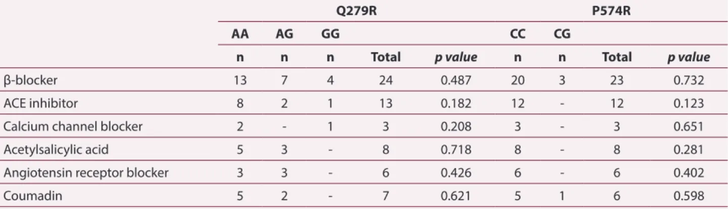
Table 3. Drugs currently used by aortic dissection patients with respect to the genotypes of Q279R and P574R
Q279R P574R
AA AG GG CC CG
n n n Total p value n n Total p value
β-blocker 13 7 4 24 0.487 20 3 23 0.732
ACE inhibitor 8 2 1 13 0.182 12 - 12 0.123
Calcium channel blocker 2 - 1 3 0.208 3 - 3 0.651
Acetylsalicylic acid 5 3 - 8 0.718 8 - 8 0.281
Angiotensin receptor blocker 3 3 - 6 0.426 6 - 6 0.402
Coumadin 5 2 - 7 0.621 5 1 6 0.598
15
relationships with the polymorphisms were analyzed. It was clear that the angiotensin-converting enzyme (ACE) inhibitor, calcium channel blocker, and acetylsalicylic acid usage was in harmony with MMP9 polymorphisms. (odds ratio=0.682 for coumadin, and odds ratio=0.952 for β-blocker).
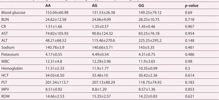
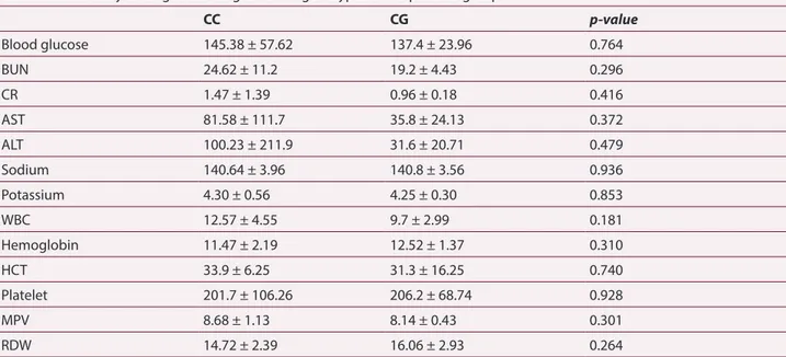
According to their blood features, the genotype distributions of the aortic dissection patients are shown in Tables 4, 5, and 6.
The patients were grouped according to their genotypes. The mean and standard deviation values of the blood features and the widespread genotype for each group based on p-values and chi-squared analysis are given in Table 4. Blood glucose and potassium levels of the AG genotype as well as the alanine aminotransferase and platelet levels of the GG genotype were relative levels of significance.
Alleles were compared with the blood features of the aortic dissection patients. The results of this comparison indicate that
MMP9 was associated with disease progenitors. There was a
distinguishable difference between alleles A and G. The allele G carriers had a risk of developing aortic dissection compared to the allele A carriers.
According to the De Bakey classification, there are three different types of aortic dissection. Type 2 involves the ascending aorta, type 3 involves the descending aorta, and type 1 aortic dissec-tion includes both. A large majority of our patients had type 1 aortic dissections. The genotype distribution of the studied sin-gle nucleotide polymorphisms across aortic dissection types is given in Table 7. Though there was no statistical significance be-tween the two polymorphisms and aortic dissection type, P574R has a higher impact probability on aortic dissection than Q279R. After these statistical analyses, MMP9 expression and methyla-tion levels had not been changed on study groups of selected datasets in comparison with control groups as we expected—all these expression data were obtained from dissected aorta tissue that has the low secretion of gelatinase proteins.
Nevertheless, circulating miRNA levels had been determined by plasma, not from aortic tissue, and MMP9 targeted miRNAs
Table 4. Laboratory findings according to Q279R genotypes in the patients group
AA AG GG p-value Blood glucose 153.04±60.98 131.53±26.38 149.25±79.12 0.69 BUN 24.62±12.58 24.06±9.09 28.25±10.75 0.718 CR 1.51±1.66 1.35±0.57 1.45±0.46 0.967 AST 74.82±105.93 90.8±124.32 83.25±76.18 0.954 ALT 48.21±68.52 115.46±270.6 225.25±295.2 0.148 Sodium 140.78±3.9 140.66±3.71 143±5.35 0.461 Potassium 4.17±0.55 4.49±0.54 4.31±0.75 0.486 WBC 12.31±4.8 12.29±3.96 11.9±3.65 0.98 Hemoglobin 11.51±2.33 11.9±1.77 10.35±0.99 0.3 HCT 34.03±6.50 33.48±10 30.42±2.36 0.614 PLT 201.34±113.7 207.13±80.29 118.75±74.92 0.183 MPV 8.51±0.92 8.8±1.20 8.57±1.36 0.853 RDW 14.66±2.53 15.35±2.57 14.22±0.83 0.621
(BUN: blood urea nitrogen, CR: creatinine, AST: aspartate aminotransferase, ALT: Alanine aminotransferase, WBC: white blood cells, HCT: hematocrit, PLT: platelet, MPV: mean platelet volume, RDW: red cell distribution width. Data are given as mean ± standard deviation.)
Table 5. Statistical comparison (p-value) of laboratory findings
according to Q279R genotypes in the patients group
AA vs. AG AA vs. GG AG-GG vs. AA Blood glucose 0.45 0.99 0.284 BUN 0.987 0.815 0.921 CR 0.93 0.996 0.734 AST 0.902 0.989 0.674 ALT 0.536 0.207 0.131 Sodium 0.996 0.562 0.762 Potassium 0.215 0.89 0.112 WBC 1 0.984 0.938 Hemoglobin 0.84 0.571 0.919 HCT 0.976 0.67 0.622 PLT 0.983 0.292 0.687 MPV 0.708 0.995 0.478 RDW 0.678 0.942 0.556
(BUN: blood urea nitrogen, CR: creatinine, AST: aspartate aminotransferase, ALT: Alanine aminotransferase, WBC: white blood cells, HCT: hematocrit, PLT: platelet, MPV: mean platelet volume, RDW: red cell distribution width.)
16
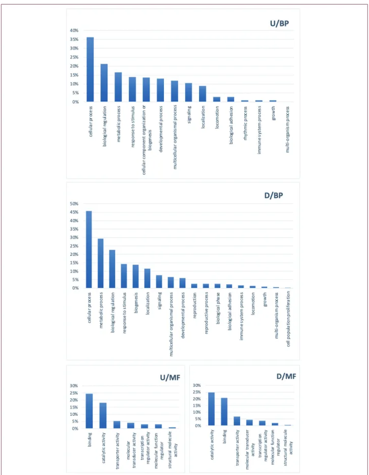
were found downregulated. All miRNAs were checked from Tar-getScanHuman (version 7.2) and miRWalk (version 2.0) (20,21). 100 genes from each dataset were analyzed to find the biolog-ical pathways they relate to. MMP9 expression did not critbiolog-ically change in studies, but regulators of MMP9, TIMP1, and TIMP2 had been shown expression differences. However, upregulated genes were found to meet in a typical biological process, mitotic cell cycle regulation, and muscle cell construction. Downregulated genes play roles in cellular adhesion and developmental process-es like angiogenprocess-esis. Also, thprocess-ese genprocess-es had a different affinity to chemicals and cofactors. Analysis results are given in Figure 1.
DISCUSSIONS
MMPs are known with their roles in collagen-based diseases, such as vascular rearrangement and devastation. They are divided into ligand subgroups, one of which is MMP9. A col-lagenase involved in extracellular matrix degradation, MMP9 directly affects collagen-based structures and has been impli-cated in several vascular diseases. Research on the relationship between MMPs and aortic dissection has primarily focused on the concentrations of matrix metalloproteinase proteins and inhibitors (TIMPs) in plasma and tissue.
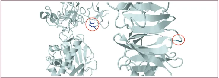
We investigated two amino acid changes, Q279R and P574R, located on different exons of the MMP9 gene. The PolyPhen-2 prediction tool classifies Q279R and P574R as benign alleles (22) . Conformational changes in protein structure which result from Q279R and P574R variations are shown in Figure 2 (23-26). Moreover, MMP9 and TIMP1/2 interactions come into existence by hemopexin-like domains of MMP9 and the C terminal of
TIMP. P574R affects a hairpin structure in its place as a result of
this docking of two proteins (27).
Insights into this structural information, we decided to focus on the proteins that bind directly to MMP9. In datasets GSE52093 and GSE84274, we ascertained proteins related to T cell regula-tion, hemopoiesis, angiogenesis, chemokine regularegula-tion, extra-cellular matrix binging, and vascular morphogenesis mission. Interestingly in the dataset GSE52093, some of the vital protein kinases which play roles in DNA damage, cell cycle, and apop-totic process are upregulated in dissected aorta tissue. There was no significant association between genotypes and the patients’ medications, which shows a tendency toward aortic dissection. Previous studies have shown that MMP9 is strongly linked to hypertension due to its vascular
remodel-Table 6. Laboratory findings according to P574R genotypes in the patients group
CC CG p-value Blood glucose 145.38 ± 57.62 137.4 ± 23.96 0.764 BUN 24.62 ± 11.2 19.2 ± 4.43 0.296 CR 1.47 ± 1.39 0.96 ± 0.18 0.416 AST 81.58 ± 111.7 35.8 ± 24.13 0.372 ALT 100.23 ± 211.9 31.6 ± 20.71 0.479 Sodium 140.64 ± 3.96 140.8 ± 3.56 0.936 Potassium 4.30 ± 0.56 4.25 ± 0.30 0.853 WBC 12.57 ± 4.55 9.7 ± 2.99 0.181 Hemoglobin 11.47 ± 2.19 12.52 ± 1.37 0.310 HCT 33.9 ± 6.25 31.3 ± 16.25 0.740 Platelet 201.7 ± 106.26 206.2 ± 68.74 0.928 MPV 8.68 ± 1.13 8.14 ± 0.43 0.301 RDW 14.72 ± 2.39 16.06 ± 2.93 0.264
(BUN: blood urea nitrogen, CR: creatinine, AST: aspartate aminotransferase, ALT: Alanine aminotransferase, WBC: white blood cells, HCT: hematocrit, PLT: platelet, MPV: mean platelet volume, RDW: red cell distribution width. Data are given as mean ± standard deviation.)
Table 7. Aortic dissection types and genotype distributions
Q279R P574R
AA AG GG Total p value CC CG Total p value
Type 1 22 13 4 39 32 4 36
Type 2 1 - - 1 0.860 1 - 1 0.497
17
Figure 1. PANTHER enrichment results of all datasetsincluding top 100 upregulated and downregulated genes. (D: downregulated18
ing properties (28). In our study, one out of every three aortic dissection patients used drugs for hypertension, a significant risk factor for aortic dissection (29). Calcium channel blockers, ACE inhibitors acetylsalicylic acid usage differed significantly among the patients carrying the homozygous ancestral alleles. The patients carrying the G alleles showed obvious differences in alanine aminotransferase, potassium, platelet levels for those with Q279R, potassium, and white blood cell levels P574R. Our blood testing and genotyping analyses also demonstrat-ed platelet differences. A correlation was found between the
MMP9 genotypes and acetylsalicylic acid, which blocks the
blood’s coagulation mechanism (30). Blood platelets and red blood cells play an essential role in coagulation. In the dataset GSE52093, JAK2 plays a role in blood coagulation expression level measured as downregulated. However, the function of
MMP9 in the coagulation mechanism is still unknown. Future
studies can be designed to explore the effects of MMPs on the ion channels of the cell membrane.
MMP9 affects cell membrane structure and cell proliferation.
The expression of the mitotic cell cycle regulator genes had different fold changes in aorta tissue. We may speculate that single nucleotide changes on MMP9 might disrupt the cell membrane’s calcium and potassium channels via binding regulatory proteins as SULF2. Calcium channels have differ-ent functions in differdiffer-ent cell types; in cardiac tissue, calcium channels are involved in excitation-contraction coupling and pace-making properties (31). Potassium channels are directly associated with the calcium ions that activate the potassium channel. Both membrane channels are related to vascular conformation (32).
Recent studies have shown that MMPs break down nonmatrix proteins related to immune processes (33-35). The cells of the immune system not only protect the body against diseases and foreign invaders, but they also repair tissue injuries by prolifer-ation and cell migrprolifer-ation. These processes are triggered by che-motactic signals that are easily modified by MMPs. For instance,
MMP9 modulates T-cell function by cleaving the CD25 receptor
of interleukin-2. In vivo experiments have shown that MMP9 and a second MMP type, MMP-2, assist in T cell migration. In
vitro experiments have indicated that high expression levels
of MMP9 and MMP-2 lead to the increased migratory capacity of type 1 T helper cells. MMP9 activity in both endothelial and T cells directly regulate the behavior of leukocytes and cyto-kine and chemocyto-kine formation. These environmental changes recruit white blood cells, an outcome supported by our study results showing that aortic dissection patients have high blood levels of leukocytes (34). In addition to these findings, aortic dissection types vary among patients. Also, MMP9 targeted proteins like MBP, RUNX2, ANGPT2, and SNAI2, which have roles in blood cell migration and immune response regulation, had a different dissected aorta tissue expression.
In conclusion, MMP9 is a collagenase that directly and/or indirectly influences vascular remodeling mechanisms. Pa-tients carrying the G allele show an increased risk of aortic dissection than the ancestral allele carriers (A for Q279R and C for P574R) in accordance with RFLP results. Our study re-vealed the probable relationship between the cell membrane and MMP9 proteins’ ion channels with the Q279R and P574R polymorphisms. Our findings confirm that MMP9 plays a role in aortic dissection so far as pharmaceuticals listed above explain how deformation occurs on the aorta. Aortic vessel deformations resulting from true or false lumens like aor-tic dissection lead to many cardiovascular diseases. Besides morphological deformation, chemicals also destroy vein structure. We can say that Q279R and P574R polymorphisms of the MMP9 gene affect the extracellular matrix and cause vessel deformation in a roundabout way. Membrane organi-zation defects might not result from MMP9 polymorphisms, but these structural changes disrupt the molecular function of matrix metalloproteinase and form a basis of aortic dissec-tion. These polymorphisms have been studied in the aortic dissection patient group for the first time in Turkey, and it will be a pioneering work for future studies.
19
Ethics Committee Approval: This study has the ethics committee approval from Istanbul University (Istanbul Medical Faculty Clinical Research Ethics Committee; 2018/1252), and an informed consent was obtained from all selected individuals.
Peer-review: Externally peer-reviewed.
Author Contributions: Concept - B.S.Y., M.S., N.A.; Data Collection - B.S.Y., M.S., A.E., M.U., N.A.; Data Analysis and/or Interpretation - B.S.Y., E.E., F.C., Ö.M.G.T., N.A.; Literature Search - B.S.Y., M.S., A.E., M.U., E.E., F.C., Ö.M.G.T., N.A.; Writing - B.S.Y., M.S., N.A.
Conflict of Interest: The authors have no conflict of interest to declare.
Financial Disclosure: This work was funded by Istanbul University Sci-entific Research Project Unit. Project number: BAP-2019K12-149071.
Etik Komite Onayı: Bu çalışma, İstanbul Üniversitesi’nden (İstanbul Tıp Fakültesi Klinik Araştırmalar Etik Kurulu; 2018/1252) etik kurul onayına sahiptir ve seçilen tüm bireylerden bilgilendirilmiş onam alınmıştır.
Hakem Değerlendirmesi: Dış bağımsız.
Yazar Katkıları: Çalışma Konsepti - B.S.Y., M.S., N.A.; Veri Toplama - B.S.Y., M.S., A.E., M.U., N.A.; Veri Analizi/Yorumlama - B.S.Y., E.E., F.C., Ö.M.G.T., N.A.; Literatür taraması - B.S.Y., M.S., A.E., M.U., E.E., F.C., Ö.M.G.T., N.A.; Yazma - B.S.Y., M.S., N.A.
Çıkar Çatışması: Yazarlar çıkar çatışması bildirmemişlerdir.
Finansal Destek: Bu çalışma İstanbul Üniversitesi Bilimsel Araştır-ma Proje Birimi tarafından finanse edilmiştir. Proje nuAraştır-marası: BAP-2019K12-149071.
REFERENCES
1. Mukherjee D, Eagle KA. Aortic dissection--an update. Curr Probl Cardiol 2005; 30: 287-325. [CrossRef]
2. Criado FJ. Aortic dissection: a 250-year perspective. Tex Heart Inst J 2011; 38: 694-700.
3. Gawinecka J, Schönrath F, von Eckardstein A. Acute aortic dissec-tion: pathogenesis, risk factors and diagnosis. Swiss Med Wkly 2017; 147: w14489. [CrossRef]
4. Ekmekçi A, Uluganyan M, Güngör B, Abacı N, Ozcan KS, Ertaş G, et al. Association between endothelial nitric oxide synthase intron 4a/b polymorphism and aortic dissection. Turk Kardiyol Dern Ars 2014; 42: 55-60. [CrossRef]
5. Salhab K, Gioia W, Rabenstein AP, Gubernikoff G, Schubach S. Medical Management of Three Patients with an Acute Type A Aor-tic Dissection: Case Series and a Review of the Literature. Aorta 2018; 6: 98-101. [CrossRef]
6. Sherifova S, Holzapfel GA. Biomechanics of aortic wall failure with a focus on dissection and aneurysm: A review. Acta Biomater 2019; 99: 1-17. [CrossRef]
7. Zitka O, Kukacka J, Krizkova S, Huska D, Adam V, Masarik M, et al. Matrix metalloproteinases. Curr Med Chem 2010; 17: 3751-68.
[CrossRef]
8. Yabluchanskiy A, Ma Y, Iyer RP, Hall ME, Lindsey ML. Matrix metal-loproteinase-9: Many shades of function in cardiovascular dis-ease. Physiology 2013; 28: 391-403. [CrossRef]
9. O'Farrell TJ, Pourmotabbed T. The fibronectin-like domain is re-quired for the type V and XI collagenolytic activity of gelatinase B. Arch Biochem Biophys 1998; 354: 24-30. [CrossRef]
10. Papazafiropoulou A, Tentolouris N. Matrix metalloproteinases and cardiovascular diseases. Hippokratia 2009; 13: 76-82.
11. Brown DL, Hibbs MS, Kearney M, Loushin C, Isner JM. Identifica-tion of 92-kD gelatinase in human coronary atherosclerotic le-sions. Association of active enzyme synthesis with unstable angi-na. Circulation 1995; 9: 2125-31. [CrossRef]
12. Pan S, Lai H, Shen Y, Breeze C, Beck S, Hong T, et al. DNA meth-ylome analysis reveals distinct epigenetic patterns of ascending aortic dissection and bicuspid aortic valve. Cardiovasc Res 2017; 113: 692-704. [CrossRef]
13. Pan S, Wu D, Teschendorff AE, Hong T, Wang L, Qian M, et al. JAK2-centered interactome hotspot identified by an integrative network algorithm in acute Stanford type A aortic dissection. PLoS One 2014; 9: e89406. [CrossRef]
14. Kimura N, Futamura K, Arakawa M, Okada N, Emrich F, Okamura H, et al. Gene expression profiling of acute type A aortic dissection combined with in vitro assessment. Eur J Cardiothorac Surg 2017; 52: 810-7. [CrossRef]
15. Dong J, Bao J, Feng R, Zhao Z, Lu Q, Wang G, et al. Circulating mi-croRNAs: a novel potential biomarker for diagnosing acute aortic dissection. Sci Rep 2017; 7: 12784. [CrossRef]
16. Barrett T, Wilhite SE, Ledoux P, Evangelista C, Kim IF, Tomashevsky M, et al. NCBI GEO: archive for functional genomics data sets--up-date. Nucleic Acids Res 2013; 41: D991-5. [CrossRef]
17. Szklarczyk D, Gable AL, Lyon D, Junge A, Wyder S, Huerta-Ce-pas J, et al. STRING v11: protein-protein association networks with increased coverage, supporting functional discovery in ge-nome-wide experimental datasets. Nucleic Acids Res 2019; 47: D607-D613. [CrossRef]
18. UniProt Consortium. UniProt: a worldwide hub of protein knowl-edge. Nucleic Acids Res 2019; 47: D506-D515. [CrossRef]
19. Mi H, Muruganujan A, Ebert D, Huang X, Thomas PD. PANTHER version 14: more genomes, a new PANTHER GO-slim and improve-ments in enrichment analysis tools. Nucleic Acids Res 2019; 47: D419-D426. [CrossRef]
20. Lewis BP, Burge CB, Bartel DP. Conserved seed pairing, often flanked by adenosines, indicates that thousands of human genes are microRNA targets. Cell 2005; 120: 15-20. [CrossRef]
21. Sticht C, De La Torre C, Parveen A, Gretz N. miRWalk: An online resource for prediction of microRNA binding sites. PLoS One 2018; 13: e0206239. [CrossRef]
22. Adzhubei IA, Schmidt S, Peshkin L, Ramensky VE, Gerasimova A, Bork P, et al. A method and server for predicting damaging mis-sense mutations. Nat Methods 2010; 7: 248-9. [CrossRef]
23. Burley SK, Berman HM, Bhikadiya C, Bi C, Chen L, Di Costanzo L, et al. RCSB Protein Data Bank: biological macromolecular struc-tures enabling research and education in fundamental biology, biomedicine, biotechnology and energy. Nucleic Acids Res 2019; 47: D464-D474. [CrossRef]
24. Elkins PA, Ho YS, Smith WW, Janson CA, D'Alessio KJ, McQueney MS, et al. Structure of the C-terminally truncated human ProM-MP9, a gelatin-binding matrix metalloproteinase. Acta Crystallogr D Biol Crystallogr 2002; 58: 1182-92. [CrossRef]
25. Cha H, Kopetzki E, Huber R, Lanzendörfer M, Brandstetter H. Struc-tural basis of the adaptive molecular recognition by MMP9. J Mol Biol 2002; 320: 1065-79. [CrossRef]
26. SIttisoponpisan S, Islam SA, Khanna T, Alhuzimi E, David A, Ster-nberg MJE. Can Predicted Protein 3D Structures Provide Reliable Insights into whether Missense Variants Are Disease Associated? J Mol Biol 2019; 431: 2197-212. [CrossRef]
20
27. Kuhlman B, Bradley P. Advances in protein structure prediction and design. Nat Rev Mol Cell Biol 2019; 20: 681-97. [CrossRef]
28. Galis ZS, Khatri JJ. Matrix metalloproteinases in vascular remodel-ing and atherogenesis: the good, the bad, and the ugly. Circ Res 2002; 90: 251-62. [CrossRef]
29. Wen D, Zhou XL, Li JJ, Hui RT. Biomarkers in aortic dissection. Clin Chim Acta 2011; 412: 688-95. [CrossRef]
30. Roth GJ, Majerus PW. The mechanism of the effect of aspirin on human platelets. I. Acetylation of a particulate fraction protein. J Clin Invest 1975; 56: 624-32. [CrossRef]
31. Catterall WA, Perez-Reyes E, Snutch TP, Striessnig J. International Union of Pharmacology. XLVIII. Nomenclature and structure-func-tion relastructure-func-tionships of voltage-gated calcium channels. Pharmacol Rev 2005; 57: 411-25. [CrossRef]
32. Sobey CG. Potassium channel function in vascular disease. Arte-rioscler Thromb Vasc Biol 2001; 21: 28-38. [CrossRef]
33. Gong Y, Hart E, Shchurin A, Hoover-Plow J. Inflammatory macro-phage migration requires MMP-9 activation by plasminogen in mice. J Clin Invest 2008; 118: 3012-24. [CrossRef]
34. Smigiel KS, Parks WC. Matrix Metalloproteinases and Leukocyte Activation. Prog Mol Biol Transl Sci 2017; 147: 167-95. [CrossRef]
35. Snitker S, Xie K, Ryan KA, Yu D, Shuldiner AR, Mitchell BD, et al. Correlation of circulating MMP-9 with white blood cell count in humans: effect of smoking. PLoS One 2013; 8: e66277. [CrossRef]