R E S E A R C H A R T I C L E
Open Access
Physiological parameters for Prognosis in
Abdominal Sepsis (PIPAS) Study: a WSES
observational study
Massimo Sartelli
1*, Fikri M. Abu-Zidan
2, Francesco M. Labricciosa
3, Yoram Kluger
4, Federico Coccolini
5, Luca Ansaloni
5,
Ari Leppäniemi
6, Andrew W. Kirkpatrick
7, Matti Tolonen
6, Cristian Tranà
1, Jean-Marc Regimbeau
8, Timothy Hardcastle
9,
Renol M. Koshy
10, Ashraf Abbas
11, Ula
ş Aday
12, A. R. K. Adesunkanmi
13, Adesina Ajibade
14, Lali Akhmeteli
15,
Emrah Ak
ın
16, Nezih Akkapulu
17, Alhenouf Alotaibi
18, Fatih Altintoprak
19, Dimitrios Anyfantakis
20, Boyko Atanasov
21,
Goran Augustin
22, Constança Azevedo
23, Miklosh Bala
24, Dimitrios Balalis
25, Oussama Baraket
26, Suman Baral
27,
Or Barkai
4, Marcelo Beltran
28, Roberto Bini
29, Konstantinos Bouliaris
30, Ana B. Caballero
31, Valentin Calu
32,
Marco Catani
33, Marco Ceresoli
34, Vasileios Charalampakis
35, Asri Che Jusoh
36, Massimo Chiarugi
37, Nicola Cillara
38,
Raquel Cobos Cuesta
39, Luigi Cobuccio
37, Gianfranco Cocorullo
40, Elif Colak
41, Luigi Conti
42, Yunfeng Cui
43,
Belinda De Simone
44, Samir Delibegovic
45, Zaza Demetrashvili
46, Demetrios Demetriades
47, Ana Dimova
22,
Agron Dogjani
48, Mushira Enani
49, Federica Farina
50, Francesco Ferrara
51, Domitilla Foghetti
52, Tommaso Fontana
40,
Gustavo P. Fraga
53, Mahir Gachabayov
54, Grelpois Gérard
55, Wagih Ghnnam
56, Teresa Giménez Maurel
57,
Georgios Gkiokas
58, Carlos A. Gomes
59, Ali Guner
60, Sanjay Gupta
61, Andreas Hecker
62, Elcio S. Hirano
53,
Adrien Hodonou
63, Martin Hutan
64, Igor Ilaschuk
65, Orestis Ioannidis
66, Arda Isik
67, Georgy Ivakhov
68, Sumita Jain
69,
Mantas Jokubauskas
70, Aleksandar Karamarkovic
71, Robin Kaushik
61, Jakub Kenig
72, Vladimir Khokha
73, Denis Khokha
74,
Jae Il Kim
75, Victor Kong
76, Dimitris Korkolis
25, Vitor F. Kruger
53, Ashok Kshirsagar
77, Romeo Lages Simões
78,
Andrea Lanaia
79, Konstantinos Lasithiotakis
80, Pedro Leão
81, Miguel León Arellano
82, Holger Listle
83, Andrey Litvin
84,
Aintzane Lizarazu Pérez
85, Eudaldo Lopez-Tomassetti Fernandez
86, Eftychios Lostoridis
87, Davide Luppi
88,
Gustavo M. Machain V
89, Piotr Major
90, Dimitrios Manatakis
91, Marianne Marchini Reitz
47, Athanasios Marinis
92,
Daniele Marrelli
93, Aleix Martínez-Pérez
94, Sanjay Marwah
95, Michael McFarlane
96, Mirza Mesic
45, Cristian Mesina
97,
Nickos Michalopoulos
98, Evangelos Misiakos
99, Felipe Gonçalves Moreira
78, Ouadii Mouaqit
100, Ali Muhtaroglu
16,
Noel Naidoo
101, Ionut Negoi
102, Zane Nikitina
103, Ioannis Nikolopoulos
104, Gabriela-Elisa Nita
105,
Savino Occhionorelli
106, Iyiade Olaoye
107, Carlos A. Ordoñez
108, Zeynep Ozkan
109, Ajay Pal
110, Gian M. Palini
111,
Kyriaki Papageorgiou
112, Dimitris Papagoras
113, Francesco Pata
114, Micha
ł Pędziwiatr
115, Jorge Pereira
116,
Gerson A. Pereira Junior
117, Gennaro Perrone
118, Tadeja Pintar
119, Magdalena Pisarska
120, Oleksandr Plehutsa
121,
Mauro Podda
122, Gaetano Poillucci
123, Martha Quiodettis
124, Tuba Rahim
9, Daniel Rios-Cruz
125, Gabriel Rodrigues
126,
Dmytry Rozov
4, Boris Sakakushev
127, Ibrahima Sall
128, Alexander Sazhin
68, Miguel Semião
23, Taanya Sharda
61,
Vishal Shelat
129, Giovanni Sinibaldi
130, Dmitrijs Skicko
131, Matej Skrovina
132, Dimitrios Stamatiou
133, Marco Stella
51,
Marcin Strza
łka
134, Ruslan Sydorchuk
135, Ricardo A. Teixeira Gonsaga
136, Joel Noutakdie Tochie
137, Gia Tomadze
138,
Lara Ugoletti
139, Jan Ulrych
140, Toomas Ümarik
141, Mustafa Y. Uzunoglu
142, Alin Vasilescu
143, Osborne Vaz
144,
Andras Vereczkei
145, Nutu Vlad
143, Maciej Wal
ędziak
146, Ali I. Yahya
147, Omer Yalkin
148, Tonguç U. Yilmaz
149,
Ali Ekrem Ünal
148, Kuo-Ching Yuan
150, Sanoop K. Zachariah
151, Justas
Žilinskas
71, Maurizio Zizzo
152,
Vittoria Pattonieri
153, Gian Luca Baiocchi
154and Fausto Catena
153© The Author(s). 2019 Open Access This article is distributed under the terms of the Creative Commons Attribution 4.0 International License (http://creativecommons.org/licenses/by/4.0/), which permits unrestricted use, distribution, and reproduction in any medium, provided you give appropriate credit to the original author(s) and the source, provide a link to the Creative Commons license, and indicate if changes were made. The Creative Commons Public Domain Dedication waiver (http://creativecommons.org/publicdomain/zero/1.0/) applies to the data made available in this article, unless otherwise stated.
* Correspondence:[email protected]
1Department of Surgery, Macerata Hospital, Macerata, Italy
Abstract
Background: Timing and adequacy of peritoneal source control are the most important pillars in the management of patients with acute peritonitis. Therefore, early prognostic evaluation of acute peritonitis is paramount to assess the severity and establish a prompt and appropriate treatment. The objectives of this study were to identify clinical and laboratory predictors for in-hospital mortality in patients with acute peritonitis and to develop a warning score system, based on easily recognizable and assessable variables, globally accepted.
Methods: This worldwide multicentre observational study included 153 surgical departments across 56 countries over a 4-month study period between February 1, 2018, and May 31, 2018.
Results: A total of 3137 patients were included, with 1815 (57.9%) men and 1322 (42.1%) women, with a median age of 47 years (interquartile range [IQR] 28–66). The overall in-hospital mortality rate was 8.9%, with a median length of stay of 6 days (IQR 4–10). Using multivariable logistic regression, independent variables associated with in-hospital mortality were identified: age > 80 years, malignancy, severe cardiovascular disease, severe chronic kidney disease, respiratory rate≥ 22 breaths/min, systolic blood pressure < 100 mmHg, AVPU responsiveness scale (voice and unresponsive), blood oxygen saturation level (SpO2) < 90% in air, platelet count < 50,000 cells/mm3, and
lactate > 4 mmol/l. These variables were used to create the PIPAS Severity Score, a bedside early warning score for patients with acute peritonitis. The overall mortality was 2.9% for patients who had scores of 0–1, 22.7% for those who had scores of 2–3, 46.8% for those who had scores of 4–5, and 86.7% for those who have scores of 7–8. Conclusions: The simple PIPAS Severity Score can be used on a global level and can help clinicians to identify patients at high risk for treatment failure and mortality.
Keywords: Acute peritonitis, Source control, Early warning score, Emergency surgery
Introduction
Peritonitis is an inflammation of the peritoneum. De-pending on the underlying pathology, it can be infec-tious or sterile [1]. Infectious peritonitis is classified into primary peritonitis, secondary peritonitis, and tertiary peritonitis. Primary peritonitis is a diffuse bacterial infec-tion (usually caused by a single organism) without loss of integrity of the gastrointestinal tract, typically seen in cirrhotic patients with ascites or in patients with a peritoneal dialysis catheter. It has a low incidence in sur-gical wards and is usually managed without any sursur-gical intervention. Secondary peritonitis is an acute peritoneal infection resulting from loss of integrity of the gastro-intestinal tract. Tertiary peritonitis is a recurrent infection of the peritoneal cavity that occurs > 48 h after apparently successful and adequate surgical source con-trol of secondary peritonitis. Secondary peritonitis is the most common form of peritonitis. It is caused by per-foration of the gastrointestinal tract (e.g. perforated duodenal ulcer) by direct invasion from infected intra-abdominal viscera (e.g. gangrenous appendicitis). It is an important cause of patient morbidity and is frequently associated with significant morbidity and mortality rates [2], despite development in diagnosis and management.
Timing and adequacy of peritoneal source control are the most important pillars in the management of pa-tients with acute peritonitis, being determinant to con-trol or interrupt the septic process [2,3].
Many peritonitis-specific scoring systems have been designed and used to grade the severity of acute periton-itis [4–7].
Patients with acute peritonitis are generally classified
into low risk and high risk. “High risk” is generally
intended to describe patients at high risk for treatment failure and mortality [6]. In high-risk patients, the in-creased mortality associated with inappropriate manage-ment cannot be reversed by subsequent modifications. Therefore, early prognostic evaluation of acute peritonitis is important to assess the severity and decide the aggres-siveness of treatment. Moreover, in emergency depart-ments of limited-resource hospitals, diagnosis of acute peritonitis is mainly clinical, and supported only by basic laboratory tests [8], making some scoring systems imprac-tical to a large part of the world’s population.
The objectives of this study were (a) to identify all clinical and laboratory predictors for in-hospital mortal-ity in patients with acute peritonitis and (b) to develop a warning score system, based on easily recognizable and assessable variables, globally accepted, so as to provide the clinician with a simple tool to identify patients at high risk for treatment failure and mortality.
Methods Study population
This worldwide multicentre observational study was performed across 153 surgical departments from 56
countries over a 4-month study period (February 1, 2018 – May 31, 2018). All consecutive patients admitted to surgical departments with a clinical diagnosis of acute peritonitis were included in the study. The following data were collected: age and gender; presence of comor-bidities, namely primary or secondary immunodeficiency (chronic treatment with glucocorticoids, with immuno-suppressive agents or chemotherapy, and patients with lymphatic diseases or with virus-related immunosup-pression; solid or haematopoietic and lymphoid malig-nancy; severe cardiovascular disease (medical history of ischemic heart disease, history of heart failure, severe valvular disease [9]); diabetes with or without organ dys-function; severe chronic kidney disease; and severe
chronic obstructive pulmonary disease (COPD) [10].
Clinical findings were recorded at admission: abdominal findings (localized or diffuse abdominal pain, localized or diffuse abdominal rigidity); core temperature (defining fever as core temperature > 38.0 °C, and hypothermia as core temperature < 36.0 °C); heart rate (bpm); respira-tory rate (breaths/min); systolic blood pressure (mmHg); alert/verbal/painful/unresponsive (AVPU) responsiveness scale [11]; and numerical rating scale (NRS) [12].
The following laboratory findings were also collected: blood oxygen saturation level (SpO2) (%) in air, white blood
count (WBC) (cells/mm3), platelet count (cells/ mm3), international normalised ratio (INR), C-reactive protein (CRP) (mg/l), procalcitonin (ng/ml), and lactate (mmol/l). Quick Sequential Organ Failure Assessment (qSOFA) score upon admission was calculated [13]. The modality and setting of acquisition of radiological investigations (abdominal x-ray, ultrasound [US], computer tomography [CT] scan) was specified. Peritonitis was classified as community-acquired or healthcare-acquired. Peritonitis was considered healthcare-associated in patients hospital-ized for at least 48 h during the previous 90 days; or those residing in skilled nursing or long-term care facility during the previous 30 days; or those who have received intraven-ous therapy, wound care, or renal replacement therapy within the preceding 30 days. Source of infection, extent of peritonitis (generalized or localized peritonitis/abscess), source control (conservative treatment, operative or non-operative interventional procedures), and its adequacy were noted. The adequacy of the intervention was defined by the establishment of the cause of peritonitis and the ability to control the source of the peritonitis [14]. Delay in the initial intervention (> 24 h of admission), and adequacy of antimicrobial therapy (if guided by antibiograms per-formed) were assessed. Reoperation during the hospital stay, laparotomy strategy (open abdomen, planned re-laparotomy, on demand re-laparotomy) and its timing, immediate (within 72 h) infectious post-operative compli-cations, delayed infectious post-operative complicompli-cations, length of hospital stay (LOS), and in-hospital mortality
were determined. All patients were monitored until they were discharged or transferred to another facility.
Study design
The centre coordinator of each participating medical insti-tution collected data in an online case report database. Differences in local surgical practice of each centre were respected, and no changes were impinged on local man-agement strategies. Each centre followed its own ethical standards and local rules. The study was monitored by a coordinating centre, which processed and verified any missing or unclear data submitted to the central database. The study did not attempt to change or modify the clinical practice of the participating physicians. Accordingly, in-formed consent was not needed and each hospital followed their ethical rules for formal research including an ethical approval if approval was needed. The data were completely anonymised. The study protocol was approved by the board of the World Society of Emergency Surgery (WSES), and the study was conducted under its supervi-sion. The board of the WSES granted the proper ethical conduct of the study. The study met and conformed to the standards outlined in the Declaration of Helsinki and Good Epidemiological Practices.
Statistical analysis
The data were analysed in absolute frequency and percent-age, in the case of qualitative variables. Quantitative vari-ables were analysed as medians and interquartile range (IQR). Univariate analyses were performed to study the as-sociation between risk factors and in-hospital mortality using a chi-square test, or a Fisher’s exact test, if the ex-pected value of a cell was < 5. All tests were two-sided, and p values of 0.05 were considered statistically significant.
To identify independent risk factors associated with in-hospital mortality, a multivariable logistic regression analysis was performed selecting independent variables that hadp value < 0.05 in the univariate analysis. Then, a backward selection method was applied to select a lim-ited number of variables, using a likelihood ratio test for comparing the nested models (α = 0.05). At each step, we removed from the previous model the variable with the highestp value greater than α, checking the fit of the
obtained model, and then stopping when all p values
were less thanα. Then, we checked the global perform-ance of the test calculating the area under the receiver operating characteristic (ROC) curve. All statistical ana-lyses were performed using the Stata 11 software pack-age (StataCorp, College Station, TX).
Results
Patients and diagnosis
During the study, 3137 patients from 153 hospitals worldwide were collected; these included 1815 (57.9%)
men and 1322 (42.1%) women, with a median age of 47 years (IQR, 28–66). Considering World Health Organization regions, 1981 (63.1%) patients were collected in countries belonging to European region, 396 (12.6%) patients were from the African region, 275 (8.8%) from the region of the Americas, 239 (7.6%) from the South-East Asia region, 173 (5.5%) from the Eastern-Mediterranean region, and 73 (2.3%) from the Western Pacific region.
Forty-one (1.3%) patients were asymptomatic, while 990 (31.6%) reported localized abdominal pain, 665 (21.2%) localized abdominal rigidity, 797 (25.4%) diffuse abdominal pain, and 592 (18.9%) diffuse abdominal rigid-ity. In 52 (1.7%) patients, abdominal findings were not re-ported. Three hundred and thirty (10.5%) patients underwent abdominal x-ray, 756 (24.1%) patients had an US, 1016 (32.4%) abdominal CT scan, 189 (6.0%) patients had both abdominal x-ray and US, 76 (2.4%) had both ab-dominal x-ray scan and CT, 199 (6.3%) patients had both CT scan and US, 93 (3.0%) patients underwent abdominal x-ray scan, US and CT, and 445 (14.3%) patient did not undergo any radiological investigation. In 33 (1.1%) pa-tients, radiological diagnosis was not specified.
Considering the setting of acquisition, 2826 (90.1%) patients were affected by community-acquired intra-abdominal infections (IAIs), while the remaining 311 (9.9%) suffered from healthcare-associated IAIs; moreover, 1242 patients (39.6%) were affected by generalized peri-tonitis, while 1895 (60.4%) suffered from localized periton-itis or abscesses. The cause of infection was acute appendicitis in 1321 (42.1%) patients, acute cholecystitis in 415 (13.2%), gastroduodenal perforation in 364 (11.6%) patients, small bowel perforation in 219 (7.0%), acute diverticulitis in 217 (6.9%), colonic perforation in 203 (6.5%), post-traumatic perforation in 79 (2.5%), acute in-fected pancreatitis in 40 (1.3%), pelvic inflammatory dis-ease (PID) in 30 (1.0%), and other causes in 249 (7.9%).
Management
Among all patients enrolled in the PIPAS Study, 377 (12%) underwent non-operative procedures, and the other 2760 (88.0%) patients underwent operative inter-ventional procedures as first-line treatment. Source con-trol was considered inadequate in 247 (247/2834, 8.7%) patients who underwent surgical procedures. In 1630 (1630/2834, 57.5%) patients the initial intervention was delayed. Among 2159 patients who received antimicro-bial therapy, in 336 (15.6%), it was considered inad-equate. During the same hospitalization, 242 (242/2760, 8.8%) patients underwent a second procedure after 4 (IQR 2–7) days because of a postoperative complication or a worsening of the initial stage. In particular, 79 (2.9%) patients underwent an open abdomen surgery, 57 (2.1%) a planned relaparotomy, and 87 (3.2%) an
on-demand relaparotomy, and in 19 (0.7%) patients, no spe-cific procedure was specified.
Immediate post-operative complications were ob-served in 339 (339/2760, 12.3%) patients who underwent a surgical procedure; among them we observed ongoing peritonitis in 174 (6.3%) patients, multi-organ failure in 33 (1.2%), bleeding in 32 (1.2%), cardiovascular compli-cations in 17 (0.6%), respiratory complicompli-cations in 15 (0.5%), sepsis or septic shock in 13 (0.5%), and other complications in 55 (2.0%). Delayed post-operative com-plications were detected in 774 (774/2760, 28.0%) pa-tients who underwent an interventional procedure; in particular, they suffered from surgical site infections in 343 (12.4%) patients, post-operative peritonitis in 132 (4.8%), post-operative abdominal abscess in 118 (4.3%), respiratory complications in 54 (2.0%),cardiovascular complications in 39 (1.4%), sepsis or septic shock in 33 (1.2%), ileus in 22 (0.8%), multi-organ failure in 18 (0.7%), renal complications in 13 (0.5%), and other com-plications in 79 (2.9%).
Outcome
The overall in-hospital mortality rate was 8.9%. The me-dian duration of hospitalization was 6 days (IQR 4–10). Bivariate analyses were performed to analyse the associ-ation between risk factors and in-hospital mortality using a two-sided chi-square test or a two-sided Fisher’s exact test where appropriate. Distribution of clinical pre-dictive variables of in-hospital mortality is reported in Table1. Distribution of laboratory predictive variables of in-hospital mortality is reported in Table2.
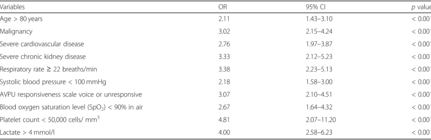
Independent variables associated with in-hospital mor-tality according to the multivariable logistic regression are reported in Table3. The model was highly significant (p < 0.0001), and the global performance of the test is explained by the area under the ROC curve, which is equals to 0.84 (95% CI).
Developing the severity score
The second aim of the study was to develop a severity score for patients with a clinical diagnosis of acute peri-tonitis that is simple and globally acceptable with a good prognostic value. Only the significant clinical variables associated with in-hospital mortality obtained from the multivariable logistic regression model were included, excluding the lactate, and platelet count. This modifica-tion was done for three reasons: (a) to simplify the score, (b) to make it more universal and globally acceptable, and (c) because of lack of facilities to obtain lactate in low-income countries. The coefficients of the variables were used to develop the score, and not the Odds Ratio. The significant clinical variables were subjected to different direct logistic regression models using either simple binomial variables or ordinal data, to arrive at a
Table 1 Distribution of clinical predictive variables of in-hospital mortality
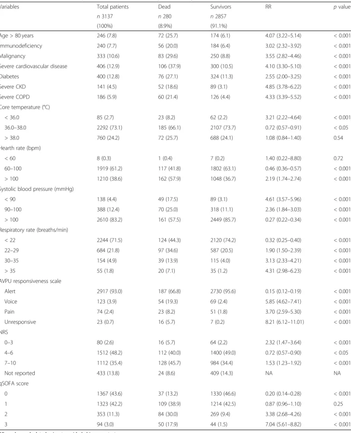
Variables Total patients Dead Survivors RR p value
n 3137 n 280 n 2857
(100%) (8.9%) (91.1%)
Age > 80 years 246 (7.8) 72 (25.7) 174 (6.1) 4.07 (3.22–5.14) < 0.001
Immunodeficiency 240 (7.7) 56 (20.0) 184 (6.4) 3.02 (2.32–3.92) < 0.001
Malignancy 333 (10.6) 83 (29.6) 250 (8.8) 3.55 (2.82–4.46) < 0.001
Severe cardiovascular disease 406 (12.9) 106 (37.9) 300 (10.5) 4.10 (3.30–5.10) < 0.001
Diabetes 400 (12.8) 76 (27.1) 324 (11.3) 2.55 (2.00–3.25) < 0.001 Severe CKD 141 (4.5) 52 (18.6) 89 (3.1) 4.85 (3.78–6.22) < 0.001 Severe COPD 186 (5.9) 60 (21.4) 126 (4.4) 4.33 (3.39–5.52) < 0.001 Core temperature (°C) < 36.0 85 (2.7) 23 (8.2) 62 (2.2) 3.21 (2.22–4.64) < 0.001 36.0–38.0 2292 (73.1) 185 (66.1) 2107 (73.7) 0.72 (0.57–0.91) < 0.05 > 38.0 760 (24.2) 72 (25.7) 688 (24.1) 1.08 (0.84–1.40) 0.54 Hearth rate (bpm) < 60 8 (0.3) 1 (0.4) 7 (0.2) 1.40 (0.22–8.80) 0.72 60–100 1919 (61.2) 117 (41.8) 1802 (63.1) 0.46 (0.36–0.57) < 0.001 > 100 1210 (38.6) 162 (57.9) 1048 (36.7) 2.19 (1.74–2.74) < 0.001
Systolic blood pressure (mmHg)
< 90 138 (4.4) 49 (17.5) 89 (3.1) 4.61 (3.57–5.96) < 0.001
90–100 388 (12.4) 70 (25.0) 318 (11.1) 2.36 (1.84–3.03) < 0.001
> 100 2610 (83.2) 161 (57.5) 2449 (85.7) 0.27 (0.22–0.34) < 0.001
Respiratory rate (breaths/min)
< 22 2244 (71.5) 124 (44.3) 2120 (74.2) 0.32 (0.25–0.40) < 0.001
22–29 684 (21.8) 97 (34.6) 587 (20.5) 1.90 (1.50–2.39) < 0.001
30–35 154 (4.9) 39 (13.9) 115 (4.0) 3.13 (2.33–4.21) < 0.001
> 35 55 (1.8) 20 (7.1) 35 (1.2) 4.31 (2.98–6.23) < 0.001
AVPU responsiveness scale
Alert 2917 (93.0) 187 (66.8) 2730 (95.6) 0.15 (0.12–0.19) < 0.001 Voice 123 (3.9) 54 (19.3) 69 (2.4) 5.85 (4.62–7.41) < 0.001 Pain 74 (2.4) 23 (8.2) 51 (1.8) 3.70 (2.59–5.30) < 0.001 Unresponsive 23 (0.7) 16 (5.7) 7 (0.2) 8.21 (6.12–11.01) < 0.001 NRS 0–3 80 (2.6) 16 (5.7) 64 (2.2) 2.32 (1.47–3.64) < 0.001 4–6 1512 (48.2) 112 (40.0) 1400 (49.0) 0.72 (0.57–0.90) < 0.05 7–10 1112 (35.4) 128 (45.7) 984 (34.4) 1.53 (1.23–1.92) < 0.001 Not reported 433 (13.8) 24 (8.6) 409 (14.3) NA NA qSOFA score 0 1367 (43.6) 37 (13.2) 1330 (46.6) 0.20 (0.14–0.28) < 0.001 1 1323 (42.2) 109 (38.9) 1214 (42.5) 0.87 (0.96–1.10) 0.25 2 353 (11.3) 84 (30.0) 269 (9.4) 3.38 (2.68–4.26) < 0.001 3 94 (3.0) 50 (17.9) 44 (1.5) 7.04 (5.61–8.82) < 0.001
All p values calculated using two-sided chi-square test
RR: risk ratio, NA: not applicable, CKD: chronic kidney disease, COPD: chronic obstructive pulmonary disease, AVPU: alert/verbal/painful/unresponsive, NRS: numerical rating scale, qSOFA: Quick Sequential Organ Failure Assessment
simplified and acceptable model. Direct logistic regres-sion model of the clinical variables affecting mortality which were used to develop the score is reported in
Table 4. The score would have become complicated if
we had to follow the model proposed by Moons et al. [15], whereby the coefficient would have to be multiplied by 10 and the value approximated to the nearest integral to get a score. This meant that the scores derived from
Table 2 Distribution of laboratory predictive variables of in-hospital mortality
Variables Total patients Dead Survivors RR p value
n 3137 n 280 n 2857
(100%) (8.9%) (91.1%)
Blood oxygen saturation level (SpO2) (%) in air
> 92 2782 (88.7) 152 (54.3) 2630 (92.1) 0.15 (0.12–0.19) < 0.001 90–91 198 (6.3) 66 (23.6) 132 (4.6) 4.58 (3.62–5.79) < 0.001 85–89 99 (3.1) 41 (14.6) 58 (2.0) 5.26 (4.04–6.85) < 0.001 < 85 21 (0.7) 9 (3.2) 12 (0.4) 4.93 (2.97–8.18) < 0.001 Not reported 37 (1.2) 12 (4.3) 25 (0.9) NA NA WBC (cells/mm3) > 12,000 1950 (62.2) 182 (65.0) 1768 (61.9) 1.13 (0.89–1.43) 0.30 4000–12,000 1043 (33.2) 63 (22.5) 980 (34.3) 0.58 (0.44–0.76) < 0.001 < 4000 94 (3.0) 29 (10.4) 65 (2.3) 3.74 (2.70–5.18) < 0.001 Not reported 50 (1.6) 6 (2.1) 44 (1.5) NA NA
Platelet count (cells/ mm3)
> 150,000 2606 (83.1) 183 (65.4) 2423 (84.8) 0.38 (0.31–0.49) < 0.001 50,000–1,500,000 387 (12.3) 73 (26.1) 314 (11.0) 2.51 (1.96–3.20) < 0.001 < 50,000 32 (1.0) 18 (6.4) 14 (0.5) 6.67 (4.81–9.24) < 0.001 Not reported 112 (3.6) 6 (2.1) 106 (3.7) NA NA INR > 3 23 (0.7) 12 (4.3) 11 (0.4) 6.06 (4.03–9.11) < 0.001 1.2–3 296 (9.4) 72 (25.7) 224 (7.8) 3.32 (2.61–4.22) < 0.001 < 1.2 1954 (62.3) 149 (53.2) 1805 (63.2) 0.69 (0.55–0.86) 0.001 Not reported 864 (27.5) 47 (16.8) 817 (28.6) NA NA CRP (mg/l) > 200 450 (14.3) 70 (25.0) 380 (13.3) 1.99 (1.55–2.56) < 0.001 101–200 462 (14.7) 51 (18.2) 411 (14.4) 1.29 (0.97–1.72) 0.08 5–100 946 (30.2) 69 (24.6) 877 (30.7) 0.76 (0.58–0.98) 0.04 < 5 258 (8.2) 3 (1.1) 255 (8.9) 0.12 (0.04–0.37) < 0.001 Not reported 1471 (46.9) 157 (56.1) 1314 (46.0) NA NA Procalcitonin (ng/ml) > 10 85 (2.7) 31 (11.1) 54 (1.9) 4.47 (3.30–6.06) < 0.001 0.5–10 260 (8.3) 42 (15.0) 218 (7.6) 1.96 (1.44–2.64) < 0.001 < 0.5 100 (3.2) 3 (1.1) 97 (3.4) 0.33 (0.11–1.01) 0.03 Not reported 2692 (85.8) 204 (72.9) 2488 (87.1) NA NA Lactate (mmol/l) >4 139 (4.4) 61 (21.8) 78 (2.7) 6.01 (4.79–7.54) < 0.001 1–4 615 (19.6) 86 (30.7) 529 (18.5) 1.82 (1.43–2.31) < 0.001 < 1 136 (4.3) 6 (2.1) 130 (4.6) 0.48 (0.22–1.07) 0.06 Not reported 2247 (71.6) 127 (45.4) 2120 (74.2) NA NA
All p values calculated using two-sided chi-square test
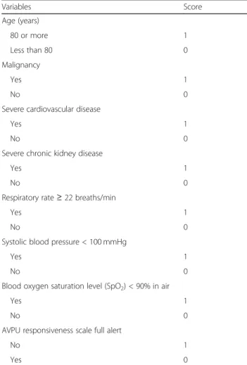
the model would be 10, 11, 9, 12, 8, 9, 9, and 14, making it very complex. Hence, it was decided to approximate the coefficient to the nearest integral number and test the model. Since the coefficients were approximated to 1, each of these variables could have a score of 1 or 0 with a maximum score of 8 and a range of 0–8. The simplified and finalized the PIPAS Severity Score is shown in theAppendix.
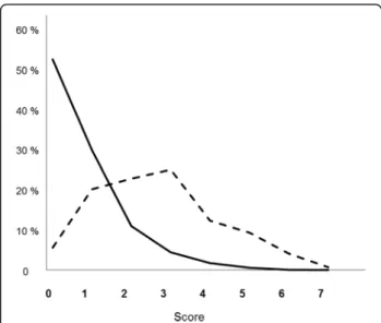
The PIPAS Severity Score had a very good ability of distinguishing those who survived from those who died
(Fig. 1). The ROC curve showed that the best cutoff
point for predicting mortality was a PIPAS Severity Score of 1.5 having a sensitivity of 74.3%, a specificity of 82.2% (Fig.2) and an area under the curve of 85.1%. The overall mortality was 2.9% for the patients who had scores of 0 and 1, 22.7% for those who had scores of 2 and 3, 46.8% for those who had scores 4 and 5, and 86.7% for those who have scores 7–8.
Discussion
Using the multivariable logistic regression, ten independent variables associated with in-hospital mortality were
identified. The model was highly significant, with a good global performance of the test. Excluding platelet count and lactate, eight bedside easy-to-measure parameters were recognized to develop an early warning score, the PIPAS Severity Score, assessing anamnestic data (age > 80 years, malignancy, severe cardiovascular disease, severe chronic kidney disease), and physiological functions (respiratory rate ≥ 22 breaths/min, systolic blood pressure < 100 mmHg, AVPU responsiveness scale voice or unresponsive, blood oxygen saturation level (SpO2) < 90% in air).
The PIPAS Severity Score, taking into account physio-logical parameters recognizable on hospital admission, immediately allows clinicians to assess the severity and decide the aggressiveness of treatment. Particularly for clinicians working in low- and middle-income countries, where diagnostic imaging is often insufficient, and in some instances completely lacking, the utility of this score system is remarkable [16].
Sometimes, the atypical clinical presentation of acute peritonitis may be responsible for a delay in diagnosis and treatment. Therefore, a triage system that quickly recog-nizes patients at high risk for mortality and allows to
Table 3 Results of multinomial logistic regression for the analysis of variables associated with in-hospital mortality
Variables OR 95% CI p value
Age > 80 years 2.11 1.43–3.10 < 0.001
Malignancy 3.02 2.15–4.24 < 0.001
Severe cardiovascular disease 2.76 1.97–3.87 < 0.001
Severe chronic kidney disease 3.33 2.12–5.23 < 0.001
Respiratory rate≥ 22 breaths/min 3.38 2.23–5.13 < 0.001
Systolic blood pressure < 100 mmHg 2.18 1.58–3.00 < 0.001
AVPU responsiveness scale voice or unresponsive 3.07 2.10–4.51 < 0.001
Blood oxygen saturation level (SpO2) < 90% in air 2.67 1.64–4.32 < 0.001
Platelet count < 50,000 cells/ mm3 4.81 2.07–11.20 < 0.001
Lactate > 4 mmol/l 4.00 2.58–6.23 < 0.001
CI: confidence interval, OR: odds ratio, AVPU: alert/verbal/painful/unresponsive
Table 4 Direct logistic regression model with clinical variables affecting mortality of patients used to develop the score
Variable Estimate SE Wald
test P OR 95% CI LL UL Age > 80 years 0.97 0.19 25.91 < 0.0001 2.63 1.81 3.89 Malignancy 1.13 0.17 42.43 < 0.0001 3.11 2.21 4.37 Severe CVD 0.88 0.17 26.09 < 0.0001 2.41 1.72 3.38 Severe CKD 1.2 0.23 26.23 < 0.0001 3.32 2.1 5.26 RR≥ 22 breaths/min 0.75 0.16 22.61 < 0.0001 2.11 1.55 2.87 SBP < 100 mmHg 0.86 0.17 27.29 < 0.0001 2.37 1.71 3.27
AVPU responsiveness scale: not completely alert. 1.35 0.2 47.98 < 0.0001 3.86 2.63 5.65
Blood oxygen saturation level: SpO2< 90% in air 0.87 0.25 12.15 < 0.0001 2.39 1.46 3.89
Constant − 3.79 0.13 834.77 < 0.0001 0.023 – –
SE: standard error, OR: odds ratio, CI: confidence interval, LL: lower limit, UL: upper limit, CVD: cardiovascular disease, CKD: chronic kidney disease, RR: respiratory rate, SBP: systolic blood pressure, AVPU: alert/verbal/painful/unresponsive
transfer them immediately to an acute care unit is a vital component of the emergency services. As a consequence, any process of improving the quality of emergency care globally should focus on simple diagnostic criteria based on physical examination findings that can recognize pa-tients needing critical care. From a global perspective, a feasible, low-cost method of rapidly identifying patients
requiring critical care is crucial. Early warning system scores utilize physiological, easy-to-measure parameters, assessing physiological parameters such as systolic blood pressure, pulse rate, respiratory rate, temperature, oxygen saturations, and level of consciousness [17].
The statistical analysis shows that the PIPAS Severity Score has a very good ability of distinguishing those who survived from those who died. The overall mortality was 2.9% for the patients who had scores of 0 and 1, 22.7% for those who had scores of 2 and 3, 46.8% for those who had scores of 4 and 5, and 86.7% for those who have scores of 7–8.
PIPAS Study has strengths and limitations. It is an observational multicentre study involving a large, but probably not representative, number of hospitals world-wide, since the majority of patients were collected in countries belonging to the WHO European region. More-over, its validity needs to be tested in future large pro-spective series before potentially serving as a template for future database and research into patient outcomes. Finally, a potential limitation may be the high rate of patients with acute appendicitis enrolled in the study (42.1%). Some authors [18], after excluding patients with perforated appendicitis, found that the cure rate among patients who had peritonitis and were enrolled in clinical trials, was much higher than that of patients who were not enrolled and that the mortality rate was much lower. Although, delineating the source of infection as accurately as possible prior to surgery is described as the primary aim and the first step in managing acute peritonitis, in emergency departments of limited-resource hospitals, diagnosis of acute peritonitis is mainly clinical, and sup-ported only by basic laboratory tests, and excluding acute appendicitis in the pre-operative phase would make the score impractical to a large part of the world’s population.
Conclusions
This worldwide multicentre observational study was per-formed in 153 surgical departments from 56 countries over a 4-month study period (February 1, 2018–May 31, 2018). All consecutive patients admitted to surgical departments with clinical diagnosis of acute peritonitis were included in the study. The most significant independent variables asso-ciated with in-hospital mortality were adjusted to clinical criteria and were used to create a new bedside early warn-ing score for patients with acute peritonitis. The simple PIPAS Severity Score for patients with acute peritonitis can be used on the global level and can help clinicians to assess patients with acute peritonitis at high risk for treatment failure and mortality. The authors created an acronym for the PIPAS Severity Score to help remember the variables “Scores Must Be Simple For Sepsis Risk Assessment” (severe cardiovascular disease, malignancy, blood oxygen saturation level, severe chronic kidney disease, fully alert, systolic blood pressure, respiratory rate, age).
Fig. 1 Distribution of the percentile PIPAS Severity Score of hospitalized peritonitis patients for those who survived (continuous line) (n = 2832) and those who died (interrupted line) (n = 268). Global data from 153 worldwide surgical departments in 56 countries, over a 4-month study period (February 1, 2018–May 31, 2018). Thirty-seven patients (1.2%) had missing data in whom the score could not be computed
Fig. 2 Receiver operating characteristic (ROC) curve for the best PIPAS Severity Score (1.5, black circle) that predicted mortality in peritonitis patients. Global data from 153 worldwide surgical departments in 56 countries, over a 4-month study period (February 1, 2018–May 31, 2018)
Abbreviations
AVPU:Alert/verbal/painful/unresponsive; COPD: Chronic obstructive pulmonary disease; CRP: C-reactive protein; CT: Computer tomography; INR: International normalised ratio; IQR: Interquartile range; LOS: Length of hospital stay; NRS: Numerical rating scale; PID: Pelvic inflammatory disease. IAIs: intra-abdominal infections; qSOFA: Quick Sequential Organ Failure Assessment; ROC: Receiver operating characteristic; US: Ultrasound; WBC: White blood count; WSES: World Society of Emergency Surgery Acknowledgements
Not applicable. Funding. Not applicable. Authors’ contributions
M Sartelli designed the study and wrote the manuscript. FM Abu-Zidan developed the severity score. FM Labricciosa performed the statistical analysis. All authors participated in the study. All authors read and approved the final manuscript.
Availability of data and materials
The authors are responsible for the data described in the manuscript and assure full availability of the study material upon request to the corresponding author.
Ethics approval and consent to participate
The data was completely anonymised, and no patient or hospital information was collected in the database. The study protocol was approved by the board of the WSES, and the study was conducted under its supervision. The board of the WSES granted the proper ethical conduct of the study.
Consent for publication Not applicable. Competing interests
The authors declare that they have no competing interests. Author details
1Department of Surgery, Macerata Hospital, Macerata, Italy.2Department of
Surgery, College of Medicine and Health Sciences, UAE University, Al-Ain, United Arab Emirates.3Global Alliance for Infections in Surgery, Porto, Portugal.4Department of General Surgery, Rambam Health Care Campus,
Haifa, Israel.5Department of Emergency Surgery, Bufalini Hospital, Cesena,
Italy.6Abdominal Center, Department of Abdominal Surgery, Helsinki
University Hospital Meilahti and University of Helsinki, Helsinki, Finland.
7General, Acute Care, Abdominal Wall Reconstruction, and Trauma Surgery,
Foothills Medical Centre, Calgary, AB, Canada.8Department of Digestive
Surgery and SSPC Research Unit, CHU Amiens-Picardie, Amiens, France.
9
Department of Trauma ICU, IALCH, University of KwaZulu-Natal, Durban, South Africa.10Department of General Surgery, University Hospital of
Coventry & Warwickshire, Coventry, UK.11Department of Surgery, Mansoura
University and Emergency Hospital, Mansoura, Egypt.12Department of
Gastrointestinal Surgery, University of Health Sciences, Elazig Training and Research Hospital, Elazig, Turkey.13Department of Surgery, College of Health
Sciences, Obafemi Awolowo University, Ile-Ife, Nigeria.14Department of
Surgery, LAUTECH Teaching Hospital, Osogbo, Nigeria.15Department of
Surgery, TSMU First University Clinic, Tbilisi, Georgia.16Department of General Surgery, Sakarya University Research and Educational Hospital, Sakarya, Turkey.17Department of General Surgery, Hacettepe University Hospital,
Ankara, Turkey.18Department of Surgical Oncology, King Fahad Medical City,
Riyadh, Saudi Arabia.19Department of General Surgery, Istinye University Faculty of Medicine, Istanbul, Turkey.20Department of Primary Care, Primary
Health Care Centre of Kissamos, Chania, Greece.21Surgical Department,
UMHAT“Eurohospital”, Medical University, Plovdiv, Bulgaria.22Department of
Surgery, University Hospital Centre Zagreb, Zagreb, Croatia.23Cirurgia Geral, Centro Hospitalar Universitário da Cova da Beira, Covilhã, Portugal.
24Department of General Surgery, Hadassah Medical Center, Jerusalem, Israel. 25Department of Surgery, Saint Savvas Anticancer Hospital, Athens, Greece. 26
General Surgery, Habib bougatfa, Bizerte, Tunisia.27Department of Surgery, Lumbini Medical College and Teaching Hospital Ltd., Tansen, Palpa, Nepal.
28Department of Surgery, Hospital San Juan de Dios de La Serena, La Serena,
Chile.29Emergency and General Surgery, SG Bosco, Torino, Italy.30Surgical
Department and ICU Department, General Hospital of Larissa, Larissa, Greece.
31General Surgery, Hospital Santo Tomas, Panama, Panama.32Department of
Surgery, Elias Emergency Hospital, Bucharest, Romania.33Dipartimento
Emergenza e Accettazione, Policlinico Umberto I, Roma, Italy.34Department
of General and Emergency Surgery, ASST Monza - Ospedale San Gerardo, Monza, Italy.35General Surgery, South Warwickshire NHS Foundation Trust,
Warwick, UK.36Department of General Surgery, Kuala Krai Hospital, Kuala Krai,
Malaysia.37U.O. Chirurgia d’Urgenza Universitaria, Azienda
Ospedaliero-Universitaria Pisana, Pisa, Italy.38U.O.C. Chirurgia Generale, PO Santissima Trinità, Cagliari, Italia.39UGC Cirugía General, Complejo
Hospitalario de Jaén, Jaén, Spain.40Department of General and Emergency
Surgery, Azienda Ospedaliera Policlinico Universitario Palermo“Paolo Giaccone”, Palermo, Italy.41General Surgery, University of Health Sciences, Samsun Training and Research Hospital, Samsun, Turkey.42Department of
Surgery, G. Da Saliceto Hospital, Piacenza, Italy.43Department of Surgery,
Tianjin Nankai Hospital, Tianjin, China.44Chirurgie Viscerale et d’Urgence,
Centre Hospitalier Regional de Perpignan, Perpignan, France.45Department of Surgery, University Clinical Center Tuzla, Tuzla, Bosnia and Herzegovina.
46Department of Surgery, Kipshidze Central University Hospital, Tbilisi,
Georgia.47Division of Trauma and Acute Care Surgery, LAC+USC Medical
Center, Los Angeles, USA.48Department of General Surgery, University Hospital of Trauma, Tirana, Albania.49Department of Infectious Diseases, King
Fahad Medical City, Riyadh, Saudi Arabia.50Chirurgia Generale, Ospedale
Versilia, La Spezia, Italy.51Department of Surgery, San Carlo Borromeo
Apenndix
Table 5 PIPAS Severity Score for patients with acute peritonitis (range 0–8) Variables Score Age (years) 80 or more 1 Less than 80 0 Malignancy Yes 1 No 0
Severe cardiovascular disease
Yes 1
No 0
Severe chronic kidney disease
Yes 1
No 0
Respiratory rate≥ 22 breaths/min
Yes 1
No 0
Systolic blood pressure < 100 mmHg
Yes 1
No 0
Blood oxygen saturation level (SpO2) < 90% in air
Yes 1
No 0
AVPU responsiveness scale full alert
No 1
Hospital, Milan, Italy.52Department of General Surgery, San Salvatore, Pesaro,
Italy.53Division of Trauma Surgery, Hospital de Clinicas, University of
Campinas, Campinas, Brazil.54Department of Abdominal Surgery, Vladimir
City Clinical Hospital of Emergency Medicine, Vladimir, Russia.55Department of Surgery, University hospital, Amiens, France.56Department of General
Surgery, Mansoura University Hospital, Mansoura, Egypt.57Department of
General Surgery, Miguel Servet, Zaragoza, Spain.582nd Department of
Surgery, Aretaieion University Hospital, National and Kapodistrian University of Athens, Athens, Greece.59Department of Surgery, Hospital Universitário
Terezinha de Jesus, Faculdade de Ciências Médicas e da Saúde de Juiz de Fora (SUPREMA), Juiz de Fora, Brazil.60Department of General Surgery,
Karadeniz Technical University, Trabzon, Turkey.61Department of General Surgery, Government Medical College and Hospital, Chandigarh, India.
62Department of General and Thoracic Surgery, University Hospital of
Giessen, Giessen, Germany.63Department of General Surgery, University and
Regional Hospital Center of Borgou, Parakou, Republic of Benin.
64Chirurgische Abteilung, Landesklinikum Hainburg, Hainburg an der Donau,
Austria.65Intensive Care Unit, Chernivtsi City Emergency Hospital, Chernivtsi,
Ukraine.664th Surgical Department, Medical School, Aristotle University of
Thessaloniki, General Hospital“G. Papanikolaou”, Thessaloniki, Greece.
67Department of General Surgery, Erzincan University Hospital, Erzincan,
Turkey.68Department of Faculty Surgery #1, Pirogov Russian National
Research Medical University, Moscow, Russia.69Department of Surgery, SMS
Hospital, Jaipur, India.70Department of Surgery, Hospital of Lithuanian University of Health Sciences Kaunas Clinics, Kaunas, Lithuania.71Faculty of
Medicine University of Belgrade Clinic for Surgery, University Clinical Center “Zvezdara”, Belgrade, Serbia.72Department of General, Oncologic and
Geriatric Surgery, Jagiellonian University Collegium Medicum, Kraków, Poland.
73Department of Emergency Surgery, City Hospital, Mozyr, Belarus. 74Department of Vascular Surgery, City Hospital, Mozyr, Belarus.
75Department of Surgery, Inje University Ilsan Paik Hospital, Goyang, Republic
of Korea.76Trauma and Acute Care Surgery, Edendale Hospital,
Pietermaritzburg, South Africa.77Department of Surgery, Krishna Hospital and
Medical Research University Karad, Karad, India.78Departament of General
Surgery, Hospital Municipal de Governador Valadares, Vale do Rio Doce University, Governador Valadares, Brazil.79Chirurgia d’Urgenza, Arcispedale Santa Maria Nuova IRCCS, Reggio Emilia, Italy.80General Surgery,
Scarborough Hospital, York Teaching Hospital NHS FT, York, UK.81Cirurgia
Geral, Hospital de Braga, Life and Health Sciences Research Institute, ICVS/ 3Bs, Universidade do Minho, Braga, Portugal.82General and Digestive Surgery, Hospital Fundación Jimenez Diaz, Madrid, Spain.83General, Visceral,
Thoracic and Vascular Surgery, University Hospital Greifswald, Greifswald, Germany.84Department of Surgical Disciplines, Regional Clinical Hospital,
Immanuel Kant Baltic Federal University, Kaliningrad, Russia.85Cirugía general y del aparato digestivo, Hospital Universitario Donostia, Donostia, Spain.
86Gastrointestinal Surgery, Hospital Insular de Gran Canaria, Las Palmas de
Gran Canaria, Spain.871st Department of Surgery, Kavala General Hospital,
Kavala, Greece.88Department of General and Emergency Surgery, ASMN Reggio Emilia, Modena, Italy.89II Catedra de Clinica Quirúrgica, Hospital de
Clinicas, Facultad de Ciencias Médicas, Universidad Nacional de Asunción, Asunción, Paraguay.902nd Department of General Surgery, Jagiellonian
University Medical College, Kraków, Poland.91Department of Surgery, Athens Naval and Veterans Hospital, Athens, Greece.92First Department of Surgery,
Tzaneio General Hospital, Piraeus, Greece.93Department of General Surgery
and Surgical Oncology, Policlinico Le Scotte, University of Siena, Siena, Italy.
94
Department of General and Digestive Surgery, Hospital Universitario Doctor Peset, Valencia, Spain.95Department of General Surgery, Post-graduate
Institute of Medical Sciences, Rohtak, India.96Department of Surgery,
Radiology, Anaesthesia and Intensive Care, University Hospital of the West Indies, Kingston, Jamaica.97Second Surgical Clinic, Emergency County Hospital of Craiova, Craiova, Romania.983rd Department of Surgery, Ahepa
University Hospital, Thessaloniki, Greece.993rd Department of Surgery,
Attikon University Hospital, Athens, Greece.100Department of Surgery, Hassan
II, Fez, Morocco.101Department of Specialist Surgery, Port Shepstone Regional Hospital, Port Shepstone, Republic of South Africa.102General
Surgery Department, Emergency Hospital of Bucharest, Bucharest, Romania.
103Toxicology and Sepsis, Riga East University Hospital, Riga, Latvia. 104
Department of General Surgery, Queen Elizabeth Hospital, London, UK.
105Chirurgia generale, Sant’Anna (AUSL Reggio Emilia), Castelnovo ne’ Monti,
Italy.106U.O. Chirurgia d’Urgenza, Arcispedale S. Anna Ferrara, Ferrara, Italy. 107Department of Surgery, University of Ilorin Teaching Hospital, Ilorin,
Nigeria.108Department of Surgery, Fundacion Valle del Lili - Universidad del
Valle, Cali, Colombia.109Department of General Surgery, University of Health
Sciences, Elazig Training and Research Hospital, Elazig, Turkey.110Department
of Surgery, King George’s Medical University, Lucknow, India.111Chirurgia Generale e d’Urgenza, Ospedale Infermi, Rimini, Italy.112Surgical Oncology,
University Hospital Heraclion Crete, Heraclion Crete, Greece.113Department
of General Surgery, General Hospital of Trikala, Trikala, Greece.114Department
of Surgery, Sant’Antonio Abate Hospital, Gallarate, Italy.115Department of General and Emergency Surgery, University Hospital, University Hospital Kraków, Kraków, Poland.116Cirurgia Geral, Centro Hospitalar Tondela-Viseu,
Viseu, Portugal.117Medicina, Base Hospital, Bauru, Brazil.118Chirurgia
d’Urgenza – Dipartimento Urgenza/Emergenza, AOU Parma, Parma, Italy.
119Department of Abdominal Surgery, UMC Ljubljana, Ljubljana, Slovenia. 120Department of Endoscopic, Metabolic and Soft Tissue Tumors Surgery,
University Hospital, Kraków, Poland.121Surgery Department, Chernivtsi City
Emergency Hospital, Chernivtsi, Ukraine.122Department of General, Emergency and Robotic Surgery, San Francesco Hospital, Nuoro, Italy.
123Department of Surgery, AO San Giovanni Addolorata, Rome, Italy. 124Department of Surgery/Trauma, Hospital Santo Tomás, Panama, Panama. 125
Department of Gastrointestinal Surgery, HGR1 IMSS, Cuernavaca, Mexico.
126Department of General Surgery, Kasturba Medical College, Manipal
Academy of Higher Education, Manipal, India.127First Clinic of General
Surgery, University Hospital St George/Medical University Plovdiv, Plovdiv, Bulgaria.128Chirurgie Générale et Viscérale, Hôpital d’instruction des Armées, Hôpital Principal de Dakar, Dakar, Senegal.129Department of General Surgery,
Tan Tock Seng Hospital, Singapore, Singapore.130Department of Surgery,
Fatebbenefratelli Hospital, Isola Tiberina, Rome, Italy.131Department of
Surgery (Department No. 10), Riga East Clinical University Hospital“Gaiļezers”, Riga, Latvia.132Department of Surgery, Hospital and Oncological Centre Novy
Jicin, Novy Jicin, Czech Republic.133General Surgery, Heartlands Hospital,
Birmingham, UK.134Department of General Surgery, Polytrauma and
Emergency Medicine, University Hospital of the Jagiellonian University Medical College, Kraków, Poland.135General Surgery Department, Bukovinian
State Medical University, Chernivtsi, Ukraine.136Trauma and Emergency
Surgery, Hospital Escola Padre Albino, Catanduva, Brazil.137Faculty of
Medicine and Biomedical Sciences, University of Yaounde I, Yaounde, Cameroon and Department of Surgery and Anaesthesiology, Yaounde Central Hospital, Yaounde, Cameroon.138Surgery Department, Tbilisi State
Medical University, Tbilisi, Georgia.139Chirurgia Generale, Ospedale Civile di
Guastalla, Reggio Emilia, Italy.140First Department of Surgery, Department of Abdominal, Thoracic Surgery and Traumatology, First Faculty of Medicine, Charles University and General University Hospital, Prague, Czech Republic.
141Upper Gastrointestinal Tract Surgery Department, North Estonia Medical
Centre, Tallinn, Estonia.142Department of General Surgery, Siirt State Hospital, Siirt, Turkey.143First Surgical Unit,“St. Spiridon” University Hospital Iasi,
University of Medicine and Pharmacy“Grigore T. Popa”, Iasi, Romania.
144Renal Transplant and General Surgery, Manchester Royal Infirmary,
Manchester, UK.145Department of Surgery, Clinical Center University of Pecs, Pecs, Hungary.146Department of General, Oncological, Metabolic and
Thoracic Surgery, Military Institute of Medicine, Warsaw, Poland.
147Department of Surgey, Zliten Teaching Hospital, Zliten, Libya. 148
Department of General Surgery, Ankara University School of Medicine, Ankara, Turkey.149Transplantation Unıt, Acibadem Atakent Hospital, İstanbul,
Turkey.150Department of Surgery, Taipei Medical University Hospital, Taipei,
Taiwan.151Department of Surgery, Mosc Medical College, Kolenchery,
Cochin, India.152Surgical Oncology Unit, Azienda Unità Sanitaria Locale -IRCCS di Reggio Emilia, Reggio Emilia, Italy.153Emergency Surgery
Department, Maggiore Parma Hospital, Parma, Italy.154Department of Clinical
and Experimental Sciences, University of Brescia, Brescia, Italy.
Received: 8 March 2019 Accepted: 3 July 2019
References
1. Sartelli M, Catena F, Abu-Zidan FM, Ansaloni L, Biffl WL, Boermeester MA, et al. Management of intra-abdominal infections: recommendations by the WSES 2016 consensus conference. World J Emerg Surg. 2017;12:22. 2. Sartelli M, Catena F, Di Saverio S, Ansaloni L, Malangoni M, Moore EE, et al.
Current concept of abdominal sepsis: WSES position paper. World J Emerg Surg. 2014;9:22.
3. Angus DC, van der Poll T. Severe sepsis and septic shock. N Engl J Med. 2013;369:840–51.
4. Wacha H, Linder MM, Feldman U, Wesch G, Gundlach E, Steifensand RA. Mannheim peritonitis index– prediction of risk of death from peritonitis: construction of a statistical and validation of an empirically based index. Theor Surg. 1987;1:169–77.
5. Bosscha K, Reijnders K, Hulstaert PF, Algra A, van der Werken C. Prognostic scoring systems to predict outcome in peritonitis and intra-abdominal sepsis. Br J Surg. 1997;84:1532–4.
6. Sartelli M, Abu-Zidan FM, Catena F, Griffiths EA, Di Saverio S, Coimbra R, et al. Global validation of the WSES sepsis severity score for patients with complicated intra-abdominal infections: a prospective multicenter study (WISS study). World J Emerg Surg. 2015;10:61.
7. Chatterjee AS, Renganathan DN. POSSUM: A Scoring System for Perforative Peritonitis. J Clin Diagn Res. 2015;9:PC05–9.
8. Sartelli M, Chichom-Mefire A, Labricciosa FM, Hardcastle T, Abu-Zidan FM, Adesunkanmi AK, et al. The management of intra-abdominal infections from a global perspective: 2017 WSES guidelines for management of intra-abdominal Infections. World J Emerg Surg. 2017;12:29.
9. Fleisher LA, Beckman JA, Brown KA, Calkins H, Chaikof E, Fleischmann KE, et al. ACC/AHA 2007 Guidelines on Perioperative Cardivascular Evaluation and Care for Noncardiac Surgery: Executive Summary: A Report of the American College of Cardiology/American Heart Association Task Force on Practice Guidelines (Writing Committee to Revise the 2002 Guidelines on Perioperative Cardiovascular Evaluation for Noncardiac Surgery): Developed in Collaboration With the American Society of Echocardiography, American Society of Nuclear Cardiology, Heart Rhythm Society, Society of
Cardiovascular Anesthesiologists, Society for Cardiovascular Angiography and Interventions, Society for Vascular Medicine and Biology, and Society for Vascular Surgery. Circulation. 2007;116:1971–96.
10. Vestbo J, Hurd SS, Agustí AG, Jones PW, Vogelmeier C, Anzueto A, et al. Global strategy for the diagnosis, management, and prevention of chronic obstructive pulmonary disease: GOLD executive summary. Am J Respir Crit Care Med. 2013;187:347–65.
11. Teasdale G, Jennett B. Assessment of coma and impaired consciousness. A practical scale. Lancet. 1974;2:81–4.
12. Farrar JT, Young JP Jr, LaMoreaux L, Werth JL, Poole RM. Clinical importance of changes in chronic pain intensity measured on an 11-point numerical pain rating scale. Pain. 2001;94:149–58.
13. Singer M, Deutschman CS, Seymour CW, Shankar-Hari M, Annane D, Bauer M, et al. The Third International Consensus Definitions for Sepsis and Septic Shock (Sepsis-3). JAMA. 2016;315:801–10.
14. Sartelli M. A focus on intra-abdominal infections. World J Emerg Surg. 2010;5:9. 15. Bickler SW, Spiegel D. Improving surgical care in low- and middle-income
countries: a pivotal role for the World Health Organization. World J Surg. 2010;34:386–90.
16. Moons KG, Harrell FE, Steyerberg EW. Should scoring rules be based on odds ratios or regression coefficients? J Clin Epidemiol. 2002;55:1054–5. 17. Kruisselbrink R, Kwizera A, Crowther M, Fox-Robichaud A, O’Shea T,
Nakibuuka J, et al. Modified early warning score (MEWS) identifies critical illness among ward patients in a resource restricted setting in Kampala. Uganda: a prospective observational study. PLoS One. 2016;11:e0151408. 18. Merlino JI, Malangoni MA, Smith CM, Lange RL. Prospective randomized trials
affect the outcomes of intraabdominal infection. Ann Surg. 2001;233:859–66.
Publisher’s Note
Springer Nature remains neutral with regard to jurisdictional claims in published maps and institutional affiliations.