Mucormycosis with Orbital Apex Syndrome in a Renal
Transplant Recipient
Bir Böbrek Transplantasyon Alıcısında Orbital Apeks Sendromu ile Seyreden
Mukormikoz
Ebru Kurşun1, Tuba Turunç1, Yusuf Ziya Demiroğlu1, Hakan Yabanoğlu2, Şenay Demir2, Kenan Çalışkan3, Gökhan Moray3, Hande Arslan1, Mehmet Haberal3
1Başkent University Medical Faculty, Department of Infectious Diseases and Clinical Microbiology, 2
Department of Radiology, 3Department of General Surgery. ADANA
Cukurova Medical Journal 2015;40(2):384-389.
ABSTRACT
Mucormycosis is a rarely encountered invasive fungal infection with high mortality.Solid organ transplantation is one of the risk factors for mucormycosis. Mucormycosis can be classified in six different groups according to the anatomical localization; rhinocerebral, pulmonary, cutaneous, gastrointestinal, disseminated, and other less common involvements. This paper presented a mucormycosis case with rhinoorbitocerebral involvementin a renal transplantation receiver, which manifested with orbital apex syndrome.
Key words: Mucormycosis, orbital apex syndrome, solid organ transplantation.
ÖZET
Mukormikozis nadir görülen mortalitesi yüksek invaziv bir mantar enfeksiyonudur. Solid organ transplantasyonu mukormikoz için risk faktörlerinden birisidir. Mukormikoz anatomik yerleşim yerleri temel alındığında; rinoserebral, pulmoner, kutanöz, gastrointestinal, dissemine ve sık rastlanılmayan diğer tutulumlar olmak üzere altı farklı grup olarak sınıflandırılabilmektedir. Bu yazıda böbrek transplantasyonu alıcısında orbital apeks sendromu ile prezente olan rino-orbito-serebral tutulumu olan mukormikoz olgusu sunulmuştur.
Anahtar kelimeler: Mukormikoz, orbital apeks sendromu, solid organ transplantasyonu.
INTRODUCTION
Mucormycosis, which is caused by the members of Mucorales team, is a fungal infection with high mortality. Mucormycosis infection occurs due to inhalation of sporangiospores of fungi or by direct inoculation in the skin and/or mucosa with impaired integrity. Nonetheless, mucormycosis is characterized by thrombus and tissue necrosis, which occur due to the invasion of fungal hypha into the vascular configurations3. Risk factors for the disease include immune suppression such as diabetic ketoacidosis, chronic renal insufficiency,
solid organ or bone marrow stem cell transplantation, burnt, and hematological cancers, as well as excessive iron load (desferroxamine use) and impaired skin integrity.
Orbital apex syndrome (OAS), which is one of the atypical clinical pictures of mucormycosis, is characterized by sudden loss of vision, ophthalmoplegia, proptosis, optic atrophy, and diplopia due to the involvement of cranial nerves II, III, IV, VII and ophthalmic branch of cranial nerve V1.
This paper presented a rhinoorbitaocerebral mucormycosis case that manifested with OAS, atypical clinical pictureand resulted in mortality, in a renal transplantation receiver.
CASE REPORT
It was determined from the anamnesis of a 35-year-old male patient, who received renal transplantation in February 2013 from his ABO-match and HLA-misABO-match brother, that he had hypertension and received dialysis for 3 times a weekin the last month. In the patient, whose panel reactive antigen was negative, cold ischemia duration was 85 minutes. B-cell cross-match was borderline positive in the tissue typing performed prior to the surgery. It was determined that the patient received antithymocyte immunoglobulin (ATG, Fresenius) therapy at a dose of 400 mg/day during surgery and in the first 3 days after renal transplantation.It was determined that methyl prednisolone was administered at a dose of 1000 mg/day on the days before and after transplantation and that steroid therapy was commenced at a dose 90 mg on the first day after transplantation and continued for a week by gradually decreasing the dose by 20 mg each day. It was also learned that mycophenolate sodium was commenced at a dose of 720 mg/day on the operation day and tacrolimus was included in the therapy at a dose of 0.1-0.2 mg/kg/day on the postoperative 4th day. It was determined that urine output was 500 ml/hour in early period after surgery and that renal functions began to be improved. Post-transplantation control revealed normal renal doppler ultrasonography and scintigraphy.
On the postoperative day 14, the patient complained about pain and swelling around the left eye, paresthesia on the left side of his face, ptosis in the left eyelid, diplopia and decreased hearing in the left ear. On his physical examination, body temperature was 36.5°C, BP was 130/90 mm/Hg, conscious was open, and he was cooperated and oriented, whereas there were left peripheral facial
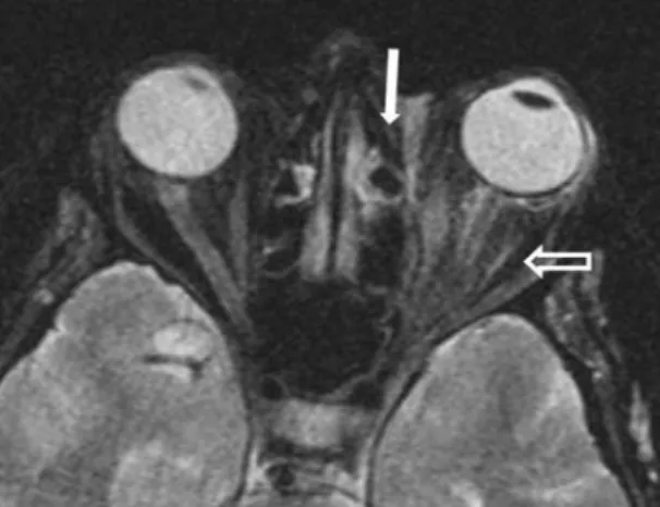
paralysis, proptosis and exophthalmos in the left eye together with chemosis, light reflex was negative, there was total ophthalmoplegia (complete paralysis of left nervus oculomotorius, n. trochlearis and n. abducens) and no gag reflex in the left soft palate. A black appearance consistent with tissue necrosis was determined in the soft and hard palates. Results of laboratory analyses were as follows; hemoglobin: 8 g/dL, leukocyte: 16.300/mm3, thrombocyte: 376.000 K/mm3, C-reactive protein > 211 mg/L, erythrocyte sedimentation rate 111 mm/hour, blood urea nitrogen 66 mg/dL, and creatinine 2.2 mg/dL. The patient, in whom mucormycosis was considered as the pre-diagnosis, underwent computed tomography (CT) of paranasal sinuses and non-contrast-enhanced cranial and orbital magnetic resonance imaging (MRI) due to elevation in renal function tests. Paranasal CT revealed soft tissue densities predominantly in the form of mucosal thickening in the left ethmoid cell extending also into the nasal cavity and an appearance consistent with mild mucosal thickening also in the left maxillary sinus (Figure 1a and 1b). No acute pathology was observed on cranial MRI. Orbital MRI revealed mild proptotic appearance in the left globe, linear signal increase in T2 sequences in the lateral aspect of globeand around the optic nerve and in the retrobulbar adipose tissue, as well as pathological signal changes in the retrobulbar adipose tissue in the medial aspect and mucosal thickening in the left ethmoid cells (Figure 2 ).
Being considered to have mucormycosis based on existing clinical and radiological findings, the case underwent endoscopic sinus surgery in the Department of Ear Nose Throat (ENT) Diseases. Black necrotic tissues filling nasal cavity and sinuses that were consistent with mucormycosis were removed. After surgery, intravenous 5 mg/kg/day liposomal amphotericin B (LAmB) and 2 g/day meropenem were commenced. Histopathological examination of necrotic tissue removed during operation revealed findings consistent with mucormycosis. 385
Zygomycetes spp. growth was detected in fungal
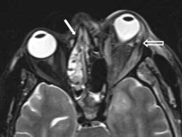
culture of tissue sample. All immunosuppressive drugs that the patient has been receiving were discontinued. On the 8th day of antifungal therapy, the patient underwent cranial and orbital MRI due to impaired consciousness. MRI demonstrated notable enhancement in the left exophthalmos, increase in the antero-posterior size of left globe, thickening in the left conal muscles as well as signal increment in the T2 A sequence, fouling increment in the retrobulbar adipose tissue, defective areas in ethmoidal cells secondary to surgery, and an appearance consistent with notable progression of sinusitis (Figure 3).
Since the disease picture showed clinical and radiological progression, the dose of LAmB was switched to 7 mg/kg/day, all immunosuppressive drugs were discontinued, and parenteral linezolid 1200 mg/kg/day was included in treatment. Because of inadequate response to treatment, left orbital exenteration was performed. On histopathological examination, the surgical tissue sample was found consistent with mucormycosis and Zygomycetes spp. was isolated from fungal culture. Despite overall supportive treatments, the patient died on the 3rd week of antifungal therapy.
Figure 1a and b. On paranasal sinus CT; mucosal thickening in the left ethmoidal cells and in the left maxillary sinus
extending into the nasal cavity (white arrow).
Figure 2. On orbital MRI; mild proptotic appearance in the left globe, linear signal increment around the optic nerve and
retrobulbar adipose tissue in T2 sequence (empty arrow); mucosal thickening in the left ethmoidal cells (filled arrow).
Figure 3. On control MRI; notable worsening in the left exophthalmos, increase in the anteroposterior size of the left
globe, thickening and signal increment in the left conal muscles, more apparent pathological signal changes in the retrobulbar adipose tissue (empty arrow), defective areas secondary to surgery in the left nasal cavity and ethmoid cells and remarkable progression of sinusitis (filled arrow).
DISCUSSION
Mucormycosis is a rarely encountered opportunist fungal infection and Rhizopus,
Lichtheimia, Mucor and Rhizomucor are the
leading causes of this infection. The prevalence of this infection is 0.2-1.2%in the patients that underwent renal transplantation4,5. Chronic immunosuppression, frequent use of broad-spectrum antibiotics, concomitant metabolic disorders such as uremia, and diabetes mellitus that is developed after transplantation enhance the risk of mucormycosis in renal transplantation patients. In such patients, pulmonary, disseminated, gastrointestinal and most commonly rhinocerebral involvements may be seen6. Mucormycosis infection begins with transportation of fungal spores to paranasal sinuses via inhalation. Spores that evade phagocytosis invade vascular endothelium and cause necrosis, thrombus and infarction7. As the consequence, necrotic lesions occur in the nasal mucosa and hard palate and then necrosis extends to orbital and cranial tissues. Spread may occur either via direct neighborhood or via embolus from surrounding vasculatures depending on necrosis. Rhinoorbitacerebral involvement is a rare but the
most important clinical manifestation of mucormycosis with high mortality rate and usually develops in the patients with diabetes mellitus or immune system defect8.
Neutrophils and macrophages play important role in defensing against mucormycosis. Whilst macrophages kill the spores in the cell by oxidative mechanism, neutrophils damage fungal hyphae by extracellular mechanisms. Therefore, inadequate neutrophil count in circulation (neutropenia) or impaired phagocytosis function (steroid therapy or diabetes) poses a risk for this infection9. We think that, immunosuppressive therapy commenced for renal transplantation might have caused mucormycosis in the present case.
OAS, which is one of the atypical clinical pictures of mucormycosis, is a rarely encountered syndrome developed due to the involvement of cranial nerves II, III, IV, VI and ophthalmic branch of the cranial nerve V, and it is characterized by sudden loss of vision, ophthalmoplegia, proptosis, optic atrophy and diplopia1. This syndrome is caused by tumors, inflammatory diseases such as SLE and sarcoidosis, fungal infections such as aspergillosis and mucormycosis, bacterial infections, iatrogenic/trauma, and vascular 387
diseases10. Mucormycosis should be suspected as the cause of OAS determined in immunosuppressed patients, particularly in diabetics, and must be promptly diagnosed and treated.
Diagnosis requires combined use of clinical, mycological, pathological and radiological methods. In the present case, endoscopic sinus surgery was performed because of mucormycosis suspected based on clinical and radiological findings, and treatment was started. Definite diagnosis of mucormycosis was made by histopathological examination of tissue removed by endoscopic sinus surgery and isolation of the agent in culture. Since surgical debridement accounts for the substantial proportion of treatment in mucormycosis, tissue samples removed from the patients must undergo histopathological examination and evaluated by culture.
Early diagnosis, elimination of the underlying predisposing condition, removal of infected tissue by surgical debridement and appropriate antifungal therapy are of great importance in the treatment of mucormycosis. The present patient case died despite discontinuation of immunosuppressive agents, commencement of antifungal therapy, surgical debridement and left orbital exenteretion.
CONCLUSIONS
Today, the number of patients that undergo solid organ transplantation is gradually increasing in line with advancement in the field of medicine and immunosuppressive drugs. Mucormycosis, which is an opportunist and life-threatening fungal infection, is frequently encountered in these patients. Early diagnosis and treatment of this disease with high mortality is life-saving. Therefore, mucormycosis should certainly be considered in the differential diagnosis in the patients that
underwent solid organ transplantation and have orbital apex syndrome.
REFERENCES
1. A Turan MN, Tatar E, Yaprak M, Arda B, Kitiş Ö, Metin DY et al. A mucormycosis case presented with orbital apex syndrome and hemiplegia in a renal transplant patient. Int Urol Nephrol. 2012.
2. Demiroglu YZ, Turunc T, Erkan NB, Alkan Ö, Alıskan HE, Colakoglu S et al. Rhinocerebral Mucormycosis Treated with Posaconazole: A Case Report. Klimik Dergisi. 2010;23:130-3.
3. Sun HY, Singh N. Mucormycosis: its contemporary face and management strategies. Lancet Infect Dis. 2011;11:301-11.
4. Chkhotua A, Yussim A, Tovar A, Weinberger M, Sobolev V, Bar-Nathan N et al. Mucormycosis of the renal allograft: case report and review of the literature. Transpl Int. 2001;14:438-41.
5. Nampoory MR, Khan ZU, Johny KV, Constandi JN, Gupta RK, Al-Muzairi I et al. Invasive fungal infections in renal transplant recipients. J Infect. 1996;33:95-101.
6. Brown J. Zygomycosis: an emerging fungal infection. Am J Health Syst Pharm. 2005;62:2593-6.
7. Wali U, Balkhair A, Al-Mujaini A. Cerebro-rhino orbital mucormycosis: an update. J Infect Public Health. 2012;5:116-26.
8. Koc Z, Koc F, Yerdelen D, Ozdogu H. Rhino-orbital-cerebral mucormycosis with different Rhino-orbital-cerebral involvements: infarct, hemorrhage, and ophthalmoplegia.Int J Neurosci. 2007;117:1677-90. 9. Spellberg B, Kontoyiannis DP, Fredricks D, Morris
MI, Perfect JR, Chin-Hong PV et al. Risk factors for mortality in patients with mucormycosis. Med Mycol. 2012;50:611-8.
10. Yeh S, Foroozan R. Orbital apex syndrome. Curr Opin Ophthalmol. 2004;15:490-8.
Yazışma Adresi / Address for Correspondence:
Dr. Ebru Kurşun
Başkent University Medical Faculty, Department of Infectious Diseases Yüreğir, ADANA
E-mail: [email protected] Geliş tarihi/Received on : 19.03.2014 Kabul tarihi/Accepted on: 21.04.2014