Abstract
Objectives: The aim of this study was to evaluate the
incidence of posttransplant malignancy in kidney transplant patients and investigate the clinical and histopathologic features of these patients.
Materials and Methods: We retrospectively reviewed
information on donor and recipient characteristics, patient and graft survival, and cancer incidence after transplant for 867 kidney transplant patients. Patients with neoplasms prior to transplant were excluded. A follow-up study estimated cancer incidence after transplant.
Results: Neoplasms were diagnosed in 59 patients
(6.8%), 41 men and 18 women; 22 (37.3%) had skin tumors, 19 (32.2%) had solid tumors, 10 (16.9%) had posttransplant lymphoproliferative disorders, and 8 (13.6%) had Kaposi sarcoma. The mean age at the time of malignant tumor diagnosis was 42.7 ± 13.6 years, and statistically significant differences were found between tumor groups (P < .01). The average latency period between transplant and diagnosis of malignant tumors was 99.8 ± 56.9 months for solid tumors, 78.4 ± 52 months for skin tumors, 64.5 ± 48.8 months for posttransplant lymphoproliferative disorders, and 13.5 ± 8.8 months for Kaposi sarcoma, with significant difference found between tumor groups (P < .01). Ten patients (16.9%) had more than 1 malignant tumor. Eighteen patients died, with a mean time to death of 31.5 ± 22.8 months after tumor diagnosis. A significant positive association was found between survival and the number of tumors (P = .001); 5-year survival after tumor diagnosis was 81% and 40% for patients with 1 malignant tumor and patients with more than 1 malignant tumor, respectively.
Conclusions: Malignancy is a common cause of death
after renal transplant. Early detection and treatment of posttransplant malignancies is an important challenge. Screening these patients for malignancies posttransplant is crucial, and efforts should be directed to define effective immunosuppressive protocols that are associated with a lower incidence of malignancy.
Key words:Allograft carcinoma, Immunosuppression, Posttransplant malignancies
Introduction
De novo malignancies in renal transplant recipients are one of the leading causes of morbidity and mortality.1 Advances in transplant surgery and new immunosuppressive agents extend overall life expectancy and quality of life for these patients. Compared with the general population, certain tumors occur more frequently in renal transplant recipients and are associated with poor prognosis.2,3 Immunosuppressive treatment, in addition to common risk factors, is thought to play a major role in the development of malignancy.4Our aim in this study was to evaluate the incidence of posttransplant malignancy and investigate the clinical and histopathologic features of these patients.
Materials and Methods
Only 867 renal transplant patients who received transplants between January 2000 and December 2015 at Başkent University Transplantation Center were included in the study. For each patient, information obtained included donor type and recipient’s demographic and clinical findings: age, sex, immunosuppressive therapy, age at transplant, time from transplant to tumor diagnosis, survival, and cancer incidence after transplant. Patients with neoplasms before the transplant were excluded. A
Eda Yılmaz Akçay,
1Merih Tepeoğlu,
1Binnaz Handan Özdemir,
1Ebru Deniz,
1Pelin Börcek,
1Mehmet Haberal
2Copyright © Başkent University 2016 Printed in Turkey. All Rights Reserved.
From the Departments of 1Pathology and 2Transplant Surgery, Faculty of Medicine, Başkent University, Ankara, Turkey
Acknowledgements: The authors declare that they have no sources of funding for this study and
no conflicts of interest to disclose.
Corresponding author: Eda Yılmaz Akçay, Baskent University Faculty of Medicine,
Department of Pathology, 79 st no:7/4, 06490, Bahçelievler, Ankara, Turkey
Phone: +90 312 212 6591 E-mail: [email protected]
Experimental and Clinical Transplantation (2016) Suppl 3: 100-105
new case of a malignant neoplasm after the trans -plant with pathologic confirmation was considered as incident cancer. The study was approved by the Başkent University Ethical Review Committee.
Statistical analyses were performed with statistical software (SPSS for Windows, version 16.0, IBM Corporation, SPSS Inc, Armonk, NY, USA). Cate -gorical data were compared using the chi-squared test. Patient survival was computed with the Kaplan-Meier method. Statistical significance was defined by P value less than < .05.
Results
Of the 867 patients, 63 neoplasms were diagnosed in 59 patients (6.8%) (41 men, 18 women) during the posttransplant period. The 59 patients were followed for a mean of 112.9 ± 34.4 months after transplant. The mean age at transplant was 36.4 ± 1.7 years. Graft source was deceased donors in 15 (25.4%) and living donors in 44 patients (74.6%). The cause of end-stage renal disease was polycystic kidney disease in 9 (15.3%), chronic glomerulonephritis in 7 (11.9%), nephrolithiasis in 6 (10.2%), tubulointerstitial nephritis in 5 (8.5%), Familial Mediterranean fever in 2 (3.4%), diabetic nephropathy in 2 (3.4%), hypertensive nephropathy in 2 (3.4%), vesicoureteral reflux disease in 2(3.4%), unknown in 20 (33.9%), and 1 (1.7%) for each of Alport syndrome, focal segmental glo -merulosclerosis, and IgA nephropathy. At the time of tumor diagnosis, 35 patients (59.3%) were receiving cyclosporine (CsA), 15 (25.4%) were receiving tacrolimus, and 9 (15.3) were receiving sirolimus-based immunosuppressive therapy.
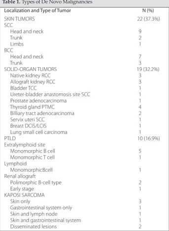
We analyzed posttransplant tumors in 4 main groups. Of the 59 patients, 22 (37.3%) had skin tumors, 19 (32.2%) had solid-organ tumors, 10 (16.9%) had posttransplant lymphoproliferative disorders (PTLD), and 8 (13.6%) had Kaposi sarcoma (KS). Of the 22 patients with skin tumors, 12 had squamous cell carcinoma (SCC), and 10 had basal cell carcinoma (BCC). Most of the skin tumors were located in the head and neck area (Table 1). Two of the SCCs were verrucous carcinoma, and 2 were carcinoma in situ; others were mostly welldif -ferentiated. Of 10 BCCs, 7 were nodular, 1 was metatypical, and 2 were mixed nodular and superficial histological subtypes. Of the 8 patients with KS, 3 had lesions limited to skin, 1 was restricted to the gastrointestinal system, 1 was skin and lymph node,
1 was skin and gastrointestinal system, and 2 had disseminated lesions to more than 2 organs. Histologic examination showed tumor stage in 1, plaque stage in 3, and patch stage in 2 patients with KS localized to skin. Immunohistochemically, only 6 of the 8 KS lesions showed positive expression of human herpes virus 8 antibody.
In the solid-organ tumor group, the most frequent neoplasms were urinary tract cancers (9 patients), followed by thyroid gland cancers (4 patients), gastrointestinal tract cancers (2 patients), and gynecological tract cancers (2 patients). Only 1 patient developed lung cancer (Table 1). The most common urinary tract cancers were located in the kidney (3 native kidneys and 3 renal allografts). Two of 3 native kidney cancers were diagnosed as clear cell type renal cell carcinoma (RCC) and the remaining 1 was diagnosed as papillary type RCC. Within renal allografts, we detected 2 clear cell RCC and 1 mucinous RCC. Nephron-sparing surgery was performed in 2 of the 3 renal allografts with RCC.
Abbreviations: BCC, basal cell carcinoma; DCIS, ductal carcinoma in situ;
LCIS, lobular carcinoma in situ; PTLD, posttransplant lymphoproliferative disorders; PTMC, papillary thyroid microcarcinoma; RCC, renal cell carcinoma; SCC, squamous cell carcinoma; TCC, Transitional cell carcinoma
Table 1. Types of De Novo Malignancies
Localization and Type of Tumor N (%)
SKIN TUMORS 22 (37.3%)
SCC
Head and neck 9
Trunk 2
Limbs 1
BCC
Head and neck 7
Trunk 3
SOLID-ORGAN TUMORS 19 (32.2%)
Native kidney RCC 3
Allograft kidney RCC 3
Bladder TCC 1
Ureter-bladder anastomosis site SCC 1 Prostate adenocarcinoma 1 Thyroid gland PTMC 4 Billiary tract adenocarcinoma 2
Servix uteri SCC 1
Breast DCIS/LCIS 1
Lung small cell carcinoma 1
PTLD 10 (16.9%) Extralymphoid site Monomorphic B cell 5 Monomorphic T cell 1 Lymphoid MonomorphicBcell 1 Renal allograft
Polimorphic B-cell type 2
Early stage 1
KAPOSI SARCOMA
Skin only 3
Gastrointestinal system only 1 Skin and lymph node 1 Skin and gastrointestinal system 1 Disseminated lesions 2
The remaining renal allograft with RCC also had chronic rejection at the time of tumor diagnosis. Thus, the patient had undergone graft nephrectomy. All of the urinary tract tumors were detected in organ-confined stages.
All of the thyroid gland cancers were papillary microcarcinoma, and 2 of 4 had lymph node me -tastases. Gastrointestinal neoplasms were biliary tract adenocarcinomas and were in stage pT4 at the time of diagnosis in all cases.
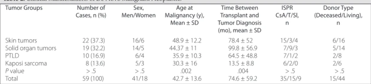
Posttransplant lymphoproliferative disorders were diagnosed in skin (2 patients), renal allograft (3 patient), and 1 patient for each of small bowel, brain, liver, peritoneum, and lymph node. Histologic diagnosis was polymorphic B-cell PTLD in 2 patients, monomorphic B-cell PTLD in 6 patients, monomorphic T-cell PTLD in 1 patient, and early lesion PTLD in 1 patient (Table 1). Epstein-Barr virus was positive in only 8 histologic specimens. Most of the patients with skin neoplasms, KS, and PTLD were treated with a CsA-based immunosuppressive regimen. However, we found that most of the patients who had solid-organ malignancy had a tacrolimus-based regimen (Table 2).
The mean age of patients at time of diagnosis of malignant tumor was 42.7 ± 13.6 years. Patients who had a diagnosis of KS were found to be younger than the other tumor groups. Statistically significant differences were found between tumor groups in regard to the age at the time of diagnosis of malignant tumor (P < .01) (Table 2). The average latency period between transplant and time of diagnosis of malignant tumors was 99.8 ± 56.9 months for solid-organ tumors, 78.4 ± 52 months for skin tumors, 64.5 ± 48.8 months for PTLD, and 13.5 ± 8.8 months for KS. Significant differences were found between tumor groups in regard to the time between transplant and tumor diagnosis (P < .01) (Table 2). However, there were no significant differences between tumor groups in regard to sex,
donor type, primary disease, or immuno suppressive treatment.
Ten patients (16.9%) had more than 1 malignant tumor, and 49 patients (83.1%) had only 1 malignant tumor. Of the 10 patients who had more than 1 tumor, 1 had BCC and gingival KS, 1 had BCC and intramucosal carcinoma in a tubular adenoma of the colon, 2 had multiple BCCs, and 6 had KS in multiple organs.
Of the 59 patients, 18 died at a mean time of 31.5 ± 22.8 months after tumor diagnosis. A significant positive association was found between patient survival and the number of tumors (P = .001). Overall 5-year survival after the diagnosis of the tumor was 81% and 40% for patients with 1 malignant tumor and patients with more than 1 malignant tumor, respectively.
Discussion
The development of posttransplant malignancies is a well-known complication after renal transplant and the third most common cause of death in transplant recipients.1 The overall incidence of neoplasms following renal transplant is reported to be 4% to 18%, with a 3- to 8-fold increase in cancer risk. However, this increase is as high as 100-fold for virus-related cancers such as KS.2,5-7In this study, we analyzed the occurrence of posttransplant de novo malignancies in our transplant center, and we found that the incidence of the development of a malignant tumor was 6.8% for our 867 patients.
Of all malignant tumors, skin cancers, specifically BCCs and SCCs, are common after transplant, and the incidence increases with time.8The incidence of posttransplant skin cancer varies from 2% to 68%.9-12 In the present study, incidence of skin cancers was found to be 37.3% and compatible with the literature. Advanced patient age is a common risk factor for skin tumors in transplant recipients; patients aged
Abbreviations: CsA, cyclosporine; ISPR, immunosuppressive regimen; PTLD, posttransplant lymphoproliferative disorders; Sl, sirolimus;
T, tacrolimus
Table 2. Clinical Manifestations of De Novo Malignant Neoplasms
Tumor Groups Number of Sex, Age at Time Between ISPR Donor Type
Cases, n (%) Men/Women Malignancy (y), Transplant and CsA/T/Sl, (Deceased/Living),
Mean ± SD Tumor Diagnosis n n
(mo), mean ± SD
Skin tumors 22 (37.3) 16/6 48.9 ± 12.2 78.4 ± 52 15/3/4 6/16 Solid organ tumors 19 (32.2) 14/5 44.37 ± 11 99.8 ± 56.9 7/9/3 5/14 PTLD 10 (16.9) 6/4 35.9 ± 10.3 64.5 ± 48.8 7/1/2 2/8 Kaposi sarcoma 8 (13.6) 5/3 30.3 ± 16 13.5 ± 8.8 6/2/0 2/6 P value > .5 > .5 .002 .004 > .5 > .5 Total 59 (100) 41/18 42.7 ± 13.6 74.6 ± 59.2 35/15/9 15/44
> 55 years were reported to have greater risk compared with patients < 35 years.9,12,13Similar to the literature, the mean age of our patients with skin cancers was 48.9 ± 12.2 years, and it is significantly higher than that of patients who developed other malignant tumors. The development of skin SCC was reported more frequently than BCC in kidney transplant recipients.10,14 Confirming these studies, we also found that SCC was the most common malignant tumor, followed by BCC. In contrast, some other studies showed higher ratios of BCC to SCC.9,11 Some cancers that are uncommon in general populations have a tendency to develop in organ transplant recipients. One of these uncommon neoplasms is KS. Human herpes virus 8 associated with KS and infection prevalence shows geographical variation ranging from 5% in North America and Northern Europe, to 30% in Mediterranean and Middle East countries, and to 50% in African regions.8,15-17 KS is reported with incidence rates between 17.6% and 70%, which is higher in the Middle East and Mediterranean countries compatible with the infection prevalence.18-21 Similar to the literature, the incidence of KS was 13.6% in our study. The age at the time of tumor diagnosis of patients with KS was significantly lower compared with patients from other tumor groups. Also, the average latency period between transplant and the diagnosis of KS was shorter than other tumor groups, and it is compatible with other centers’ reports that showed KS devel -opment in the first 2 years following transplant.15,22 Kaposi sarcoma is quite often limited to the skin in transplant recipients. However, visceral involvement tends to be associated with worse outcome and occurs in 10% of patients.23Contrary to the literature, most of our patients with the diagnosis of KS had visceral involvement. In the KS group, 75% of our patients had CsA-based immunosuppressive regimen. It has been reported that patients who received CsA were at a higher incidence of developing KS.24Penn also suggested similar reports that explained the increased incidence of KS with the use of CsA.25 Barrett and associates reported higher frequencies of KS and PTLD for recipients treated with CsA compared with patients who were not receiving CsA treatment.26 Piselli and associates reported that recipients treated with mammalian target of rapamycin (mTOR) inhibitors had a 45% reduction in overall cancer risk, and they suggested that this drug can control KS evolution.15In our center, the
initial step to treating KS patients was to decrease the immunosuppressive dosage while maintaining graft function.
PTLD is a heterogeneous group of diseases characterized by abnormal lymphoid proliferation after organ transplant and mostly manifested as B-cell neoplasia.27In this study, a higher percentage of PTLD was found to be the B-cell origin and monomorphic type. Sixty percent of our cases were found at higher stages at the time of diagnosis. The mean time of the development of PTLD was 64.5 ± 48.8 months after kidney transplant, and this is not compatible with the reports that state an average length of 32 months.1,28 Patients who developed PTLD had mostly CsA-based regimens. There are conflicting results on whether CsA had a higher influence on the development of malignant neoplasms.4,26,29The rate of allograft involvement by PTLD was reported to be 17% to 33% in renal allografts.30Similarly, we also found 30% of renal allograft involvement in our patients with PTLD.
The most frequently occurring malignant tumors in the general population, such as carcinomas of the colon, breast, and prostate, are not seen frequently after renal transplant.31In the review by Kauffman and associates, percentages of solid nonlymphoma cancers varied between 32.8% and 80%, and higher percentages of malignant tumors were seen in the genitourinary tract, and the lowest were in the endocrine system.7Apel and associates reported that urinary tract malignancies represented the most frequent malignancies among the nonskin tumors in their series of 1882 kidney transplant patients.32 Similarly, we detected solid-organ tumors in 32.2% of our patients, and the most common solid-organ tumor was urinary tract cancer. Renal cell carcinoma was reported to be the most frequent posttransplant urologic tract cancer.33,34In this study, of the 9 urinary tract cancers, 3 of them were kidney-graft RCC and 3 were native kidney cancer. Renal allograft carcinoma was reported in a few case reports.35It is not entirely clear whether these allograft tumors occur because of malignant transformation after transplant or if they are present at the time of transplant. However, early diagnosis is necessary to save the graft with limited surgery.36We detected the second most common solid-organ tumor as thyroid papillary microcar cinoma in our patients. Two of 4 patients with thyroid carcinoma had metastatic lymph nodes at the time of tumor diagnosis. In the English literature, post
-transplant thyroid cancers were reported as individual cases.32
Advanced patient age was reported to be a conventional risk factor for posttransplant malig -nancies, particularly for SCC.9,37At the time of tumor diagnosis, the patients with solid-organ tumors were older than those with KS and PTLD, but they were nearly the same age as the skin tumor group patients in our study. Apel and associates showed similar findings in which they found skin and nonskin solid organ tumors at the same age.32Between the tumor groups, solid tumors occurred later than the others, with an average latency period of 99.8 ± 56.9 months. Based on immunosuppressive regimens, the solid tumor group differs from others with tacrolimus-based treatment, but the difference was not statistically significant.
The overall mortality associated with post -transplant de novo malignancies is high and progressively increases with time. Pedotti and associates reported that the 10-year survival in kidney recipients without cancer is 92.8% and with cancer is 56.6%.3In the 58 patients who developed skin cancer, there was 50% mortality from cancer.6 Overall 5-year survival after the diagnosis of the tumor was 81% and 40% for patients with 1 malignant tumor and patients with more than 1 malignant tumor, respectively, in this study. However, we did not detect a statistically significant difference in tumor-specific survival between the 4 tumor groups.
In conclusion, regular screening for neoplasms must be done for all renal allograft recipients for early diagnosis and treatment.
References
1.Venyo A, Al-Hammadi A. Malignancy after renal transplantation: a review of the literature. Webmed Central Transplantation. 2012; 3(3):WMC003186. https://www.webmedcentral.com/ wmcpdf/ Article_with_review_WMC003186.pdf.
2. Penn I. Posttransplant malignancies. Transplant Proc. 1999;31(1-2):1260-1262.
3. Pedotti P, Cardillo M, Rossini G, et al. Incidence of cancer after kidney transplant: results from the North Italy transplant program. Transplantation. 2003;76(10):1448-1451.
4. Vogt P, Frei U, Repp H, Bunzendahl H, Oldhafer K, Pichlmayr R. Malignant tumours in renal transplant recipients receiving cyclosporin: survey of 598 first-kidney transplantations. Nephrol Dial Transplant. 1990;5(4):282-288.
5. Penn I. Cancers in renal transplant recipients. Adv Ren Replace Ther. 2000;7(2):147-156.
6. Tremblay F, Fernandes M, Habbab F, deB Edwardes MD, Loertscher R, Meterissian S. Malignancy after renal transplantation: incidence and role of type of immunosuppression. Ann Surg Oncol. 2002; 9(8):785-788.
7. Kauffman HM, Cherikh WS, McBride MA, Cheng Y, Hanto DW. Post-transplant de novo malignancies in renal Post-transplant recipients: the past and present. Transplant Int. 2006;19(8):607-620. 8. Chapman JR, Webster AC, Wong G. Cancer in transplant recipient.
Cold Spring Harb Perspect Med. 2013;3(7):a015677.
9. Fuente MJ, Sabat M, Roca J, Lauzurica R, Fernández-Figueras MT, Ferrándiz C. A prospective study of the incidence of skin cancer and its risk factors in a Spanish Mediterranean population of kidney transplant recipients. Br J Dermatol. 2003;149(6):1221-1226. 10. Karczewski M, Stronka M, Karczewski J, Wiktorowicz K. Skin cancer following kidney transplantation: a single-center experience. Transplant Proc. 2011;43(10):3760-3761.
11. Bernat García J, Morales Suárez-Varela M, Vilata JJ, Marquina A, Pallardó L, Crespo J. Risk factors for non-melanoma skin cancer in kidney transplant patients in a Spanish population in the Mediterranean region. Acta Derm Venereol. 2013;93(4):422-427. 12. Moosa, MR, Gralla J. Skin cancer in renal allograft recipients:
experience in different ethnic groups residing in the same geographical region. Clin Transplant. 2005;19(6):735-741. 13. Otley CC, Cherikh WS, Salasche SJ, McBride MA, Christenson LJ,
Kauffman HM. Skin cancer in organ transplant recipients: effect of pretransplant end-organ disease. J Am Acad Dermatol. 2005; 53(5):783-790.
14. Falsarella PM, Alves-Filho G, Mazzali M. Skin malignancies in renal transplant recipients: a Brazilian center registry. Transplant Proc. 2008;40(3):767-768.
15. Piselli P, Serraino D, Segoloni GP, et al. Risk of de novo cancers after transplantation: results from a cohort of 7217 kidney transplant recipients, Italy 1997-2009. Eur J Cancer. 2013;49(2):336-344. 16. Rama I, Grinyó JM. Malignancy after renal transplantation: the role
of immunosuppression. Nat Rev Nephrol. 2010;6(9):511-519. 17. Campistol JM, Schena FP. Kaposi’s sarcoma in renal transplant
recipients: the impact of proliferation signal inhibitors. Nephrol Dial Transplant. 2007;22(suppl1):i17-i22.
18. Samhan, M, Al-Mousawi M, Donia F, Fathi T, Nasim J, Nampoory MR. Malignancy in renal recipients. Transplant Proc. 2005;37(7): 3068-3070.
19. Moosa MR. Kaposi’s sarcoma in kidney transplant recipients: a 23-year experience. QJM. 2005;98(3):205-214.
20. Wong G, Chapman, JR. Cancers after renal transplantation. Transplant Rev. 2008;22(2):141-149.
21. Trofe J, Beebe TM, Buell JF, et al. Posttransplant malignancy. Prog Transplant. 2004;14(3):193-200.
22. Veroux M, Puliatti C, Fiamingo P, et al. Early de novo malignancies after kidney transplantation. Tranplant Proc. 2004;36(3):718-720. 23. Moosa MR. Racial and ethnic variations in incidence and pattern
of malignancies after kidney transplantation. Medicine. 2005;84 (1):12-22.
24. Haberal M, Moray G, Karakayali H, et al. Effect of immuno -suppressive treatment protocol on malignancy development in renal transplant patients. Transplant Proc. 2002;34(6):2133-2135. 25. Penn I. The changing pattern of posttransplant malignancies.
Transplant Proc.1991;23(1, pt 2):1101-1103.
26. Barrett WL, First MR, Aron BS, Penn I. Clinical course of malignancies in renal transplant recipients. Cancer. 1993;72(7): 2186-2189.
27. Pascual J. Post-transplant lymphoproliferative disorder—the potential of proliferation signal inhibitors. Nephrol Dial Transplant. 2007;22(suppl 1):i127-i35.
28. Caillard S, Agodoa LY, Bohen EM, Abbott KC. Myeloma, Hodgkin disease, and lymphoid leukemia after renal transplantation: characteristics, risk factors and prognosis. Transplantation. 2006; 81(6):888-895.
29. Shuttleworth D, Marks R, Griffin PJ, Salaman JR. Epidermal dysplasia and cyclosporine therapy in renal transplant patients: a comparison with azathioprine. Br J Dermatol. 1989;120(4):551-554.
30. Cohen JI. Epstein-Barr virus lymphoproliferative disease associated with acquired immunodeficiency. Medicine. 1991;70(2):137-160. 31. Penn I. De novo cancers in organ allograft recipients. Curr Opin
32. Apel H, Walschburger-Zorn K, Häberle L, Wach S, Engehausen DG, Wullich B. De novo malignancies in renal transplant recipients: experience at a single center with 1882 transplant patients over 39 yr. Clin Transplant. 2013;27(1):E30-E36.
33. Tsaur I, Karalis A, Probst M, et al. Development of urological cancers in renal transplant recipients: 30-year experience at the Frankfurt Transplant Center. Cancer Sci. 2010;101(11):2430-2435. 34. Hevia V, Gómez V, Díez Nicolás V, et al. Development of urologic de novo malignancies after renal transplantation. Transplant Proc. 2014;46(1):170-175.
35. Zhou M, Zhu Y, Wang L, Wang Y, Fu S, Min Z. Urological malignancy as a complication of renal transplantation: a report of twelve clinical cases. Clin Transpl. 2006;395-398.
36. Besarani D, Cranston D. Urological malignancy after renal transplantation. BJU Int. 2007;100(3):502-505.
37. Danpanich E, Kasiske BL. Risk factors for cancer in renal transplant recipients. Transplantation. 1999; 68(12):1859-1864.