Ankara Üniv Vet Fak Derg, 59, 151-153, 2012
Short Communication / Kısa Bilimsel Çalışma
Mesothelioma in a dog
Kübra A. TERİM KAPAKİN1, Rıfkı HAZIROĞLU2, Nesrin GÜRSAN3, Gözde YÜCEL2 1Department of Pathology, Faculty of Veterinary Medicine, Atatürk University, Erzurum; 2Department of Pathology, Faculty of
Veterinary Medicine, Ankara University, Ankara; 3Department of Pathology, Faculty of Medicine, Atatürk University, Erzurum,
Turkey.
Summary: In this study a mesothelioma case observed in a four–year-old female Rottweiler dog was defined. Macroscopically,
on the serosal surfaces of the stomach and intestines, numerous masses showing diffuse distribution, greyish yellow in colour and hard consistency were observed. Microscopically, tumour cells were predominantly spindle-shaped with moderate amounts of eosinophilic cytoplasm and ovoid nuclei. These cells was not stained with epithelial membrane antigen (EMA) staining, the epithelial marker, while stained with the epithelial marker pancytokeratin and mesenchymal marker vimentin in the avidin-biotin-peroxidase (ABC-P) method. As a result, tumor case seen in a dog's stomach and intestinal serosal surface was defined as a mesothelioma according to the histopathological and immunohistochemical findings.
Key words: Dog, immunohistochemistry, mesothelioma.
Köpekte mezotelioma olgusu
Özet: Bu çalışmada 4 yaşlı Rottweiler ırkı, dişi bir köpekte gözlenen mezotelioma olgusu tanımlandı. Makroskobik olarak
mide ve bağırsakların serozal yüzeyinde diffuz dağılım gösteren, değişik büyüklükte ve sert kıvamda, grimsi sarı renkte kitleler görüldü. Mikroskobik olarak tümör hücreleri çoğunlukla iğ şeklinde, eozinofilik sitoplazmalı ovoid çekirdekliydi. Bu hücreler uygulanan avidin biyotin peroksidaz (ABC-P) immunohistokimyasal boyama yönteminde, epiteliyal belirteç pansitokeratin ve mezenkimal belirteç vimentin ile boyanırken epiteliyal belirteç olan epitheliyal membran antijeni (EMA) ile boyanmadı. Sonuç olarak bir köpeğin mide ve bağırsak serozal yüzeyinde karşılaşılan tümör olgusu, histopatolojik ve immunohistokimyasal bulgularına göre mesotelioma olarak tanımlandı.
Anahtar sözcükler: İmmunohistokimya, köpek, mezotelioma.
Mesothelioma is a tumour of low malignancy, which either covers surfaces including the pericardium, pleura and peritoneum, or originates from the mesothelial cells in the tunica vaginalis of the testes. Three main histological types are described in domestic animals: the epithelioid, the sarcomatoid (spindle cell), and the biphasic (or mixed) type. But several subtypes have been described in human pathology, such as lymphohistiocytoid, desmoplastic, microcystic, clear cell, signet ring, small cell, deciduoid, solid, and tubulopapillary. Among the mesothelial tumours, sarcomatoid type has been less frequently reported. Mesotheliomas are rarely documented tumours (7, 10) which may develop in different animal species (4, 5, 15). However, these tumours are most frequently observed in cattles (8) and dogs (1, 3, 12, 14). In dogs, like in humans, the pleura is the main site of mesotheliomas development after the pericardial and the peritoneal cavity (7, 10).
The present study describes the case of a sarcomatoid mesothelioma in Rottweiler dog and its characterisation by pathological and immunohistochemical finding.
The stomach and intestines of a four-year-old female Rottweiler dog, submitted by a private veterinary clinic to the Department of Pathology of Ankara University Faculty of Veterinary Medicine for examination, constituted the material of the study.
The samples were fixed in 10% buffered formalin and routinely processed for histological examination by embedding in paraffin wax. Sections were cut 4 μm in thickness, afterwards stained by the haematoxylin and eosin (HE) (13).
Four µm sections from all the tissue samples were cut and processed for immunohistochemical examination by a standard avidin-biotin-peroxidase (ABC-P) method as described by the producer. By using the primer antibodies were specific for cytokeratin (pan cytokeratin
Kübra A. Terim Kapakin - Rıfkı Hazıroğlu - Nesrin Gürsan - Gözde Yücel 152
antibody, clone AE1/AE3, DAKO, Hamburg, Germany, diluted 1:500), vimentin (clone Vim 3B4, DAKO, Hamburg, Germany, diluted 1: 40) and epithelial membrane antigen (EMA, clone E23, DAKO, Hamburg, Germany, diluted 1: 200).
According to the anamnesis given, the dog died following the sudden development of inappetence, after inactivity and progressive weight loss.
On the serosal surfaces of the stomach and intestines, numerous masses showing diffuse distribution, having distinct borders, with varying sizes of around 2 mm- 3 cm in diameter, greyish yellow in colour and hard consistency were observed.
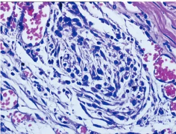
Tumour cells were arranged in sheets and streams supported by a prominent fibro-vascular stroma. These cells were predominantly spindle-shaped with moderate amounts of eosinophilic cytoplasm and ovoid nuclei which were irregular and not uniform in shape (Figure 1). Furthermore, it was determined that the blood vessels within the serosa were enlarged and filled with erythrocytes.
Immunohistochemically, tumoral cells were reacted by anti cytokeratin and anti vimentin sera (Figure 2), but not reacted by epithelial membrane antigen (EMA) (Figure 3).
It is reported that mesotheliomas occur mostly in dogs of a mean age above 8 years (1, 3, 9, 10). But, these tumours may also be observed in younger animals (11, 14), and even congenitally, as indicated in cattles (8) in the case. Among the many dog breeds, the Bouvier des Flandres, Irish Setters and German Shepherds are predisposed to mesotheliomas, and the occurrence of these tumours is higher in male dogs, compared to females (7). The present case concern an old female Rottweiler dog (four-year old) and is in agreement with previous report.
Exposure to chemicals, in particular, asbestos, iron and silicate play a major role in the development of mesotheliomas (7-10). However, it is reported that genetic factors and viruses also lead to the development of these tumours (6, 10). Moreover, idiopathic and spontaneous tumours have also been reported in animals (15). Based on the anamnesis and the information obtained to the through live of the animal, if expected out of the probability of the animal being exposed to any chemical.
Sarcomatoid type mesothelioma cases in animals are rare and a few histopathological and immunohistochemical studies have been reported on this subject. Macroscopically, sarcomatoid mesotheliomas are diffuse nodular multifocal masses that cover body cavities. Microscopically, sarcomatoid cells are known for their elongated spindle shape (4, 7, 10). The macroscopic and microscopic findings obtained in the present case are in agreement with the sarcomatoid mesothelioma characteristics found in the literature.
Figures 1. Spindle-shaped mesothelial cells, (HE, x400). Şekil 1. İğ şeklinde mezotel hücreleri, (HE, x400).
Figure 2. Immunohistochemically, cytoplasm of the tumour cells stained as dark red in colour with anti vimentin sera. (arrow) (ABC-P, x200).
Şekil 2. İmmunohistokimyasal olarak anti vimentin serumu ile sitoplazması koyu kırmızı renkte boyanan tümör hücreleri. (oklar) (ABC-P, x200).
Figure 3. Immunohistochemically, the EMA staining was negative for tumour cells. (arrow) (ABC-P, x400).
Şekil 3. İmmunohistokimyasal olarak, EMA negatif tümör hücreleri. (oklar) (ABC-P, x400).
Ankara Üniv Vet Fak Derg, 59, 2012 153
Histological mesothelioma should be differentiated from carcinomas, adenocarsinomas, or sarcomas depending on the type of mesothelioma. In general, the differential diagnosis for mesothelioma depends on its basic histological category: the differential diagnosis for epithelioid mesotheliomas includes carcinomas and other epithelioid cancers, the differential diagnosis for sarcomatoid mesothelioma includes sarcomas and other spindle cell neoplasms, and the differential diagnosis of mixed mesothelioma includes other mixed or biphasic tumours such as synovial. Mesothelioma immunohistochemistry can be most useful in differentiating. Vimentin positivity in epithelial mesothelioma may be useful in distinguishing it from pulmonary adenocarcinoma, which is usually vimentin-negative. In our case, the tumor cells reacted positively for vimentin.
Similarly, the positivity of sarcomatous mesotheliomas for broad-spectrum pancytokeratin is useful in distinguishing this subtype from sarcomas, which are usually cytokeratine negative (2, 4). The expression of both pancytokeratin and vimentin markers is a constant feature in mesotheliomas, attributable to the origin of mesothelial cells from the mesoderm, which can differentiate into both epithelial and mesenchymal cells (4). In this case, tumour cells expressed both by pancytokeratin, as an epithelial marker, and vimentin as a mesenchymal marker.
EMA reaction can be used to distinguish epithelioid type mesothelioma from sarcomatoid type mesothelioma. EMA gives a positive staining for epithelioid type mesothelioma (2). In order to distinguish whether tumor cells showing positive reaction for both vimentin and pancytokeratin were epithelial or mesenchymal origin, the epithelial marker EMA was used. However, the EMA staining, as an epithelial marker, was negative for tumour cells.
According to the gross and microscopic appearance of this tumors, together with its immunohistochemical staining characteristics, (positive reaction for mesenchymal marker and negative reaction for epithelial marker EMA) are consistent with a diagnosis of sarcomatoid type mesothelioma.
In conclusion, in this case of sarcomatoid mesothelioma, rarely reported in animals, was observed in a Rottweiler dog and examined histopathologically and immunohistochemically.
References
1. Amaya JO, Arevalo GH (2008): Invasive Mesothelioma
of Canine: Cytology, Clinical and Pathology’s Findings.
Int J Morphol, 1, 103-112
2. Attanoos RL, Dojcinov SD, Webb R, Gibbs AR (2000):
Anti-mesothelial markers in sarcomatoid mesothelioma and other spindle cell neoplasms. Histopathology, 37,
224–231.
3. Avakian A, Alroy J, Rozanski E, Keating J, Rosenberg A (2008): Lipid-rich pleural mesothelioma in a dog. J Vet Diagn Invest, 5, 665-667.
4. Bacci B, Morandi F, De Meo M, Marcato PS (2006): Ten cases of feline mesothelioma: an immunohistochemical and ultrastructural study. J Comp Pathol, 134, 347–354. 5. Bollo E, Scaglione FE, Tursi . M, Schröder C, Degiorgi
G, Belluso E, Capella S, Bellis D (2010): Malignant pleural mesothelioma in a female Lion (Panthera leo) Res Vet Sci, doi:10.1016/ jrvsc. 2010.08.005
6. Cacciotti P, Libener R, Betta P, Martini F, Porta C, Procopio A, Strizzi L, Penengo L, Tognon M, Mutti L, Gaudino G (2001): SV40 replication in human
mesothelial cells induces HGF/Met receptor activation: a model for viral-related carcinogenesis of human malignant mesotelioma. Proc Nati Acad Sci, 98, 12032- 12037.
7. Caswell J, Wiliams K (2007): Respiratory system M. Maxie (ed) 5th (Ed), Jubb, Kennedy, and Palmer’s Pathology of Domestic Animals, 5th edition, Elsevier, New York, NY, USA, p. 523–655.
8. Croft W (1983): Environmental asbestos and mesotheliomas
in dairy calves. Proc Am Assoc Cancer Res, 24, 188.
9. Gibbs GW, Berry G (2008): Mesothelioma and asbestos. Regul Toxicol Pharmacol, 52, 223–31.
10. Head KW, Else RW, Dubielzig RR (2002): Tumours of
the alimentary tract. D.J. Meuten (ed.) Tumours In
Domestic Animals, p. 401-481.
11. Kim JH, Choi YK, Yoon HY, Kweon OK, Kim DY (2002): Juvenile malignant mesotelioma in a dog. J Vet Med Sci, 64, 269-271.
12. Ledecka K, Sevcikova Z, Mihaly M, Hajurka J, Pavuk V, Hluchy M, Skurkova L, Lackova M, Ledecky V (2010) Mesothelioma of the pericardium in a Bernese
Mesothelioma of the pericardium in a Bernese mountain dog. Veterinarski Arhıv, 80, 797-806.
13. Presnell JK, Schreibman MP (1997): Humason’s Animal
Tissue Techniques.. In: Presnell J., Schreibman M.P (ed.),
5th (Ed), The Johns Hopkins University Press. Ltd, London, p. 269-312.
14. Vural SA, Ozyildiz Z, Ozsoy SY (2007): Pleural
mesothelioma in a nine month-old dog. Irish Vet J, 1, 30- 33.
15. Yamate J, Tomita A, Kuwamura M, Mitsunaga F, Nakamura S (2007): Spontaneous peritoneal malignant
mesothelioma in a geriatric japanese macaque (Macaca fuscata). Exp Anim, 56, 155–159.
Geliş tarihi: 14.12.2010 / Kabul tarihi: 05.07.2011
Address for correspondence:
Dr. Kubra Asena Terim Kapakin University of Ataturk
Faculty of Veterinary Medicine, Department of Pathology, Erzurum-Turkey