Ankara Üniversitesi Tıp Fakültesi Mecmuası 2006; 59:45-47
CERRAHİ BİLİMLER / SURGICAL SCIENCES
Olgu Bildirisi / Case Report
Corresponding author Dr. Selçuk Güven
Selcuk University Meram Faculty of Medicine, Department of urology 42080 Meram, Konya, Turkey
Tel :+90 (533) 2516339 Fax :+90 (332) 2236181 E-mail adress : [email protected] Received: 14.04.2005 • Accepted: 31.08.2005
45
P
osterior urethral polyps are benign polypoid lesions arising from veru-montanum or somewhere of prostatic urethra and cause bladder outlet obstruction, hematuria and urinary infection (1-7). The widespread use of high-quality excretory urography, cystourethrography, ultrasound and cystoure-throscopy has led to an increased recognation of these rare lesions (3). Posterior urethral polyps should be considered and investigated in boys and young adults presenting with hematuria and nonspesific voiding disturbances. Conventional treatments of posterior urethral polyps are open cystotomy, transurethral resec-tion or fulguraresec-tion of the polyps and removing of the polyp via laser (8). In view of the rarity of these lesions here in we report our experience with three cases of urethral polyps and outline the diagnostic evaluation and treatment.Case reports
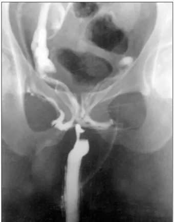
Between 1992-1999, 3 patients with posterior urethral polyps were seen at our clinic. All of them presented with obstructive voiding complaints and pain-less gross hematuria. Two of the patients had normal excretory urograms but one of them showed the filling deffect in the base of the bladder (Fig. 1) and antegrade urethrogram showed well-defined urethral filling deffect and venous leakage in the adult (Fig. 2).
Cystourethroscopy revealed three polyps, 2 of them arising from verumon-tanum and one of them in the prostatic urethra. Polyps were resected transure-thrally. In one boy after transurethral resection the polyp migrated into the bladder but we retreived it with a broken resectoscope loope (like a fish-hook) avoiding open cystotomy. All of the specimens of histopathologic examination showed fibroepithelial benign polyps.
Posterior urethral polyps may cause hematuria, various voiding disorders, out flow obstruction and urinary infections in boys and adults. We report three case of posterior urethral polyps two of them in boys and one of them in an adults. These rare lesions should be considered in the diffe-rential diagnosis of hematuria and lower obstructive urinary symptoms in boys and adults.
Key Words: Posterior urethral polyp, obstruction
Posterior ürethral polipler çocuklarda ve yetişkinlerde hematüri, işeme bozuklukları, çıkış obstrük-siyonu ve idrar yolu enfeksiyonlarına neden olabilirler. Burada ikisi çocukluk çağı, biri yetişkin dönemde görülen üç posterior ürethral polip olgusu sunulmaktadır. Ender görülen bu lezyonlar hematüri ve alt üriner sistem obstrüksiyonlarının ayırıcı tanısında akılda tutulmalıdır.
Anahtar sözcükler: Posterior urethral polip, obstrüksiyon
Posterior urethral polyps
Posterior ürethral polipler
Recai Gürbüz, Selçuk Güven, Esat Mehmet Arslan, Tahsin Özdemir
Selcuk University Meram Faculty of Medicine, Department of Urology Konya, Turkey
Ankara Üniversitesi Tıp Fakültesi Mecmuası 2006; 59(1)
Posterior urethral polyps 46
Case 1
A 4-year-old male child presented with a history of intermittan voiding difficulties and painless gross hema-turia for a few months. Excretory urography and ultra-sound were normal but cystourethroscopy revealed a 1 cm polyp arising from verumontanum. The polyp resected via transurethrally and histopathologic examination showed a benign polyp of connective tissue covered by transitional epithelium. Subsequently, the complaints of the child have disappeared and the patient has voided well.
Case 2
A 7-year- old male was referred to our clinic because of hesitancy, a decreased stream and terminal hematuria. Excretory urography showed a filling deffect at the base of the bladder (Fig. 1). Cystourethroscopy revealed a 2 cm polyp arising from verumontanum. After the transurethral resection of the polyp, it migrated into the bladder. But the specimen removed with a broken resectescope loop (like a fish-hook) without suprapubic cystotomy. Histopatho-logic examination showed fibrovasculer core and overlying epithelium. The child has done well during follow-up.
Case 3
A 23-year-old male was referred to our clinic because of nocturia, frequency and a poor stream for a few years. Uri-Figure1. Excretory urogram showes a 2 cm bladder filling
deffect at the base.
Figure 2. Antegrade urethrogram showes a 2 cm urethral filling
deffect and venous leakage.
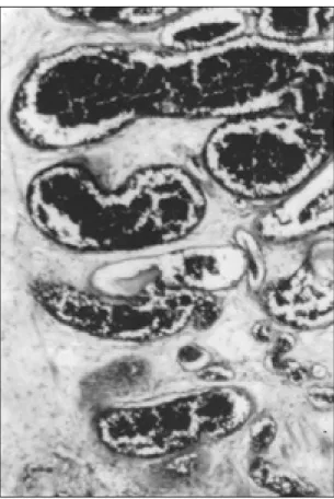
nanalysis showed microscopic hematuria. Excretory urog-raphy and ultrasonogurog-raphy were normal but antegrade urethrogram showed a 2 cm filling deffect in the mem-braneus urethra and accompanied venous leakage (Fig. 2). Cystourethrography revealed a 2 cm polyp occluded whole the urethral lumen. The polyp was resected transurethrally and histopathologic examination showed a 2 cm polyp has a fibrous core, consisting glanduler tissue covered by pseu-dostratified columnar epithelium (Fig. 3). Postoperativelly the patient voided with an excellent stream.
Discussion
Posterior urethral polyps are uncommon lesions and up to 1989, 66 cases of urethral polyps are found in the literature (9). Nearly all of them originated from posterior urethra and all reported examples have occured in boys or men as in our cases. The reported age range of the patients newborn to 79 years and 82 % of all cases occur in younger than twenty years old (9). Our cases are four, seven and twenty-three years old.
Symptoms of bladder outlet obstruction, hematuria or urinary infection are the most common complaints of polyps and the diagnosis is made by excretory urography, voiding cystourethrography, ultrasound and cystourethros-copy. Excretory urography showes the typical filling deffect
Journal of Ankara University Faculty of Medicine 2006; 59(1)
R. Gürbüz, S. Güven, E.M. Arslan, T. Özdemir 47
at the base of the bladder as one of our cases (Fig. 1). Ret-rograde urethrography is advocated by some authors (2) and retrograde urethrography showed a 2 cm filling deffect and venous leakage in the adults (Fig. 2). Cystourethros-copy revealed the polyps in all of our cases.
Urethral polyps were managed successfully by transure-thral resection in our cases without suprapubic cystotomy. Histopathologic examination of the specimens showed benign polyps consisting of fibrovascular core covered by transitional epithelium and the aduft polyps composed of prostatic type glands as in our cases (Fig. 3).
The prognosis of the posterior urethral polyps are ex-cellent with the complete transurethral resection. No re-currence was seen during follow-up in our cases as in the literature (3).
As a conclussion posterior urethral polyps may cause obstructive urinary symptoms and hematuria in pediatric age group and adults. These rare lesions should be con-sidered in the differantial diagnosis of lower urinary tract obstruction, and diagnosis is made by excretory uragraphy, voiding cystourethrography and also antegrade urethrogra-phy and cystourethroscopy. Transurethral resection is the best choice of the treatment.
Figure 3. High power view of polyp covered by
pseudostratified epithelium and consisting of connective tissue core with numerous glands.
References
1. Youssif M. Posterior urethral polyps in infants and children. Eur Urol 1985;11:69-70.
2. Raviv G, Leibowitch I, Hanani J et al. Hematuria and voiding disorders in children caused by congenital urethtal polyps. Eur Urol 1993;23:382-385.
3. Gleason PE, Kramer SE. Genitourinary polyps in children. Urology 1994;44:106-109.
4. Eglen DE, Pontius EE. Benign prostatic epithelial polyp of the urethra. J Urol 1984;131:120-122.
5. Kimche D, Lask D. Congenital polyp of the prostatic urethra. J Urol 1982;127:134.
6. Caro PA, Rosenberg HK, Synder HMC. Congenital urethral polyp. AJR 1986;147:1041-1042.
7. Foster C, Gavrett RA. Congenital posterior urethral polyps. J. Urol 1986 136:670-672.
8. Gentle DL, Kaufman RP, Mandel J. Use of neodymium. Yttrium-Aluminium-Garnet laser for removal of a congenital posterior urehral polyp in a 3-year old child. A case report and review of the literature. Urology 1996; 47:445-447.
9. Peterson RO. Urethral polyp. In: Urologic Pathology. Philedelphia: JB Lipp. Comp. Second Edition, 1992 pp 404.