Original Article
Prospective analysis of skin findings in
medical critically ill patients in intensive care units
Suzan Demir Pektas¹, Arzu Kahveci Demir2
¹Department of Dermatology, Mugla Sitki Kocman University Faculty of Medicine, 48000, Mugla, Turkey; 2Department of Anesthesia and Reanimation, Aydin State Hospital, 09000, Aydin, Turkey
Received August 29, 2016; Accepted October 27, 2016; Epub October 15, 2017; Published October 30, 2017 Abstract: We aimed prospectively to investigate skin disorders developing in critically ill medically treated patients during their ICU stay. Critically ill medically treated patients were analyzed for factors affecting the rate and clinical properties of skin disorders. We recorded age, sex, comorbidities, skin disorders, time to consultation, ICU admis-sion, duration of ICUs stay, and mortality rate. Among 1276 patients admitted to ICUs, 11.8% (n:151) had a der-matology consultation requested. The mean age of the study population was 68.9 ± 15.2 years (18-96 years), and 56% of the subjects were female. Infective skin lesions were detected in 47.7% of patients, dermatoses in 36.4%, and drug reactions in 15.9%. The mean age of patients with the skin disease was significanlty higher than without skin disease group (P<0.05). The frequency of skin disorders increased in rate in patients having diabetes mel-litus, chronic renal failure, and malignancy (P<0.05). The hospitalization period of patients with skin disease was significantly longer than without skin disease group (P<0.05). The rate of skin disorders increased with prolonged ICUs stay, and they were associated with increased mortality (P<0.05). There was an increased rate of skin infec-tion in patients of internal medicine ICU, while the rates of drug reacinfec-tions and dermatoses were significantly higher in patients admitted to other ICUs (P<0.05). The time to consultation was longest for patients diagnosed with skin infections and shortest for patients with dermatoses (P<0.05). Skin disorders that developed in patients treated at ICU were correlated to age, comorbid conditions, duration of ICU stay and mortality.
Keywords: Dermatology consultation, skin disorders, comorbidity, ICU admission, intensive care unit, duration of ICU stay
Introduction
Intensive care units (ICUs) are places designed for the management of life-threatening organ failures secondary to acute disorders or acute exacerbations of chronic conditions, where ne- cessary equipment and personnel are continu-ously kept available for close patient monitor-ing and rapid intervention [1]. In addition to the primary condition, new pathological condi-tions may emerge during ICU stay, depending on pre-existing morbidities and iatrogenic or environmental factors [2, 3]. Such pathologies also include skin disorders [2]. Previous studies have shown that skin disorders involving ICU patients reduce quality of life, prolong ICU stay, and augment mortality [4-8]. Simple measures such as changing patients’ position and regular skin cleansing using appropriate cleansing and care products have been shown to help protect
the skin barrier [9]. On the other hand, organ failures, electrolyte disorders, comorbid condi-tions, adverse medication effects, and infec-tions caused by opportunistic pathogens can lead to skin disorders. Unfortunately, the num-ber of studies on skin disorders developing at ICUs is limited [2, 3].
Herein, we aimed to investigate skin disorders in medically treated critically ill patients and thereby to contribute to the existing literature. Materials and methods
This study was approved by the local ethics committee at Adnan Menderes University Fa- culty of Medicine (Approval date 20.07.2012, Approval No 2012/87) and was prospectively conducted in medically treated critically ill pati- ents admitted to various ICUs at Aydin State
Hospital between 20.07.2012 and 20.8.2014. The patients staying at the internal diseases, pulmonary, neurological, coronary and reani-mation ICUs were included in the study. Study data were accessed via written medical records and hospital’s automation system.
Patients for whom dermatology consultation was requested formed Group A, and those for whom no dermatology consultation was requested formed Group B. Age, gender, name of ICU, comorbidities, skin disorders, time to consultation, duration of ICU stay, and mort- ality rate were recorded. Factors affecting the rate and types of skin disorders were analyzed. Patients who refused to participate in our study, who had missing medical or dermatological consultation data, who were younger than 18 years or pregnant, and who had a history of trauma or surgery were excluded.
Study data were analyzed in SPSS Windows version 18. The distribution of continuous and discrete numeric variables was evaluated by the Kolmogorov-Smirnov test. The continuous and discrete numeric variables were reported
Results
Among 1276 patients enrolled by our study, 151 (11.8%) had a dermatology consultation and were grouped in Group A. The rest of the patients (n:1125) were classified as Group B. The mean age of the patients with skin disorders was 68.9 ± 15.2 years (18-96 years). Fifty-six percent of the patients (n:86) were female and 44% (n:65) were male. Among the patients, 47.7% had infective skin lesions, 36.4% had dermatoses, and 15.9% had drug reactions (Table 1). The subgroups of skin dis-orders detected in the study subjects are pre-sented in Table 1.
The mean age of patients with the skin disease was significanlty higher than which (P<0.05, Table 2). No correlation was found between gender and skin disorders (P>0.05, Table 2). Among consulted patients, skin disorders were significantly more frequent in patients with DM, CRF, and malignancy (P<0.05 for all, Table 2), but not in those with hypertension (HT), heart failure (HF), coronary artery disease (CAD), ch- ronic obstructive pulmonary disease (COPD), Table 1. The classification of skin diseases
n (%) n (%) Infection (72) Fungal 54 (75.0) Candida 50 (92.7) Tinea cruris 3 (5.5) Tinea fascia 1 (1.8) Bacterial 11 (15.3) Folliculitis 7 (63.6) Cellulite 3 (27.3) Erysipelas 1 (9.1) Viral 7 (9.7) Herpes 5 (71.4) Zoster 2 (28.6) Drugs (n:24)
Maculopapular drug eruption 16 (66.7) Acneiform drug eruptions 6 (25.0)
Fix drug eruption 2 (8.3)
Dermatoses (n:55)
Friction blisters 18 (32.7) Allergic contact dermatitis 13 (23.7)
Urticaria 11 (20.0)
Seborrheic dermatitis 8 (14.5)
Vasculitis 4 (7.3)
Pemphigus vulgaris (recurrence) 1 (1.8)
as mean ± standard deviati-on, the nonparametric conti-nuous and discrete numeric variables: mean (the smal-lest-the largest) and interqu-artile range (IQR) and cate- gorical variables: number of cases and percentile (%). To evaluate the significance of the difference in parametric data, Student’s t test; signi- ficance of the difference in nonparametric data, Mann-Whitney and Kruskal-Wallis test data; significance of dif-ferences in categorical vari-ables, Pearson’s chi-square and Fisher’s exact tests were used. The parameters affec-ting the development of der-matological lesions variabl- es were evaluated by Binary Logistic Regression Analysis. The results were presented at a confidence interval of 95%, and the significance level was set at P<0.05.
3). The time to consultation was longest for patients diagnosed with skin infections and shortest for patients with dermatoses (P<0.05, Table 3). There was no significant correlation between skin disorders, duration of ICU stays, and mortality (P>0.05, Table 3). There was an increased rate of skin infections in patients in internal medicine ICUs, while the rates of drug reactions and dermatoses were signifi-cantly higher in patients admitted to other ICUs (P<0.05, Table 3). It was detected that the fac-tors significantly affecting development of der-matological lesions in ICU patients was dura-tion of ICU hospitalizadura-tion, the presence of CRF, COPD and malignacy (P<0.05, Table 4).
Discussion
During stay at intensive care unit, prolonged immobilization, malnutrition, impaired tissue perfusion, immune system dysfunction, incre- ased edema, fluctuations in body tempera- ture, inadequate hygiene, hyperpyrrexia, medi-cations, and skin injuries may cause the disrup-Alzheimer’s disease, parkinson, or stroke (P>
0.05 for all, Table 2). There was no significant association between ICU type and the rate of skin disorders (P>0.05). Median duration of ICU stay was 8 days (IQR:12, min:2, max:89) ; median time to dermatology consultation was 5 days (IQR:5, min:0, max:35); and the over- all mortality rate was 22.4%. The hospitaliza-tion period of patients with skin disease was significantly longer than without skin disease group (P<0.05, Table 2). The rate of skin disor-ders increased with prolonged ICU stay, and they were associated with increased mortality (P<0.05, Table 2).
Gender and age were not correlated to the rates of skin disorder subtypes (P>0.05, Table 3). It was shown that diabetics most commonly had skin infections; patients with CRF mostly had drug reactions; and patients with Parkin- son disease mostly had dermatoses (P<0.05, Table 3). No significant correlation was found between lesion type and HT, HF, CAD, COPD, Alzheimer’s disease, or stroke (P>0.05, Table
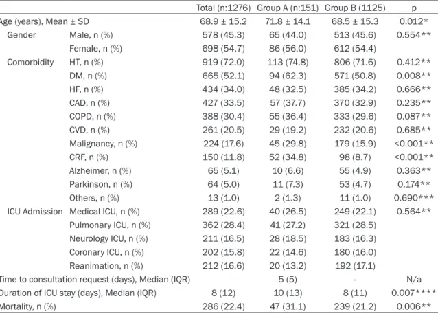
Table 2. The comparison of demographic properties of the study groups
Total (n:1276) Group A (n:151) Group B (1125) p Age (years), Mean ± SD 68.9 ± 15.2 71.8 ± 14.1 68.5 ± 15.3 0.012* Gender Male, n (%) 578 (45.3) 65 (44.0) 513 (45.6) 0.554** Female, n (%) 698 (54.7) 86 (56.0) 612 (54.4) Comorbidity HT, n (%) 919 (72.0) 113 (74.8) 806 (71.6) 0.412** DM, n (%) 665 (52.1) 94 (62.3) 571 (50.8) 0.008** HF, n (%) 434 (34.0) 48 (32.5) 385 (34.2) 0.666** CAD, n (%) 427 (33.5) 57 (37.7) 370 (32.9) 0.235** COPD, n (%) 388 (30.4) 55 (36.4) 333 (29.6) 0.087** CVD, n (%) 261 (20.5) 29 (19.2) 232 (20.6) 0.685** Malignancy, n (%) 224 (17.6) 45 (29.8) 179 (15.9) <0.001** CRF, n (%) 150 (11.8) 52 (34.8) 98 (8.7) <0.001** Alzheimer, n (%) 65 (5.1) 10 (6.6) 55 (4.9) 0.363** Parkinson, n (%) 64 (5.0) 11 (7.3) 53 (4.7) 0.174** Others, n (%) 13 (1.0) 2 (1.3) 11 (1.0) 0.690*** ICU Admission Medical ICU, n (%) 289 (22.6) 40 (26.5) 249 (22.1) 0.564**
Pulmonary ICU, n (%) 362 (28.4) 41 (27.2) 321 (28.5) Neurology ICU, n (%) 211 (16.5) 28 (18.5) 183 (16.3) Coronary ICU, n (%) 202 (15.8) 22 (14.6) 180 (16.0) Reanimation, n (%) 212 (16.6) 20 (13.2) 192 (17.1)
Time to consultation request (days), Median (IQR) 5 (5) - N/a Duration of ICU stay (days), Median (IQR) 8 (12) 10 (13) 8 (11) 0.007**** Mortality, n (%) 286 (22.4) 47 (31.1) 239 (21.2) 0.006**
*Student t test, **Pearson chi-square test, ***Fisher exact test, ****Mann whitney U test, n: number of cases, IQR: inrequantile range, DM: diabetes mellitus HT: hypertension. CF: heart failure, CAD: coronary artery disease, COPD: chronic obstructive pulmonary disease, CVD: cerebrovascular disease, CKF: chronic renal failure, ICU: intensive care unit.
tion of skin barrier, leading to skin disorders [4, 6, 8].
In our study, skin disorders that developed in ICU patients were associated with age, comor-bid conditions, duration of ICU stay, and mortal-ity. The mean age of patients with the skin dis-ease was significanlty higher than without skin disease group. There was no significant correla-tion between the incidence of skin disease and gender. The rate of skin disorders increased as ICU stay prolonged, and they were associat-ed with increasassociat-ed mortality. The frequency of skin disorders increased in patients with DM, CRF, and malignancy. There was no association between mortality and dermatological disorder subtypes.
Literature data suggest that the rate of skin pathologies developed at mixed intensive care units ranges between 2.2% and 21.5% [6, 10]. As medical problems with elevated death risk are at the center of focus in patients admitted
to ICU, dermatology consultations are fairly un- common at these units [2]. In agreement with the literature, our study demonstrated a rate of 11.8% for dermatology consultation requests for patients admitted to ICU.
Fischer et al reported that skin infections occurred at a rate of 29% [10]. In a study con-ducted by Emre et al at the internal disease and anesthesiology ICUs, 38.9% of patients developed skin infections, with the most com-mon skin infections being candidal infections followed by viral infections [4]. Candida species are the most common fungal subgroup, which infect patients as a result of temperature and humidity swings, as well as due to the adminis-tration of broad spectrum antibiotics, paren-teral nutrition, immunosuppressive agents, as well as having comorbidities [11].
Candida infections tend to be located in body regions that are hardly ever kept clean and dry when patients are immobile (e.g inguinal, axil-Table 3. Comparison of patients demographic properties between subgroups of skin disorders
Infections
(n:72) n (%) Drug Reactions (n:24) n (%) Dermatoses (n:55) n (%) p Age (years), Mean ± SD 75.5 (15) 75.5 (10) 78 (16) 0.599* Gender Male, n (%) 36 (50.0) 10 (41.7) 19 (34.5) 0.217** Female, n (%) 36 (50.0) 14 (58.3) 36 (65.5) Comorbidity HT, n (%) 56 (77.8) 20 (83.3) 37 (67.3) 0.232** DM, n (%) 59 (81.9) 9 (37.5) 26 (74.3) <0.001** HF, n (%) 20 (27.8) 7 (29.2) 22 (40.0) 0.322** CAD, n (%) 29 (40.3) 8 (33.3) 20 (36.4) 0.803** COPD, n (%) 19 (26.4) 11 (45.8) 25 (45.5) 0.050** CVD, n (%) 16 (22.2) 2 (8.3) 11 (20.0) 0.412*** Malignancy, n (%) 21 (29.2) 10 (41.7) 14 (25.5) 0.345** CRF, n (%) 21 (29.2) 14 (58.3) 17 (30.9) 0.027** Alzheimer, n (%) 7 (9.7) 1 (4.2) 2 (3.6) 0.194*** Parkinson, n (%) 3 (4.2) 0 8 (14.5) 0.047*** Others, n (%) 1 (1.4) 0 1 (1.8) >0.999***
ICU Admission Medical ICU, n (%) 28 (38.9) 6 (25,0) 6 (10.9) 0.002*** Pulmonary ICU, n (%) 16 (22.2) 9 (37,5) 16 (29.1)
Neurology ICU, n (%) 11 (15,3) 2 (8,3) 15 (27.3) Coronary ICU, n (%) 10 (13.9) 4 (16,7) 8 (14,5) Reanimation, n (%) 7 (9.7) 3 (12.5) 10 (18.2)
Time to consultation request days), Median (IQR) 7 (6) 6.5 (3) 5 (5) 0.010* Duration of ICU stay (days), Median (IQR) 10.5 (11) 10 (9) 10 (16) 0.693*
Mortality, n (%) 26 (36.1) 5 (20.8) 16 (29.1) 0.345**
*Kruskal Wallis test, **Pearson chi-square test, ***Fisher exact test, n: number of cases, IQR: inrequantile range, DM: diabe-tes mellutus HT: hypertension, HF: heart failure, CAD: coronary artery disease, COPD: chronic obstructive pulmonary disease, CVD: cerebrovascular disease, CRF: chronic renal failure, ICU: intensive care unit.
lary regions) [6]. It has been reported that vul-vovaginal candididiasis is a serious risk factor for infection in diabetic patients admitted to ICU [3].Our study detected infective skin disor-ders at a rate of 47.6%, among which fungal infections were the most common. We consider that a higher rate of infectious lesions in the present study compared to prior studies likely resulted from medically treated ICU patients who were more prone to infectious complica-tions due to having a more advanced age, more prolonged immobility and debiliation, and a hig- her number of comorbidities. To our opinion, a lower bacterial infection rate despite a higher fungal infection rate resulted from the admi- nistration of broad-spectrum antibiotics, exclu-sion of decubitis leexclu-sions from our study, and the tendency of the candida species to be located at regions difficult to be kept clean. Furthermore, it should be remembered that immunosuppression caused by the primary dis-order, comorbidities, immunosuppressive med-ications, and steroids may give rise to these opportunistic pathogens.
Emre et al reported that dermatoses are obs- erved at intensive care units at a rate of 46.6%,
gasser ganglion [14].The prevalence of derma-toses was 36.4% in our study, and as reported previously, the most common dermatosis sub-groups were frictional bullae (32.7%) and aller-gic contact dermatitis (23.7%). To our opinion, the rate of dermatoses was lower in our study compared to other studies, possibly because of a greater quality of nursing care and use of air beds and quality antiseptics at our hospital. However, we also believe that the emergence of dermatosis is inevitable due to comorbidi-ties, chlorhexidine used as the most common antiseptic, edema, and friction occurring during change of body position.
Prior studies have reported that drug reactions occur at a rate of 9.3% to 21.6% at intensive care units [2, 4, 6, 10, 15]. Genetic factors and inflammatory reactions have been implicated in the genesis of drug reactions [15]. Emre et al reported that drug reactions occurred in 14.5% of patients staying at intensive care units, and maculopapular rash was the most common reaction against antibiotics [4]. Trivalle et al stated that drug reactions create a risk for skin disorders in patients admitted to intensive care units [16]. Antibiotics and chemotherapeutics Table 4. Logistic regression analysis of the factors affecting
the dermatological lesions
Variables in the equation
B S.E. Wald df Sig. Exp (B) Age 0.011 0.007 2.669 1 0.102 1.011 Gender 0.120 0.184 0.426 1 0.514 1.128 HT -0.297 0.258 1.321 1 0.250 0.743 DM 0.311 0.226 1.889 1 0.169 1.364 HF 0.020 0.216 0.008 1 0.928 1.020 CAD -0.003 0.200 0.000 1 0.989 0.997 COPD 0.443 0.207 4.588 1 0.032 1.558 CVD -0.168 0.239 0.494 1 0.482 0.845 Malignancy 0.616 0.220 7.833 1 0.005 1.852 CRF 1.753 0.243 51.826 1 0.000 5.770 Alzheimer 0.033 0.395 0.007 1 0.934 1.033 Parkinson 0.331 0.373 0.790 1 0.374 1.393 Others 0.579 0.813 0.507 1 0.476 1.784 ICU Admission 0.152 0.078 3.809 1 0.051 1.165 Duration of ICU stay 0.014 0.007 4.872 1 0.027 1.014 Constant -4.159 0.649 41.100 1 .000 0.016
Binary logistic regresyon, DM: diabetes mellutus HT: hypertension, HF: heart failure, CAD: coronary artery disease, COPD: chronic obstructive pul-monary disease, CVD: cerebrovascular disease, CRF: chronic renal failure, ICU: intensive care unit.
and the most common pathologies were frictional bullae and allergic contact dermatitis [4]. Fischer et al reported a dermatitis rate of 49.4% in the same setting [10]. Kara et al reported that the most common skin disorders were stasis dermati- tis (25%) and diaper dermatitis (25%) in medically treated critically ill pati- ents [2]. Skin becomes more fragile and edematous as a result of aging, primary disorder, immobilization, co- morbidities, and medications used. When frequent friction is added on top of these structural alterations, frictional bullae develop [4, 12].An- tiseptic agents, especially chlorhexi-dine, medicated plasters, skin care creams, and monitoring electrodes are reportedly associated with aller-gic reactions [4, 6, 13]. The inciden- ce of seborrheic dermatitis is repor- tedly increased by neurological dis- orders such as Parkinson disease, facial paralysis, supraorbital injury, poliomyelitis syringomyelia, epilepsy, quadriplegia, and unilateral injury to
are the most common medications causing this type of drug reactions [15]. Anticonvulsive agents, allopurinol, diuretics, and non-steroidal antiinflammatory drugs may also cause drug reactions, albeit more rarely [15]. Badia et al reported that vasoactive medications increased the clinical severity of skin disorders [6]. The rate of drug reactions was 16% in our study, a figure that was consistent with figures report-ed in other studies. In our study, maculopa- pular drug eruptions were the most common drug-associated lesions, with a rate of 66.7%. In ICU patients, drug reactions may commonly take place as a result of intensive pharmaco-logical treatment of both the primary condition and comorbid conditions. We believe that the rate of maculopapular rash is higher at ICUs due to frequent antibiotic use to treat both the primary infectious disorder and nosocomial infections.
Former studies conducted in ICU patients have reported that skin disorders increase with older age [5, 8].As skin ages, the number of dermal collagen and elastin fibers is reduced. In addi-tion, total skin thickness is reduced as a result of dermal papillary flattening at the epidermal-dermal junction [17, 18]. Wollina and Nowak reported that dermatitis and other irritating skin disorders increase in number with aging, and they facilitate drug reactions and other skin disorders [8].We found that skin patholo-gies became more prevalent as patients get older, although age was not correlated to the type of skin pathology. We believe that the prevalence of skin disorders increase with aging as a result of disrupted skin architecture, comorbidities, and medication use. We obse- rved no significant differences between the types of skin disorders since patients had simi-lar primary disorders and comorbidities, and they were treated according to similar treat-ment protocols.
Emre et al found a higher rate of skin disorders in female ICU patients [4].Badia et al, however, failed to establish a link between sex and skin disorders [6].We could not find any correlation between sex and skin disorder type. This may have occurred as a consequence of sex-inde-pendence of primary conditions, comorbidities, and administered medications.
Some studies have established a causal link between some comorbidities (DM, CRF; car- diovascular disorders, and immunosuppresive
agent use etc) and dermatological disorders [4, 6, 7, 15].It has been stated that bullae become more common as a result of increased edema, and dermatoses increased in preva-lence as a result of increased vitamin levels in epidermis as a result of the actions of seba-ceous glands and sweat glands [4, 19]. Fischer et al reported an increased rate of skin infec-tions in ICU patients using immunosuppres-sives [15].Badia et al reported that diabetes and corticosteroid use increased the rate of skin infections [6].Dunnill et al reported that the rate of vulvovaginal candidiasis increased in ICU patients with DM [3].Saçar T and Saçar H demonstrated that the rate of seborrheic dermatitis increased as a result of immobiliza-tion, neurotransmitter abnormalities, and med-ication use [14]. Our study also showed that the rate of dermatological disorders increased in cases with DM, CRF, and malignancy. Further- more, dermatoses were found to increase in parkinsonism, infections in DM, adverse drug reactions in CRF. We believe that the rate of infective cutaneous signs increased in number as a result of increased rates of candidal and bacterial infections in diabetic patients. In pati- ents with CRF, drug reactions and dermatoses may have increased in frequency secondary to increased edema and reduced drug elimina-tion. We suggest that the rate of dermatoses increased in patients with Parkinson disease due to immobilization, neurotransmitter abnor-malities, and medication use.
Our literature review indicated that no study has compared different ICUs for skin disorders. In this study, it was shown that skin infections were relatively more common in patients admit-ted to medical ICU; the rates of drug reactions and dermatoses were significantly higher in patients admitted to other ICUs. The rate of skin infections may have been increased due to metabolic factors responsible for admis- sion of these patients to internal medicine ICU and having comorbidities such as CRF and DM may increase the likelihood of skin infec-tions. We believe that the rate of drug reactions increased in patients admitted to reanimation and chest diseases ICUs since infected patients such as those with sepsis or pneumonia are admitted to these ICUs, and intensive antibiotic therapy is administered to these patients. Detmatoses may have increased at neurologi-cal ICU and anesthesiology and reanimation units as a result of more prolonged immobility,
as well as an increased prevalence of sebor-rheic dermatitis in neurological disorders. We found no difference between ICUs with respect to the rate of skin disorders.
Our literature review did not show any study investigating the time to consultation request and the type of dermatological disorder. The time to consultation was longest for patients diagnosed with skin infections and shortest for patients with dermatoses. In our study, the time to consultation request was longest for skin infections and shortest for dermatoses, which, to our opinion, was due to the fact that dermatoses directly become clinically evident whereas infective lesions become manifest only after the expiration of an incubation peri-od. Moreover, skin infections may have also become clinically evident late in disease course since steroids and immunosuppressives impair body immunity, as well as infections are added to main clinical picture after a certain period of time.
Prior studies have indicated that skin disorders are frequent in patients with fatal conditions and/or prolonged ICU stay [4-6, 8]. Dermato- logical disorders may prolonged ICU stay and vice versa [2, 7].It has been shown that elimi-nating skin disorders added to pre-existing dis-orders prolongs survival [8]. In line with litera-ture reports, our study revealed that patients with fatal conditions and prolonged ICU stay had an increased rate of skin disorders al- though the rates of different subtypes of skin disorders were similar. We believe that the rate of dermatological conditions increases as patients receive more aggressive therapies due to a worse general status, and due to im- mobility. Moreover, we also think that skin dis-orders prolong ICU stay and increase mortality. In conclusion, skin disorders in patients treated at ICU were associated with increased age, comorbid conditions, duration of ICU stay, and mortality rate. We believe an improved coo- peration between ICU physicians and derma- tology department will increase the quality of patient care, shorten hospital stay, and reduce mortality rate.
Acknowledgements
The authors thank Assoc. Prof. Dr. Neriman Defne Altıntas, Ankara University, Faculty of Medicine, Ankara, Turkey for her contributions.
Disclosure of conflict of interest None.
Address correspondence to: Dr. Suzan Demir Pektas, Department of Dermatology, Mugla Sitki Kocman University Faculty of Medicine, 48000, Mugla, Turkey. Tel: +902522115219; Fax: +90252- 3116768; E-mail: [email protected] References
[1] Uysal N, Gündoğdu N, Börekçi Ş, Dikensoy Ö, Bayram N, Uyar M, Bayram H, Filiz A, Ekinci E and Mutlu GM. Üçüncü basamak merkezde dahili yoğun bakım hastalarının prognozu. Yoğun Bakım Derg 2010; 1: 1-5.
[2] Kara A, Ortaç E, Hapa A, Öcal S and Topeli A. Yoğun bakım ünitesinde karşılaşılan derma-tolojik sorunlar ve dermatoloji konsültasyonu. Turkish Journal of Medical & Surgical Intensive Care Medicine/Dahili ve Cerrahi Bilimler Yogun Bakim Dergisi 2015; 6.
[3] Dunnill M, Handfield-Jones S, Treacher D and McGibbon D. Dermatology in the intensive care unit. Br J Dermatol 1995; 132: 226-235. [4] Emre S, Emre C, Akoglu G, Demirseren DD and
Metin A. Evaluation of dermatological consul-tations of patients treated in intensive care unit. Dermatology 2013; 226: 75-80.
[5] Cox J. Predictors of pressure ulcers in adult critical care patients. Am J Crit Care 2011; 20: 364-375.
[6] Badia M, Serviá L, Casanova JM, Montserrat N, Vilanova J, Vicario E, Rodriguez A and Trujillano J. Classification of dermatological disorders in critical care patients: a prospective observa-tional study. J Crit Care 2013; 28: 220, e1-8. [7] George SM, Harrison DA, Welch CA, Nolan KM
and Friedmann PS. Dermatological conditions in intensive care: a secondary analysis of the intensive care national audit & research centre (ICNARC) case mix programme database. Crit Care 2008; 12: 1.
[8] Wollina U and Nowak A. Dermatology in the in-tensive care unit. Our Dermatol Online 2012; 3: 298-303.
[9] Elias PM and Feingold KR. Does the tail wag the dog?: role of the barrier in the pathogene-sis of inflammatory dermatoses and therapeu-tic implications. Arch Dermatol 2001; 137: 1079-1081.
[10] Fischer M, Soukup J, Wohlrab J, Radke J and Marsch W. Key dermatological symptoms in the intensive care unit. Int J Dermatol 2004; 43: 780-782.
[11] Acar A, Oncül O, Kuecuekardali Y, Ozyurt M, Haznedaroğlu T and Cavuşlu S. [Epidemiologi-cal features of candida infections detected in
intensive care units and risk factors affecting mortality]. Mikrobiyol Bul 2008; 42: 451-461. [12] Del Mar Campos-Fernández M,
Ponce-de-León-Rosales S, Archer-Dubon C and Orozco-Topete R. Incidence and risk factors for cuta-neous adverse drug reactions in an intensive care unit. Rev Invest Clin 2005; 57: 770-774. [13] Liippo J, Kousa P and Lammintausta K. The
rel-evance of chlorhexidine contact allergy. Con-tact Dermatitis 2011; 64: 229-234.
[14] Saçar T and Saçar H. Seboreic dermatitis. J Clin Anal Med 2011; 2: 57-60.
[15] Fischer M, William T and Wohlrab J. Skin dis-eases in intensive care medicine. J Dtsch Der-matol Ges 2009; 7: 108-115.
[16] Trivalle C, Cartier T, Verny C, Mathieu AM, Dav-rinche P, Agostini H, Becquemont L and Demo-lis P. Identifying and preventing adverse drug events in elderly hospitalised patients: a ran-domised trial of a program to reduce adverse drug effects. J Nutr Health Aging 2010; 14: 57-61.
[17] Waller JM and Maibach HI. Age and skin struc-ture and function, a quantitative approach (I): blood flow, pH, thickness, and ultrasound echogenicity. Skin Res Technol 2005; 11: 221-235.
[18] Farage MA, Miller KW, Berardesca E and Mai-bach HI. Clinical implications of aging skin. Am J Clin Dermatol 2009; 10: 73-86.
[19] Onelmis H, Sener S, Sasmaz S and Ozer A. Cutaneous changes in patients with chronic renal failure on hemodialysis. Cutan Ocul Toxi-col 2012; 31: 286-291.