1 Department of Pediatric, Faculty of Medicine, Dicle University, Diyarbakir, Turkey
2 Department of Clinical Microbiology and Infectious Diseases, Faculty of Medicine, Dicle University, Diyarbakir, Turkey 3 Department of Plastic Surgery, Faculty of Medicine, Dicle University, Diyarbakir, Turkey
4 Department of Pediatric Surgery, Faculty of Medicine, Dicle University, Diyarbakir, Turkey Yazışma Adresi /Correspondence: İlyas Yolbaş,
Dicle University, Medical School, Dept. Pediatric, Faculty of Medicine, Diyarbakır, Turkey Email: [email protected] ORIGINAL ARTICLE / ÖZGÜN ARAŞTIRMA
Common pathogens isolated from burn wounds and their antibiotic resistance
patterns
Yanık yaralarından izole edilen patojenler ve antibiyotik direnç durumları
İlyas Yolbaş1, Recep Tekin2, Selvi Kelekçi1, Cafer Tayyar Selçuk3, M Hanefi Okur4, İlhan Tan1, Ünal Uluca1
ABSTRACT
Objective: Burn wound infections are the most severe cause of mortality in patients in the burn units. The aim of this study is to determine the bacteriological profile and their antibiotic resistance patterns in burn unit of Dicle University Hospital.
Methods: Medical records of 151 burn patients admitted to the burn unit of Dicle University Hospital between June, 2008 and June 2010 were reviewed retrospectively. Results: Our study included 70.2% (n=106) male and 29.8% (n=45) female patients. The mean age of cases was 10.9±14.7 years. The rate of isolated microorgan-isms were 62.3% (n=94) Acinetobacter baumannii, 25.8% (n=39) Pseudomonas aeruginosa, 7.3% (n=11) Esch-erichia coli and 4.6% (n=7) Staphylococcus aureus. The most effective antibiotic against A. baumannii was colistin (95%) followed by levofloxacin (84%) and trimethoprim-sulfamethoxazole (87%). The most effective antibiotics against P. aeruginosa were amikacin (82%), ciprofloxacin (71%) and levofloxacin (71%). The most effective antibi-otics against E. coli were amikacin (91%), meropenem (73%) and imipenem (82%).
Conclusion: The prevalence of burn wound infection caused by A. baumannii and multiple drug resistant A. baumannii are increasing worldwide by time. The preva-lence of multiple drug resistant P. aeruginosa and E. coli are rising also. So, new strategies of infection prevention should improve as soon as possible.
Key words: Burn units, wound infection, multiple drug resistance, antibiotics, Acinetobacter baumannii, Pseu-domonas aeruginosa
ÖZET
Amaç: Yanık yara enfeksiyonları yanık ünitelerindeki hastaların en sık ölüm nedenleridir. Amacımız Dicle Üni-versitesi Hastanesi yanık ünitesindeki bakteri profilini ve antibiyotik direnç paternlerini belirlemekti.
Yöntemler: Dicle Üniversitesi Hastanesi yanık ünitesinde Haziran 2008 ve Haziran 2010 tarihleri arasında yatarak takip edilen 151 hastanın dosyaları geriye dönük olarak incelendi.
Bulgular: Çalışmamız 106 (%70,2) erkek ve 45 (%29,8) kadın hastadan oluşuyordu. Olguların yaş ortalaması 10.9±14.7 yıl olarak bulundu. İzole edilen mikroorganiz-malar; %62,3 (n=94) Acinetobacter baumannii, %25,8 (n=39) Pseudomonas aeruginosa, %7,3 (n=11) Escheri-chia coli and %4,6 (n=7) Staphylococcus aureus olarak saptandı. A. baumannii’ye karşı en etkili antibiyotik kolis-tin idi, ikinci sırada levofloksasin (%84) ve üçüncü sıra-da trimethoprim-sülfametoksazol (%87) takip ediyordu. P. aeruginosa’ya karşı en etkili antibiyotikler; amikasin (%82), siprofloksasin (%71) ve levofloksasin (%71) idi. E. coli’ye karşı en etkili antibiyotikler ise amikasin (%91), meropenem (%73) ve imipenem (%82) idi.
Sonuç: A. baumannii nedeniyle oluşan yanık yara enfek-siyonları ve çoklu ilaç direncine sahip A. baumannii’nin yaygınlığı dünyada giderek artmaktadır. Çoklu ilaç diren-cine sahip P. aeruginosa ve E. coli patojenleri de artmak-tadır. Bu nedenle en kısa zamanda yeni enfeksiyon önle-me stratejileri geliştirilönle-melidir.
Anahtar kelimeler: Yanık üniteleri, yara enfeksiyonu, çoklu ilaç direnci, antibiyotikler, Acinetobacter baumannii, Pseudomonas aeruginosa
INTRODUCTION
Wound infections are especially very typical in se-vere burns cause microbial invasion and destruction of skin, necrosis of tissues. Wound infections which are the most severe mortal cause in burn victims pro-vide an excellent environment for proper microbial growth [1-3]. Nosocomial wound infection rate and pathogen spectrum vary with the time spent in the specialized burn units of different hospitals. In the recent years, Acinetobacter baumannii and Pseu-domonas aeruginosa have emerged as important nosocomial pathogens that have intrinsic resistance against many antibiotics and gained a remarkable ability to develop novel resistance mechanisms dur-ing treatment [4,5]. Also they continue to be impor-tant pathogens in wound infections and especially lead to complications in patients with burn injuries contributing high mortality rates [6,7]. The urging challenge of antibiotic resistance has required the similar urgency for development of effective anti-microbial agents and alternative strategies for strug-gling with wound infections. Unfortunately, the production of new pharmaceutical antibiotic market has been insufficient.
Addition to that, extended spectrum of beta-lactamase producing strains among the clinical isolates which are A. baumannii and P. aeruginosa has further limited the therapeutic options for the increasing of the multiple drug resistance [8].
Burn wounds infected by P. aeruginosa, A. bau-mannii and Escherichia coli should be considered as a potential risk and this microorganisms’ sensitivity pattern should be précised [9]. So for the preventive and therapeutic purposes, all burn units need to car-ry out periodic reviews about isolation patterns and the susceptibility profile of infected burn wounds. Thus, this precise study was arranged over a year to state the bacteriological profile and antibiotic re-sistance patterns of burn unit of Dicle University Hospital.
METHODS
Burn unit of Dicle University Hospital which is lo-cated in Diyarbakir in the Southeast Anatolian Re-gion of Turkey is the one and only burn unit of local area and also the largest one with a capacity of 18 beds in Turkey. Besides, it provides service
approxi-mately 6,000,000 persons from both Diyarbakir and nearby provinces like Mardin, Siirt, Batman, Sir-nak, Sanliurfa, Elazig, Bitlis, Hakkari, Van.
All medical records of burn patients (range from two months to 85 years) admitted to the burn unit of Dicle University Hospital between June 2008 and June 2010 reviewed retrospectively. Patients’ age, gender and infection findings were recorded. The treatment protocol of burn was established in accordance with the main international standards of treatment including antibiotherapy, daily bath wound care with topical antimicrobial such as sil-ver sulfadiazine, fluid resuscitation, nutritional sup-port, resuscitative regimens and surgical operations like eschar excision and grafting. Basic measures of our burn unit for burn care and infection control are staff hygiene, room isolation, periodic cultures from various parts of the ward, limitation of visitors etc.
The wound swap samples inoculated directly onto the 5% sheep blood agar and Eosine Methy-lene Blue agar. These agar plates are incubated at 35±2°C for 18-24 hours aerobically after inocula-tion. The bacterial growth seen samples are record-ed and the isolatrecord-ed bacteria identifirecord-ed with the con-ventional methods and BD PhoenixTM 100 (Becton Dickinson, MD, USA) fully automatic microbiolog-ical system.
The findings were presented as numerical and percentile. The mean age of the patients were pre-sented as mean plus/minus the standard deviation. Data entry and analysis was made by SPSS version 16.0 (Chicago, IL, USA) statistical package pro-gram.
RESULTS
Our study included 151 cases with positive wound culture. The mean age of cases was 10.9±14.7 years (range from 2 months to 85 years). The age distribu-tion of the cases were 60.3% (n=91) two months-five years, 15.2% (n=23) 6-15 years, 22.5% (n=34) 16-50 years and 2% (n=3) older than 51 years. The gender distribution of the cases was 70.2% (n=106) male and 29.8% (n=45) female.
Commonly isolated microorganisms were A. baumannii and P. aeruginosa and the least often isolated microorganisms were E. coli and Staphylo-coccus aureus (Table 1).
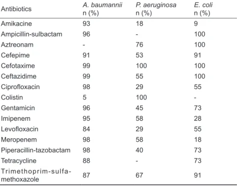
A. baumannii, P. aeruginosa and E. coli have very high antibiotic resistance rates (Table 2). The most effective antibiotic against A. baumannii was found as colistin (95%). The most effective antibiot-ics against P. aeruginosa were found as amikacine (82%) and ciprofloxacin (71%). The most effective antibiotics against E. coli were found as amikacine (91%) and imipenem (82%).
Table 2. Distribution of isolated microorganisms’ antibiotic
re-sistances Antibiotics A. baumannii n (%) P. aeruginosan (%) E. colin (%) Amikacine 93 18 9 Ampicillin-sulbactam 96 - 100 Aztreonam - 76 100 Cefepime 91 53 91 Cefotaxime 99 100 100 Ceftazidime 99 55 100 Ciprofloxacin 98 29 55 Colistin 5 100 -Gentamicin 96 45 73 Imipenem 95 58 28 Levofloxacin 84 29 55 Meropenem 98 58 18 Piperacillin-tazobactam 98 40 73 Tetracycline 88 - 73 Trimethoprim-sulfa-methoxazole 87 67 91 DISCUSSION
In the consequence, the increasing quality of burn units and improving treatment opportunities do not lead a remarkable decrease in the mortality rates. All the related causes are the combination of the rapidly changing microorganisms dominating dif-ferent burn wound infections, their great antibiotic resistances and the huge cost of treatments.
One of the most capable bacteria for develop-ing resistance is A. baumannii and it has become widespread in all intensive care units in a decade. If the required cautions will not be taken seriously against these microorganisms, the number of effec-tive antibiotics will reduce dramatically and both the mortality rates and the cost of treatment will rise up worldwide by time.
The pathogen microorganisms may easily in-vade into the burn injury site and cause infection and
serious sepsis in the case of the injury because of degradation of the integrity of the skin tissue, dehy-dration and weakening of the body resistance. Type of microorganisms may change by time depending on the flora of burn unit, the type of drugs used in the care of burn wounds and hygiene compliance of health-care workers. Using effective strict isola-tion techniques and infecisola-tion control policies may significantly decrease the occurrence of burn wound infection [10].
Interestingly a pathogen can spread between separate units, hospitals and also hospitals in the other countries. A. baumannii is one of the best examples for this situation. Its prevalence and the deduction of nosocomial agent have risen in the last decade. As a matter of fact, nowadays it is the most common cause of infection in the intensive care units and burn units [11,12]. The studies from different countries indicate the rate of pathogens
Table 1. Distribution of isolated microorganisms
Microorganisms n % A. baumannii 94 62.3 P. aeruginosa 39 25.8 E. coli 11 7.3 S. aureus 7 4.6 Total 151 100.0
in burn wound infections as P. aeruginosa (21.6-37.5%), A. baumannii (0-10.4%), Staphylococcus aureus (8.3-30.4%), E. coli (2.3%) between 2003-2004 [10,13-15]. Two other studies from Turkey indicate the rate of pathogens in burn wound infec-tions as P. aeruginosa (12.5-46.2%), A. baumannii (0.6-24.2%), Staphylococcus aureus (19-22%), E. coli (0-13%) between 1998-2007 [16,17]. A study made in our burn unit reported the rate of pathogens in burn wound infections as P. aeruginosa (58%), A. baumannii (0%), E. coli (22%) in 2000. In our study, the rate of pathogens in burn wound infections were found out as A. baumannii (62.3%), P. aeruginosa (25.8%), Staphylococcus aureus (4.6%), E. coli (7.3%). This result reveals that A. baumannii and P. aeruginosa have emerged as important nosocomial pathogens worldwide.
Antibiotic resistance of microorganisms iso-lated from burn wound infections may change from hospital to hospital, region to region and the usage pattern of antibiotics effect this situation. Especial-ly multiple drug resistant A. baumannii and other Gram-negative pathogens such as P. aeruginosa and E. coli have high rates of antibiotic resistance. A. baumannii resistance significantly increased by time [18]. The resistance rates of A. baumannii against antibiotics were ampicilin-sulbactam (76%), amika-cine (64-92%) and meropenem (7.7-71%), cephalo-sporins (3-95.9%) [17,19,20]. The resistance rates of P. aeruginosa against antibiotics were amikacine (57.1-68%), meropenem (18.5-54%) and cephalo-sporins (39.3-96.3%) [17,19].
In our study, the resistance rates of A. bauman-nii against antibiotics were colistin (5%), ampicil-lin-sulbactam (96%), amikacine (93%) and me-ropenem (98%), cephalosporins (91-99%) and the resistance rates of P. aeruginosa against antibiot-ics were amikacine (18%) and meropenem (58%), cephalosporins (53-100%). These results show that A. baumannii incredibly improve resistance against antibiotics. If the required cautions will not be taken seriously against these microorganisms, the num-ber of effective antibiotics will reduce dramatically A. baumannii will cause serious health problems worldwide by time.
Finally, the prevalence of A. baumannii infec-tions causing the majority of burn wound infecinfec-tions is rising and the resistance against antibiotics is un-preventable globally.
Also the prevalence of P. aeruginosa and E. coli are changing and they have high resistance against antibiotics. This case requires immediate effective measures and new efficient infection control strate-gies. In addition, each center should determine their patients’ profile, hospital flora and their antibiotic resistance. Thus, we believe these results will con-tribute the prevention strategy of infection.
REFERENCES
1. Manson WL, Pernot PC, Fidler V, et al. Colonisation of burns and the duration of hospital stay of severely burned patients. J Hospital Infect 1992;22:55-63.
2. Tekin R, Yolbas I, Selcuk CT, et al. An evaluation of pedi-atric burn patients over a 15-year period. Turk J Trauma & Emergency Surg 2012;18:514-518.
3. Revathi G, Puri J, Jain BK. Bacteriology of burns. Burns 1998;24:347-349.
4. Davis SC, Pisanni F, Montero RB. Effects of commonly used topical antimicrobial agents on Acinetobacter baumannii: an in vitro study. Mil Med 2008;173:74-78.
5. Peleg AY, Seifert H, Paterson DL. Acinetobacter baumannii: emergence of a successful pathogen. Clin Microbiol Rev 2008;21:538-582.
6. Tredget EE, Shankowsky HA, Rennie R, et al. Pseudo-monas infections in the thermally injured patient. Burns 2004;30:3-26.
7. Sengupta S, Kumar P, Ciraj AM, Shivananda PG. Acineto-bacter baumannii-an emerging nosocomial pathogen in the burns unit Manipal, India. Burns 2001;27:140-144. 8. Ananthakrishnan AN, Kanungo R, Kumar K, Badrinath S.
Detection of extended spectrum beta lactamase producers among surgical wound infections and burns patients in JIP-MER. Ind J Med Microbiol 2002;18:160-165.
9. Greenhalgh DG, Saffle JR, Holmes JH, et al. American Burn Association consensus conference to define sepsis and in-fection in burns. J Burn Care Res 2007;28:776-790. 10. Ekrami A, Kalantar E. Bacterial infections in burn patients
at a burn hospital in Iran. Indian J Med Res 2007;126:541-544.
11. Frame JD, Kangesu L, Malik WM. Changing flora in burn and trauma units: experience in United Kingdom. J Burn Care Rehabil 1992;13:281-286.
12. Amin M, Kalantar E. Bacteriological monitoring of hospital borne septicemia in burn patients in Ahvaz, İran. Burn Sur-gical Wound Care 2004;3:4-8.
13. Nasser S, Mabrouk A, Maher A. Colonization of burn wounds in Ain Shams University Burn Unit. Burns 2003;29:229-233.
14. Singh NP, Goyal R, Manchanda V, et al. Changing trends in bacteriology of burns in the burns unit, Delhi, İndia. Burns 2003;29:129-132.
15. Revathi G, Puri J, Jain BK. Bacteriology of burns. Burns 1998;24:347-349.
16. Erol S, Altoparlak U, Akcay MN, et al. Changes of micro-bial flora and wound colonization in burned patients. Burns 2004;30:357-361.
17. Aksaray S, Cesur S. Microorganisms isolated from wound and blood culture of burn patients and their antibiotic sus-ceptibility. Turkish J İnfect 2003;17:293-296.
18. Babik J, Bodnarova L, Sopko K. Acinetobacter-serious dan-ger for burn patients. Acta Chir Plast 2008;50:27-32.
19. Rezaei E, Safari H, Naderinasab M, Aliakbarian H. Com-mon pathogens in burn wound and changes in their drug sensitivity. Burns 2011;37:805-807.
20. Albrecht MC, Griffith ME, Murray CK, et al. Impact of Aci-netobacter infection on the mortality of burn patients. J Am Coll Surg 2006;203:546-550.