INVESTIGATION OF THE EFFECTS OF SUPRAMOLECULAR STRUCTURES OF CATIONIC PEPTIDES ON ANTIBACTERIAL ACTIVITY
A THESIS SUBMITTED TO
GRADUATE SCHOOL OF ENGINEERING AND SCIENCE OF BILKENT UNIVERSITY
IN PARTIAL FULFILLMENT OF THE REQUIREMENTS FOR THE DEGREE OF
MASTER OF SCIENCE IN
MATERIALS SCIENCE AND NANOTECHNOLOGY
By
MUSTAFA BETER
II
INVESTIGATION OF THE EFFECTS OF SUPRAMOLECULAR STRUCTURE OF CATIONIC PEPTIDES ON ANTIBACTERIAL ACTIVITY
By Mustafa Beter, May, 2017
We certify that we have read this thesis and that in our opinion it is fully adequate, in scope and in quality, as a thesis for the degree of Master of Science.
Ayşe Begüm Tekinay (Advisor)
Tamer Uyar
Ayşe Gül Gözen
Approved for the Graduate School of Engineering and Science:
Ezhan Karaşan
III
ABSTRACT
INVESTIGATION OF THE EFFECTS OF SUPRAMOLECULAR STRUCTURES OF CATIONIC PEPTIDES ON ANTIBACTERIAL ACTIVITY
Mustafa Beter
MSc in Materials Science and Nanotechnology Supervisor: Ayşe Begüm Tekinay
May, 2017
Many organisms including mammalians use Antimicrobial Peptides (AMPs) which are also called Host Defense Peptides against microbial organisms. AMPs are among one of the ancient and successful strategies for both plant and animal kingdoms.
Even though AMPs vary among closely related species and despite they have different sequences, many of the natural AMPs share similar properties. They are mostly short sequenced, structurally amphipathic and they carry overall net positive charge.
Cationic AMPs target bacterial membranes because of the electrostatic attractions between positively charged peptides and negatively charged membranes. Due to the electrostatic attractions, cationic AMPs might work on membrane disruption by passing a certain threshold concentration for hydrophobic groups to penetrate into membrane.
Noncovalent interactions and electrostatic interactions can create molecular attractions and may cause molecular self-assembly which is a common mechanism used by nature for several tasks. Self-assembling peptide amphiphiles are a group of molecules which can form nanofibrous
IV
structures and may contain bioactive epitopes depending on the target of the peptide amphiphile molecule.
This thesis describes the presentation of antimicrobial sequences on supramolecular nanofibers which are formed by self-assembling peptides. The comparison of self-assembling peptides and single soluble peptides without self-assembling capacity, resulting significant improvement for peptide nanofiber systems for antimicrobial therapeutic purposes is reported.
Keywords: Antimicrobial Peptides, Peptide Nanofibers, Self-Assembling Peptide Amphiphiles, Supramolecular Nanostructures, Multivalent Presentation
V
ÖZET
KATYONİK PEPTİTLER VE
SUPRAMOLEKÜLER NANOYAPILARININ ANTİMİKROBİYAL AMAÇLA KULLANILMASI
Mustafa Beter
Malzeme Bilimi ve Nanoteknoloji, Yüksek Lisans Tez Danışmanı: Ayşe Begüm Tekinay
May, 2017
Memeli canlılar da dahil birçok organizma, “Antimikrobiyal Peptitleri” diğer adıyla “Konak Savunma Peptitlerini” kullanırlar. Antimikrobiyal peptitler, hem bitkiler hem de hayvanlar aleminde uzun evrimsel süreçlerden geçerek başarılı olmuş savunma stratejilerinden biridir. Antimikrobiyal peptitler yakın türler arasında bile farklılık göstermelerine ve farklı amino asit sekanslarına sahip olmalarına karşın, çoğunlukla kısa amino asit sekanslı olmak, yapısal olarak amfipatik özellikte olmak ve net pozitif yük taşımak gibi benzer özellikler gösterirler.
Bakteri membranını hedef alan antimikrobiyal peptitler, peptidin taşıdığı pozitif yük ile bakteri membranının negatif yükü arasındaki elektrostatik etkileşim sebebiyle hedef alma işlevlerini yerine getirirler. Elektrostatik etkileşimin ardından, antimikrobiyal peptitler belli bir eşik konsantrasyonunu aşıp, hidrofobik grupların bakteri membranından içeri penetre etmesini sağlayabilirler.
VI
Kovalent olmayan etkileşimler ve elektrostatik etkileşimler, moleküler çekim oluşturarak kendiliğinden bir araya gelmeyi sağlar. Bu mekanizma doğada birçok işlevi yerine getirmek için kullanılmaktadır. Bu mekanizmaya örnek olan kendiliğinden bir araya gelen peptit amfifiller, nanofiber yapılar oluşturabilen ve hedef moleküle uygun biyoaktif sekanslar içerebilen bir peptit grubudur.
Bu tez antimikrobiyal sekansların, kendiliğinden bir araya gelen peptit amfifillerin oluşturduğu supramoleküler nanofiber yapılar üzerinde sunumunu tanımlar. Kendiliğinden bir araya gelebilen peptit amfifil grup ile kendiliğinden bir araya gelebilme özelliği taşımayan peptitlerin arasında yapılan karşılaştırma ve peptit nanofiber sistemin antimikrobiyal özellikleri arttırmasının altı çizilmektedir.
Anahtar kelimeler: Antimikrobiyal Peptit, Peptit Nanofiber, Kendiliğinden Bir Araya Gelen Peptit Amfifiller, Supramoleküler Nanoyapılar, Çok Valanslı Sunum
VII
ACKNOWLEDGEMENTS
First, I would like to express my gratitude to my advisor Prof. Ayşe Begüm Tekinay. She opened her laboratory to me and has provided me independence during my master studies. I am grateful for her patience which helped me develop interdisciplinary research skills. I also would like to thank BML members and Prof. Mustafa Özgür Güler for their support during my thesis project.
I would like to acknowledge the graduate scholarship from TÜBİTAK (The Scientific and Research Council of Turkey) project 113S552 and 114S913 for financial support.
I also would like to thank my friends from İYTE: Merve ŞEN for her cheerfulness, constant selfies and her friendship; Nurcan HAŞTAR for her warm personality, our shared jokes and her friendship; İdil UYAN for accompanying me to several art events and her friendship; Gökhan GÜNAY for our endless discussions about very different subjects and his friendship; Zeynep ORHAN for her sincere supports, our group travels and her friendship. They were just like friends from Friends and I am very glad to meet them. Canelif YILMAZ is from Koç but I want to add her here also to thank her for her atars, adding a nice color to the rainbow and her friendship.
I also would like to thank Melike SEVER for sharing her two schools with me, her smile and support whenever I asked a question and her friendship; Nuray GÜNDÜZ for her dıs dı dıs dıs musics, making us smile, her support and friendship; Çağla EREN for her warm personality (which can melt ice therefore she went to Canada for a challenge) and her friendship; Gözde UZUNALLI for being my travelling companion once a week, her support on many things and her friendship; Özüm Şehnaz ÇALIŞKAN for being a nice neighbor in Çankaya and her friendship; Burak
VIII
DEMİRCAN for being a weird (good weird) desk mate, his healthy diets and his friendship; Oğuz TUNCAY for his joyfulness, his cooking skills, amazing personality and his friendship; İbrahim ÇELİK for öhö öhö and his friendship; Fatih YERGÖZ for mutual suffering to finish the thesis and his friendship.
I also would like to thank former BML members: Özlem EROL for being my travelling companion for a year and her friendship; Aref Khalily for telling his past experiences on our road to Marmara, chemistry support and his friendship; Melis Şardan EKİZ for sharing her knowledge in a very nice way whenever I asked a question and her friendship; Seren HAMSİCİ for her nice personality, her help and friendship.
I also would like to thank Hatice Kübra KARA and Ahmet Emin TOPAL for their supports in our experiments and their friendship. Kübra helped me a lot with my thesis project, synthesis of our peptides and characterizations and Ahmet also helped a lot with AFM experiments and conducted them. And of course also I would like to thank Alper Devrim ÖZKAN for his amazing stories about so many weird stuff, his knowledge and his friendship.
Moreover I would like to thank Özge UYSAL, Gülistan TANSIK, Elif ARSLAN and Begüm DİKEÇOĞLU. We did not just share foods, we also shared our memories and personalities. I had the chance to know you more and I am very glad for that. I will always be a member and you will always be my friends.
Moreover I would like to thank my family: my mom Sevda and my dad Muzaffer for their love and support throughout my life, without them I would not be here, obviously. My brother Deha taught me many things despite his age and he is one of my most favourite people in the world. My aunt Sibel KAYA was the person who introduced me to art, my uncle İrfan KOÇAK introduced
IX
me to science, my uncle Hasan Hüseyin BETER introduced me to folklore and I thank them for those. Also I would like to thank my grandmother Döndü KOÇAK for her cheerfulness, my childhood years, all my memories in Ankara Kalesi and Demetevler, and also my grandfather Ali KOÇAK for introducing me to stories and museums.
My special thanks for one of my best friends, my life partner, my love Özgür Sezgi DUMAN. She helped me in so many ways that if I wanted to write them here it would at least double the total page number of my thesis.
I wish all the best for my friends who will finish their works and write their thesis.
X
CONTENTS
LIST OF FIGURES ... XIII
ABBREVIATIONS ... XVII
1. Objective ... 1
2. Introduction: Multivalent Presentation of Antimicrobial Peptides ... 3
2.1. Antibiotic Resistance of Bacteria ... 3
2.2. Antimicrobial Peptides ... 8
2.3. Bacterial Membranes and Mechanism of AMPs Action ... 12
2.4. Cationic Antimicrobial Peptides Against Bacterial Resistance ... 16
2.5. Multivalent Designs for AMPs ... 20
2.6. Self-Assembling Peptide Amphiphiles ... 24
3. Experimental Section ... 26
3.1. Materials ... 26
3.2. Methods ... 26
XI
3.2.2. Structural Characteristics of Peptides, Circular Dichroism (CD) Spectroscopy . 27
3.2.3. Morphological Characteristics of Peptides, Transmission Electron Microscopy
(TEM) Imaging ... 28
3.2.4. Antibacterial Analysis ... 28
3.2.4.1 Minimum Inhibitory Concentration of Peptides ... 28
3.2.4.2 Live/Dead Assay for Bacteria ... 29
3.2.4.3 Bacterial Survival Assay ... 30
3.2.5. Cytotoxicity of Peptides ... 30
3.2.6. Visualization of Peptide Nanofibers by Confocal Microscopy Imaging ... 30
3.2.7. Morphological Changes of Bacteria by Scanning Electron Microscopy (SEM) Imaging ... 31
3.2.8. Morphological Changes of Bacteria by Atomic Force Microscopy (AFM) Imaging ... 31
3.2.9. Zeta Potential Measurements ... 32
3.2.10. Statistical Analysis ... 32
4. Results and Discussion ... 33
4.1. Synthesis of Peptides and Characterization of Peptide Amphiphile Self-assembly into Nanofibers ... 33
XII
4.2. Structural and Mechanical Characteristics of Peptides ... 35
4.3. Critical Micelle Concentration ... 40
4.4. Antibacterial Activity of Peptides ... 42
4.4.1. Determining Minimum Inhibitory Concentrations (MICs) ... 42
4.4.2. Bacterial Live/Dead Assay ... 43
4.4.3. Bacterial Survival Assay ... 47
4.5. Cytotoxicity of Peptides ... 49
4.6. Localization of Peptide Nanofibers on Bacteria ... 50
4.7. Morphological Changes of Bacteria After Peptide Treatment ... 53
4.8. Previous Designs ... 59
4.8.1. Lauryl-VVAGK with Quaternary Ammonium ... 60
4.8.2. (RW)3-PA ... 65
5. Conclusion ... 70
6. Future Perspectives ... 71
XIII
LIST OF FIGURES
Figure 1 Discovery void of antibiotics with reported initial discovery/patent dates ... 4
Figure 2 Examples of bacterial resistance associated with porins ... 5
Figure 3 Antibiotic inhibition of bacteria ... 7
Figure 4 Biological roles of antimicrobial peptides ... 9
Figure 5 Different structures and sequences of antimicrobial peptides ... 10
Figure 6 Proposed mechanisms of AMP mediated membrane disruption ... 15
Figure 7 Co-evolution of cationic AMPs (CAMPs or CAPs) and bacterial resistance ... 18
Figure 8 Schematics of dendrimeric/branched and polymeric multivalent peptides ... 21
Figure 9 Examples for conjugation of antimicrobial reagents into multivalent structures ... 23
Figure 10 Schematics of self-assembly of peptide amphiphiles ... 25
Figure 11 Chemical structures of peptide amphiphile molecule (Lauryl-VVAGKKKGRW) and chemical structure of soluble peptide molecule (KKKGRW) ... 34
Figure 12 Structural characterizations of peptides by using transmission electron microscopy . 36 Figure 13 Liquid chromatograms and mass spectra of peptides ... 37
Figure 14 Secondary structure characterization of peptides by using circular dichroism ... 38
Figure 15 Secondary structure characterization of antibacterial peptide amphiphile at different pH values ... 39
Figure 16 Nile red analysis of critical micelle concentration of PA ... 40
XIV
Figure 18 Nanofiber treated and soluble peptide treated E. coli stained with BacLight live/dead bacterial viability kit ... 44 Figure 19 Nanofiber treated and soluble peptide treated B. subtilis stained with BacLight
live/dead bacterial viability kit ... 45 Figure 20 Higher concentration of nanofiber treated E. coli and B. subtilis stained with BacLight live/dead bacterial viability kit ... 46 Figure 21 Time course of bacterial killing by peptides ... 48 Figure 22 Fluorescent images of HUVECs with peptides after 24 h incubation using live/dead assay ... 49 Figure 23 Confocal microscopic study of localization of peptide nanofibers onto E. coli RSHM 888 and B. subtilis ATCC 6633 ... 51 Figure 24 Time lapsed confocal microscopy imaging of peptide nanofibers onto E. coli and B. subtilis ... 52 Figure 25 AFM amplitude image of B. subtilis cells treated with peptide nanofibers ... 53 Figure 26 AFM phase image of B. subtilis cells treated with peptide nanofibers ... 54 Figure 27 AFM amplitude and phase images of E. coli and B. subtilis cells treated with peptide nanofibers and soluble peptides ... 56 Figure 28 AFM amplitude and phase images of control E. coli cells in water ... 56 Figure 29 Scanning electron micrographs of E. coli cells ... 57 Figure 30 Chemical structure of peptide amphiphile molecule Lauryl-VVAGK with quaternary ammonium compound ... 61 Figure 31 Liwuid chromatograms and mass spectra of Lauryl-VVAGK with quaternary
XV
Figure 32 Structural characterizations of peptides by using transmission electron microscopy . 63 Figure 33 Secondary structure characterization of peptide by using circular dichroism ... 64 Figure 34 Charge properties of peptide amphiphiles were analyzed with zeta potential
measurements ... 64 Figure 35 Chemical structure of peptide amphiphile molecule Lauryl-VVAGRWRWRW ... 66 Figure 36 Liquid chromatograms and mass spectra of (RW)3-PA ... 67 Figure 37 Structural characterizations of peptides by using transmission electron microscopy . 68 Figure 38 Secondary structure characterization of (RW)3-PA by using circular dichroism ... 69
XVI
LIST OF TABLES
Table 1 Differences between gram positive and gram negative bacteria related to AMP design 12 Table 2 Minimum inhibitory concentrations of peptides ... 43 Table 3 Tryptophan containing antimicrobial peptides ... 65
XVII
ABBREVIATIONS
AFM : Atomic force microscopy
AMP : Antimicrobial peptide
ATCC : American type culture collection
ATP : Adenosine triphosphate
CAP : Cationic antimicrobial peptide
CAMP : Cationic antimicrobial peptide
CD : Circular dichroism
CFU : Colony forming unit
CI : Confidence interval
DCM : Dichloromethane
DMF : N, N-Dimethylformamide
DMEM : Dulbecco’s modified Eagle’s medium
DMSO : Dimethylsulfoxide
FDA : U.S. Food and Drug Administration
XVIII
HUVEC : Human umbilical vein endothelial cell
HPLC : High performance liquid chromatography
IM : Inner membrane
LC-MS : Liquid chromatography-mass spectrometry
LB : Luria-Bertani
LPS : Lipopolysaccharide
MIC : Minimum inhibitory concentration
MMP : Matrix metalloproteinase OM : Outer membrane PA : Peptide amphiphile PBS : Phosphate-buffered saline PI : Propidium iodide PP : Periplasmic membrane
Q-TOF : Quadrupole time of flight
RNA : Ribonucleic acid
RSHM : Refik Saydam Hıfzısıhha Merkezi National Type Culture Collection
XIX SD : Standard deviation
SEM (S) : Standard error of the mean
TCP : Tissue culture plate
TEM : Transmission electron microscope
TFA : Trifluoroacetic acid
1
Investigation of the effects of supramolecular structures of cationic peptides on antibacterial activity
This work is partially described in the following publication:
Mustafa Beter *, †, Hatice Kubra Kara *, †, Ahmet Emin Topal *, †, Aykutlu Dana*, †, §, Mustafa O. Guler *, †, §, Ayse B. Tekinay *, †, § (Manuscript under Submission)
1. Objective
Bacteria can be intrinsically resistant or can acquire resistance to antibiotics and this is becoming one of the biggest health problems. Bacteria can resist antibiotics in different ways. For example: they can efficiently remove antibiotics by efflux proteins, prevent antibiotics to cross the outer membrane, change the structure of the target or modify the drug directly. Organisms which fight against bacteria for survival, have several defense strategies against them. Protection by Antimicrobial Peptides is one of these methods. In this work, we designed and synthesized a peptide amphiphile molecule with a potential antimicrobial sequence and its single subunit counterpart and we analyzed the difference between the antibacterial efficiency of supramolecular structure and the single soluble subunit. We first, characterized the peptides, and showed differences in the secondary structures and supramolecular structures. Peptide amphiphile molecules create nanofiber networks by self-assembling and this trait is used for multivalent presentation of antibacterial sequence against bacteria. Following the characterizations, we evaluated the inhibitory concentrations and capacity of peptides to kill both gram positive and gram
2
negative bacteria. Our results suggest that multivalent presentation of antimicrobial sequences by self-assembled nanofibrious structures could be beneficial for using against bacteria for preventing drug resistance. This work demonstrates for the first time that the antibacterial activity of KKKGRW sequence is improved when self-assembled into supramolecular nanofibrious structure.
3
2. Introduction: Multivalent Presentation of Antimicrobial Peptides
2.1. Antibiotic Resistance of Bacteria
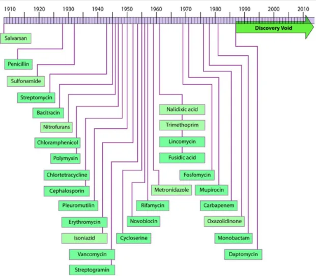
In the last century people have been using antibiotics to cure illnesses in humans and food producing animals. However, partly because of the misuse, resistance among bacteria is now causing the antibiotics either working inefficiently or not working at all. This is a very serious problem, because new ways of antibiotic drugs are not developed as fast as we expected. Figure 1 shows the “discovery void” of antibiotic drugs1.
Reports of World Health Organization suggest improving hygiene, sanitation, access to clean water and vaccination, while development of new treatment ways are continuing. Especially for gram negative enteric bacteria, this “void” is causing serious health problems. For example, recently E. coli resistance to third generation cephalosporins has been reported2. Even though human intestine is the natural environment for this bacteria, it is a common causative pathogen for nosocomial urinary tract infections and foodborne diseases.
Bacteria can be either intrinsically resistant or can acquire resistance to antibiotic drugs. There are different ways through which bacteria can resist a certain drug. In order for an antibiotic to work, it needs to be in a certain concentration in the environment. Drug release studies are based on this knowledge and it is an important part of drug research to give enough amount of doses to the target, while not causing cytotoxicity for other parts of the body. However, bacteria evolved ways of resistance. For instance, bacteria can control influx and efflux of antibiotics by preventing or decreasing drug entrance to keep the drug levels lower than the working concentration (Figure 2).
4
Figure 1 Discovery void of antibiotics with reported initial discovery/patent dates. Adapted from Silver et al. 2011 with permission of the American Society of Microbiology Journals Department.
5
Figure 2 Examples of bacterial resistance associated with porins3. Blue circles indicate antibiotic molecules, and the red cross indicates that the antibiotic cannot cross the outer membrane. Abbreviations: IM: inner membrane, OM: outer membrane, PP: periplasmic space.
6
As an intrinsic bacterial resistance mechanism, efflux pumps can pump the drug out efficiently for both gram positive and gram negative bacteria. In addition, bacteria may have multidrug resistance efflux pumps, which can pump many different molecular structured antibiotic drugs4-6. Another way of bacterial resistance is by changing the drug target molecule. Generally, antibiotic drugs bind to bacterial targets with high affinity and by doing that they prevent binding of other molecules which are necessary for bacterial survival, and therefore, cause death of bacteria. However, some mutations may cause slight structural changes which does not affect the metabolic pathway of bacteria but lower the affinity of antibiotics to the target7. Another way of doing this is using other molecules to protect target structures. For example, in order to protect the lipopolysaccharide targets on the membrane, bacteria add phosphoethanolamine to its membrane structure, and prevent targeting by protecting the target site8.
7
Figure 3 Antibiotic inhibition of bacteria9. a) A host with vulnerable target protein inhibited by antibiotics. b) Hydrolyzing the antibiotics by host enzymes. c) Transferring chemical groups to inhibit antibiotics.
Degrading or modifying the antibiotic drugs for resistance is of course widely used by bacteria to inhibit the effects of antibiotics. Bacteria can break bonds of antibiotics by targeting certain parts such as esters and amides. Also, by transferring chemical groups onto the drug, bacteria can inhibit drug activity. Acylation, phosphorylation, glycosylation of drugs are some of the examples, which bacteria use against antibiotic drugs10.
8 2.2. Antimicrobial Peptides
Even though individual organisms fail in certain environments, the species as a hole may find a beneficial mutation and continue its existence. They use various defense strategies against their parasites, pathogens in their food or their competitors which occupy the same niche. Some of these strategies become successful and evolutionarily conserved so that we can see them in many different species today. Antimicrobial peptides (AMPs) which are also called host defense peptides are one of these successful, ancient strategies displayed in both plant and animal kingdoms11.
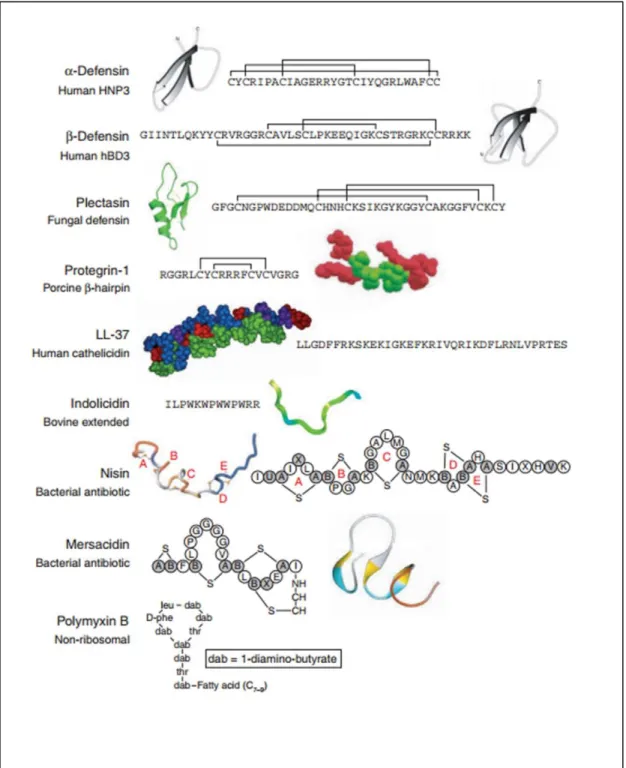
9 Figure 4 Biological roles of AMPs12.
AMPs were first discovered more than thirty years ago while studying insect immunity13. Since then, thousands of AMPs have been found in a wide variety of organisms. They are being used by multicellular organisms as a part of innate immune system, the first line of defense. Although it is hard to categorize them since they can be categorized in many different ways according to several different criteria, they can be studied according to their secondary structures which are mostly α-helical or ß-sheet for known AMPs14. Some of
10
11
AMPs vary even among closely related species, because they change according to the microbes the organism faces. On the other hand, despite they have different sequences, many of them share similar properties. They are mostly short sequenced (10-50 amino acid residues), structurally amphipathic (consisting both hydrophobic (usually >30%) and hydrophilic parts) and they carry overall net positive charge because of lysine, arginine and histidine amino acids in their sequence15.
Natural and synthetic AMPs are very useful against bacteria. Several studies showed that natural and synthetic AMPs such as cathelicidin present even anti-biofilm effects16. In addition, even though most of the AMPs show cytotoxicity at higher concentrations or sometimes even at minimum working concentrations17, there are also strong candidates which do not show cytotoxicity at these concentrations18.
Long cationic peptides face some problems for therapeutic use. They usually work at higher concentrations, therefore, they show cytotoxicity with increased concentrations. Proteolytic enzymes can degrade them and especially larger peptides are vulnerable to proteases. Also, production cost increases with the size. Small cationic peptides on the other hand, can escape from proteolytic degradation easier than long peptide sequences and the production cost is less expensive.
12
2.3. Bacterial Membranes and Mechanisms of AMP Action
Bacterial membrane components differ among gram positive and gram negative bacteria. Gram positive bacteria have a single membrane, which contains acidic polysaccharides (teichoic acids). Gram negative bacteria have an inner membrane of similar properties; however, does not contain teichoic acids. On the other hand, gram negative bacteria also have an outer membrane. Even though both gram positive and gram negative membranes are composed of negatively charged phospholipids, gram negative bacteria have more lipid content on their membrane, and they have lipopolysaccharides. Teichoic acids and lipopolysaccharides increase the net negative charge of the membrane structure of bacteria. Since both gram positive and gram negative bacteria have phospholipids, they can both be affected by cationic AMPs, which cause membrane disruption by binding to negatively charged phospholipids.
Table 1 Differences between gram positive and gram negative bacteria related to AMP designs
Gram positive bacterial membrane Gram negative bacterial membrane Lipid and lipoprotein
content
Low High
Lipopolysaccharide (LPS) content
Very low High
Teichoic acid Present in many -
Outer membrane and periplasmic space
- Present
13
Bacterial membrane phospholipids such as cardiolipin and phosphatidylglycerol are mostly acidic. However, the reason mammalian cells do not get affected in most cases is that there are phosphatidylcholine and sphingomyelin phospholipids in the outer part of the membrane which makes it zwitterionic, and negatively charged phosphatidylserine in the inner part of the mammalian membrane. Although this is the case, if hydrophobicity of AMPs are high then, they can interact with phosphatidylcholine of the mammalian cellular membrane and can affect the membrane integrity19.
First interaction of AMPs with bacterial membrane is electrostatic attractions between positively charged peptides and negatively charged membranes. Experimentally, L- or D- enantiomers of amino acids do not change antimicrobial activity. Therefore, it is thought that the first interaction is non-specific20.
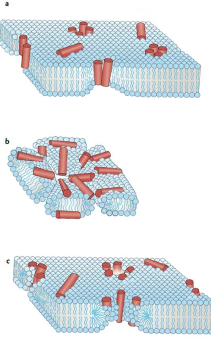
While some AMPs are internalized, others work on membrane disruption. Although passing a threshold concentration of AMPs is necessary for membrane disruption, there are different biophysical models to explain AMPs’ behavior21.
In Barrel Stave Model, peptides create transmembrane pores similar to barrel staves. Hydrophobic side faces to phospholipid acyl chains. By doing that, bacteria lose control of ion gradients and its ability to synthesize ATP. Then, it cannot control the water movement and it dies because of osmolysis22.
14
In Carpet Model, peptides cover the surface of microbial lipid bilayer membrane, and unlike pore models they do not create porous structures. Instead, after reaching a certain treshold concentration, they act like detergants and shatter the membrane structure23.
In Toroidal Pore Model, peptides elongate the lipid head groups of the bilayer and bend them by integrating into the system. After the bending, the outer and the inner sides of the bilayer connect and this creates pores in the membrane structure24.
15
Figure 6 Proposed mechanisms of AMP mediated membrane disruption: Barrel Stave Model (a), Carpet Model (b), Toroidal Pore Model (c). Edited from Melo et al. 2009.
16
AMPs can also be internalized and affect the target microbial cells in ways other than membrane disruption (such as Mersacidin, Buforin II and Histatins). Mersacidin inhibits peptidoglycan synthesis by preventing glycosylation25. Buforin II penetrates cell membrane and binds to DNA or RNA to kill microbial organisms26. Other than cationic and hydrophobic residues of AMPs there are other properties which are important. For Buforin II, the substitution of proline with leucine decreases the effect of Boforin II peptide, because the internalization of the peptide into microbial cytoplasm is decreased after the substitution27. Histatins act by binding to mitochondrion and cause ATP loss and promote the generation of reactive oxygen in order to kill the microbial cell28.
2.4. Cationic Antimicrobial Peptides Against Bacterial Resistance
Bacterial resistance to drugs is not a new problem which just happened because of misuse of antibiotic drugs – even though, of course this mistake certainly enhanced the problem. Bacterial resistance is probably also as ancient as host defense systems. This can be thought as a co-evolutionary process of bacterial resistance and host defense strategies. So, in this context, it can be said that bacterial resistance and AMPs acted as driving force for evolution of each other. Since AMPs use various strategies such as the examples given in the previous section, bacterial resistance strategies also differ. That is one of the reasons which makes it hard to develop new effective drugs against drug resistance of bacteria. However, in the case of AMPs, this resistance is a driving force for their evolution, therefore, we can get very useful information from analyzing this ancient battle.
17
Bacteria can show resistance against AMPs by reversible adaptations or inheritable mutations. In experimental conditions, bacteria developing resistance to AMPs were shown previously29. By gradually increasing AMP concentration, it seems bacteria can become resistant to AMPs just like current antibiotic resistance schemes. However, for in vivo conditions, there might be multiple AMPs working at the same time and certainly in vivo conditions are quite different than in vitro conditions. Therefore, it is hard to determine how much time it takes for a bacterium to become resistant to a certain AMP.
18
Figure 7 Co-evolution of cationic AMPs (CAMPs or CAPs) and bacterial resistance30. Adapted from Peschel et al. 2006 with permission of Nature Publishing Group.
19
Bacteria use protease activity against AMPs. By doing this, they can degrade AMPs before they act. Especially α-helical and linear structured AMPs are the targets of this bacterial resistance tactic31. On the other side, AMPs create disulfide bridges in their structure in a way which does not affect their functional activities, but provides protection against protease degradation. In one study, it was shown that when α-defensin family Crp4 AMP does not carry disulfide bridges, matrix metalloproteinase-7 (MMP-7) enzymes degraded these substituted peptides32. Also, there are different studies which shows that by adding intermolecular disulfide bonds, it is possible to increase stability of proteins33.
Another mechanism bacteria use to inactivate AMPs is directly interfering with the target peptides. Some pathogenic bacteria secrete certain molecules for complementary interference to target AMPs and therefore inhibit the antimicrobial activities of those AMPs34. On the host side, this resistance mechanism corresponds to multiple AMPs with small sequence differences35. Since the secreted molecule is target specific and works with direct sequence recognition, by evolutionarily increasing the variance of AMPs, host species inhibit bacterial development.
A broad mechanism which bacteria can use in order to inhibit cationic antimicrobial peptide action could be changing the membrane anionic structure into a more neutral one, just like mammalian zwitterionic cell membrane. However, changing important structures of the membrane, such as teichoic acid and peptidoglycans, is not an easy strategy, and actually, evolutionarily, might not be possible at all for many bacterial species. Nevertheless, bacteria use this strategy to at least partly change membrane charge, and therefore, resist against cationic AMPs. For example, Peschel et al. showed the D-alanine modification of bacteria in order to increase partial positive charge of the membrane36.
20
Despite this ability, cationic AMPs with higher positive charges still interact with bacterial membranes and kill bacteria. So, high positive charge seems an effective way of fighting bacterial resistance. However, higher positive charge brings some problems of course. An important problem for antibacterial efficiency would be the change of positive charge and hydrophobicity balance for AMPs. Increased positive charge certainly increases bacterial membrane targeting but, might decrease membrane penetration ability of AMPs30.
2.5. Multivalent Designs for AMPs
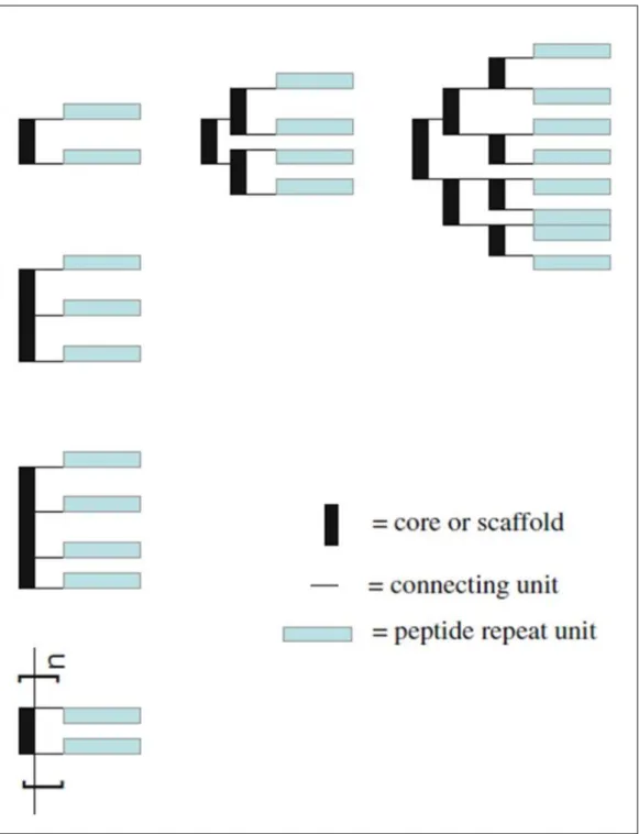
Multivalent systems especially multivalent bindings for biological organisms have been shown as examples of multivalency in nature almost twenty years ago37. Bacterial binding to the surface of urethral endothelial cells, viral binding on the surface of bronchial epithelial cells, neutrophil binding on arterial endothelial cells were given as examples of multivalent binding in natural systems.
For multivalent designs, more than one bioactive part binds to the target molecule(s). Therefore, cationic interactions which would cause AMPs to target and get closer to bacterial membranes would increase for the multivalent design. Since peptides work in groups rather than single units, required time for binding would decrease because of binding multiple sites by a single assembly38.
21
Figure 8 Schematics of dendrimeric/branched and polymeric multivalent peptides39. Adapted from Liu et al. 2010 with the permission of Springer.
22
Compared to most antibiotic drugs, one of the disadvantages of AMPs is that because of enzymatic degradation, the half-life of AMPs are significantly shorter than most antibiotics. Furthermore, both antibiotics and AMPs share a common problem that, by synthesizing proteases and peptidases, bacteria inhibit the activity of antimicrobial molecules. Another disadvantage of AMPs is their low affinity to bacterial membranes. Especially short peptides with lower cationic interactions suffer from this problem. In order for AMPs to destabilize the bacterial membranes, they need to congregate and pass a local concentration threshold to disrupt the bacterial membrane. However, multivalent display can increase the stability and half-life of peptides by intramolecular self-assembly
or unnatural scaffold linkers40. Also, since multivalent design makes AMPs work in close proximity, AMPs can pass the threshold value easier. There are studies (Figure 9) showing the increased effectivity of AMPs with dendrimeric designs41,42.
23
Figure 9 Examples for conjugation of antimicrobial reagents into multivalent structures Edited from Chamorro et al. 2012. Biochimica et Biophysica Acta Biomembranes with permission.
As a disadvantage, multivalent designs are usually harder or more expensive to synthesize compared to single peptides. However, self-assembling systems enable easier assembly to larger molecules, because single subunits which are capable of self-assembling by molecular interactions can create a multivalent system which is easier to synthesize and
24
economical for production. Self-assembling peptides form β-sheet structures through hydrophobic forces caused by hydrophobic interfaces and hydrophilic forces caused by hydrophilic interfaces which improve the stability of the overall structure43.
2.6. Self-Assembling Peptide Amphiphiles
Noncovalent interactions such as van der Waals, π-π, hydrogen bonding, and electrostatic interactions can create molecular attractions and induce molecular self-assembly44. Molecular self-assembly is a common mechanism which is used by nature for several tasks. Peptide self-assembly constitutes one common example for this phenomenon.
Amino acids have different roles in the self-assembling of peptide molecules. Aspartic acid (D), glutamic acid (E), histidine (H), ornithine, lysine (K) and arginine (R) amino acids and phosphorylation or de-phosphorylation of serine (S) or threonine (T) residues can cause electrostatic interactions. In addition alanine (A), valine (V), leucine (L), isoleucine (I), methionine (M), phenylalanine (F), tyrosine (Y) and tryptophan (W) can create hydrophobic effects45,46.
Self-assembling peptide amphiphiles are a group of peptides which contain hydrophobic tails which form nanofibrous structures through hydrophobic collapse, β-sheet forming sequence which forms intermolecular hydrogen bonding, hydrophilic sequence which increases solubility of the peptide in water and possible bioactive epitopes which can be used for electrostatic interactions with the target47,48.
25
Figure 10 Schematics of Self-Assembly of Peptide Amphiphiles49. Adapted from Xu et al. 2010 Biointerfaces with permission.
Based on the length of the hydrophobic alkyl tail and the charge of the hydrophilic amino acid sequences, peptide amphiphiles create supramolecular structures in different pH values.
26 3. Experimental Section
3.1. Materials
9-Fluorenylmethoxycarbonyl (Fmoc) and tert-butoxycarbonyl (Boc) protected L-amino acids, lauric acid, Rink amide MBHA resin, and uronium hexafluorophosphate (HBTU) were purchased from NovaBiochem. N,N-diisopropylethylamine (DIEA), dichloromethane (DCM), dimethylformamide (DMF), trifluoroacetic acid (TFA), and all other chemicals used for peptide synthesis and material characterizations were purchased from Merck, Fisher, Alfa Aesar, Sigma-Aldrich. Live/Dead BacLight bacterial viability kit L-7012 was purchased from Life Technologies. Luria-Bertani bacterial culture medium was purchased from Merck, and LB broth with agar was purchased from Sigma. Calcein-AM and other cell culture materials were purchased from Invitrogen. Other chemicals and materials were purchased from either Sigma-Aldrich or Thermo Scientific. All chemicals and solvents used in this study were analytical grade.
3.2. Methods
3.2.1 Peptide Synthesis, Purification and Characterization
Cationic antibacterial peptide amphiphile Lauryl-VVAGKKKGRW-Am and cationic antibacterial soluble peptide KKKGRW-Am were synthesized on Rink Amide MBHA resın (0.46 mmol g-1). Amino acid couplings were performed with 2 : 1.95 : 3 equivalents
of Fmoc protected amino acid, HBTU and N,N-diisopropylethylamine (DIEA), respectively, in dimethylformadmide (DMF) for 5 h. For removing Fmoc protecting groups, 20% piperidine/DMF solution was used for 25 minutes. A mixture of
27
trifluoroacetic acid (TFA), triisopropylsilane (TIS) and water (95 : 2.5 : 2.5 ratio respectively) was used for cleavage of the peptides from the resin for 2 h. Excess TFA was removed by rotary evaporation. The remaining viscous peptide solution was treated with ice-cold diethyl ether at -20 °C and the resulting white pellet was freeze-dried. The peptides were identified and analyzed by reverse phase HPLC on an Agilent 6530 accurate-Mass Q-TOF LC/MS equipped with an Agilent 1200 HPLC. A phenomenex Luna 3µ (micro) C8 100A (50 x 3.00 mm) column as the stationary phase and water-acetonitrile gradient with 0.1% volume formic acid as the mobile phase were used to identify the peptides. Peptides were purified by using 1200 Agilent HPLC on Zorbax 300SB C8 4.6 100 mm column with a water-acetonitrile (0.1%TFA) gradient. Then, peptides were treated with HCl.
3.2.2 Structural Characterization of Peptides by Circular Dichroism (CD) Spectroscopy
Spectra was recorded by using a J-815 Jasco spectrophotometer at room temperature under a constant flow of nitrogen gas during the experiment. 125 μM aqueous solutions were diluted from 2 mM stock solutions of peptides. CD spectra were obtained within the data interval of 300 to 190/min, with a bandwith of 1.0 nm, and a scanning speed of 500 nm/min. Scans were repeated three times and averaged. The results were converted to molar ellipticity per amino acid residue.
28
3.2.3 Morphological Characterization of Peptides by Transmission Electron Microscopy (TEM)
Peptide amphiphile was diluted from 2 mM stock solution into 125 μM for sample preparation. Peptide nanofibers were placed on a TEM grid. After 7 min incubation, uranyl-acetate (2% wt) staining was performed for 1 min. Fei Tecnai G2 F30 TEM at 200 kV was used for imaging.
3.2.4 Antibacterial Activity Analysis
In order to see the antibacterial activities of both peptides, minimum inhibitory concentrations (MICs) were determined. Then, to show the effectivity of both peptides in the given concentrations, we used Live/Dead analysis. After this, we used survival assay for both Escherichia coli and Bacillus subtilis bacteria to compare the efficiencies of nanofiber structures and single unit peptides,
3.2.4.1 Minimum Inhibitory Concentration (MIC) of Peptides
Escherichia coli (RSHM 888, National Type Culture Collection Laboratory, Ankara, Turkey) as gram negative and Bacillus subtilis (ATCC 6633) as gram positive bacteria were cultured at 37 °C in shaking incubator at 200 rpm for 14 h to achieve mid-logarithmic growth phase. Luria-Bertani (LB) culture medium was used for bacterial growth and broth dilution was used after bacteria reached mid-logarithmic growth phase. By using fresh LB, both bacteria were diluted to 5x10⁶ CFU mL-1 in order to use in MIC experiments.
29
Before doing this, both bacterial CFU calculations were checked by broth dilution and agar plate colony counting. OD600 = 0.1 value was accepted to be 107 – 108 CFU mL-1 of bacteria as in McFarland Standard No.1 and 5x10⁵ CFU mL-1 of bacteria were used in MIC experiments. 100 μL of bacteria (106 CFU mL-1) in LB were added into 100 μL of peptide solution in 1X PBS (peptides were prepared in 96-well plate 1 h before this part). After incubation for 18 h in shaking incubator at 37 °C, 20 μL from each sample was seeded onto agar plates. MIC was measured to be the lowest peptide concentration which inhibits bacterial growth. Bacterial cells in 100 μL LB and 100 μL 1X PBS mixture without peptides were used as positive control. For each concentration of peptides, three replicates were used.
3.2.4.2 Live/Dead Assay for Bacteria
Bacterial viability kit contains SYTO9 for staining of living bacterial cells and propidium iodide for staining of dead bacterial cells. In 1X PBS solution, 6 μM SYTO9 and 30 μM propidium iodide were mixed. Previously prepared bacteria-peptide mixtures were stained for 15 min in dark at room temperature. 5 μL of samples from each group were visualized with Zeiss LSM 510 laser scanning confocal microscope. Excitation/emission values of the dyes are SYTO9: 480/500 nm, and propidium iodide: 490/635 nm.
30 3.2.4.3 Bacterial Survival Assay
Survival assay was performed with both peptide nanofiber and soluble peptide treated bacteria at each peptide’s minimum inhibitory concentration, respectively. 10 μL of samples were taken at 1 h, 6 h, 24 h and incubated at 37 °C on agar plates. Number of bacteria prior to peptide addition was used in order to draw % survival bar graph.
3.2.5 Cytotoxicity Analysis of Peptides
Human umbilical vein endothelial cell (HUVEC) line was used in order to check the cytotoxicity of the peptides by Live/Dead assay (Invitrogen). HUVECs were donated by Yeditepe University, Istanbul, Turkey. 100 μL peptide solutions in 1X PBS were mixed with 5000 cells/well in 96-well plates. After 24 h incubation, the medium was discarded and wells were washed with 1X PBS. Then, wells were incubated with 2 μM calcein-AM and 2 μM EthD-1 in PBS for 30 min at room temperature. Three random images were taken for every well with fluorescent microscope Zeiss, Axio Scope A1 at 10x magnification. ImageJ software was used for counting.
3.2.6 Visualization of Peptide Nanofibers by Confocal Microscopy
First, peptide amphiphile stock solution was prepared (2 mM) and after 1 h incubation at room temperature, fluorescent (FITC) tagged peptide amphiphiles were mixed with it in 5% ratio compared to antibacterial peptide amphiphile50. Bacteria were incubated with the peptide solution for 30 min at 37 °C in shaker incubator. 5 μL of samples were taken between glass coverslips. Imaging was performed with Zeiss LSM 510 laser scanning
31
confocal microscope. FITC configurations were 488 nm ex. laser / 505-530 nm emission filter.
3.2.7 Analyses of Morphological Changes of Bacteria by Scanning Electron Microscopy (SEM) Imaging
A FEI Quanta 200 FEG scanning electron microscope with an ETD detector was used in order to visualize the morphological changes caused by both soluble peptides and peptide nanofibers on bacterial membranes. Bacterial cells were fixed with 2.5% gluteraldehyde in PBS for 1 h. For post-fixation, 1% osmium tetroxide was used for 1 h. Then, for step by step dehydration, 20%, 40%, 60%, 80%, 100% EtOH were used for 2 min at each step. After final 100% EtOH step, the samples were kept in fresh 100% EtOH for at least 1 h.
3.2.8 Analyses of Morphological Changes of Bacteria by Atomic Force Microscopy (AFM) Imaging
Before AFM imaging, mica surface was prepared. We cut the mica into suitable sizes to hold 300-400 μL of samples on the surface during the imaging process. Outer layer was
removed by using tape. Gelatin coating was used in order to immobilize the bacteria for capturing the images51. Briefly, 0.5 g gelatin was solved in 100 mL dH2O and the mica surfaces were coated with it at 60-70 °C. Samples were imaged using an Asylum Research MFP-3D AFM under tapping mode, on mica surfaces covered with a layer of dH2O. Soft silicon nitride tips with nominal spring constants of 0.05 N/m were used for imaging. AFM micrographs were taken at a resolution of 512 x 512 pixels.
32 3.2.9 Zeta Potential Measurements
Zeta potential measurements were done at 25 °C and at neutral pH. Peptides from 2 mM stock solutions were diluted with double distilled water to 250 µM final concentration prior to the measurements. Nano-ZS Zetasizer (Malvern) was used for measurement and conversion of zeta potential was done according to Smoluchowski equation52:
Electrophoretic mobility = electric permittivity of the liquid x zeta potential / viscosity
3.2.10 Statistical Analysis
Results were expressed as mean ± standard deviation or with 95% Confidence Intervals. One-way analysis of variance (ANOVA) with Tukey’s Multiple Comparison Test (GraphPad Prism v5) was used to compare values between the experimental groups. p ≤ 0.05 was considered as statistically significant.
33 4 Results and Discussion
4.1 Synthesis of Peptides and Characterization of Peptide Amphiphile Self-assembly into Nanofibers
In order to study antibacterial activity of peptides, Lauryl-VVAGKKKGRW (peptide amphiphile - PA) and KKKGRW (soluble peptide) molecules were designed (Figure 11) and synthesized. PA had an antimicrobial sequence, a hydrophobic alkyl tail consisting of lauric acid and a β-sheet forming peptide sequence, VVA. On the other side soluble peptide molecule did not have an alkyl tail or a β-sheet forming peptide sequence, VVA. Both molecules were synthesized by solid phase peptide synthesis method. Hydrophobicity of the alkyl tail and hydrogen bonding of the middle part of the PA molecule induce the peptide amphiphiles to create aggregates and finally nanofibrious structures53.
34
35
4.2 Structural and Mechanical Characteristics of Peptides
Antibacterial peptide amphiphiles were observed to form a nanostructured fiber morphology via the help of β-sheets driven by VVAG sequence. Alkyl tail causes hydrophobic interaction whereas VVA causes hydrogen bonding and G is used as spacer between hydrophobic and hydrophilic regions54. The antibacterial soluble peptide on the other hand does not contain an alkyl tail or VVAG sequence, therefore, there is no β-sheet packing or hydrophobic alkyl tail collapsing inside to create a fiber structure for this soluble peptide. To visualize the nanofibrous structures, transmission electron microscopy (TEM) was used (Figure 12). While peptide amphiphile molecules self-assembled into nanofiber structures, soluble peptide molecules did not aggregate.
36
Figure 12 Structural characterizations of peptides by using Transmission Electron Microscopy. (Left) Representative TEM image of peptide amphiphiles (Lauryl-VVAGKKKGRW) self-assembled into supramolecular nanofibers, (Right) on representative TEM image of soluble peptides (KKKGRW) showing no aggregate formation.
37
Figure 13 Liquid chromatograms and mass spectra of peptides. (Upper figures) PA, [M+H]+ (calculated) =1309.72, [M+H]+ (observed) = 1309.8932 (observed [M/2+H]+ = 655.4494. (Lower figures) soluble peptide, [M-H]- (calculated) = 843.05, [M-H]- (observed) = 843.5375.
38
Peptides were purified with HPLC and characterized by LC/MS (Figure 13). Mass Spectrometry and High Performance Liquid Chromatography confirmed the successful synthesis and purification of both peptides.
We characterized the secondary structures of the molecules by using Circular Dichroism (CD) spectra (Figure 14). Peptide amphiphile molecule gave local maximum point around 200 nm and local minimum point around 220 nm, which demonstrates predominantly
β-sheet structure, whereas soluble peptide gave a local minimum point around 195 nm, which demonstrates predominantly random coil structure.
39
We also checked the secondary structure of peptide amphiphile at different pH values. At pH 7.4, it shows predominantly β-sheet structure, however when environment was more
acidic, it did not show β-sheet structure. Since our peptide was positively charged for antibacterial activity purposes, at acidic pH, when environmental positive charge increased because of hydrogens, proper β-sheet formations were not observed. Therefore, we used pH 7.4 for further experimentations.
Figure 15 Secondary structure characterization of antibacterial peptide amphiphile at different pH values.
40 4.3 Critical Micelle Concentration
In order to determine the critical micelle concentration of antibacterial peptide amphiphile molecule, we conducted Nile Red assay55. The experiment was done at pH 7.4. Since our PA molecule is amphipathic, after certain concentration, hydrophobic tails get inside and trap the Nile Red dye and create micelles. It can be seen from the Figure 16 that the critical micelle concentration for PA molecules seems to be lower or equal to 50 μM.
41
For antibacterial assays, it was important to see if lower concentrations would show antibacterial activity. Therefore, after determining a relatively rough estimation, we looked at the fiber formation with a more sensitive method, Atomic Force Microscopy, to see if the synthesized peptide amphiphile molecules make fibers at lower concentrations (Figure 17).
42
AFM images showed the distinct fibrious morphology of peptide amphiphiles at as low as 10 μM concentrations. Therefore, we determined that PA molecules make fibers at as low as 10 μM concentrations. This information was used to further investigate the antibacterial efficiency of peptide amphiphile molecule using this concentration as our lower limit for investigating the minimum inhibitory concentrations of peptide nanofibers.
4.4 Antibacterial Activity of Peptides
4.4.1 Determining Minimum Inhibitory Concentrations (MICs)
Inhibitory concentration difference in antibacterial activity of peptides against gram negative (E. coli) and gram positive (B. subtilis) bacteria is summarized in Table 2. Because of their supramolecular structural difference, which is caused by self-assembling peptide amphiphile molecules organizing into nanofibers, antibacterial activity of peptide nanofibers were increased dramatically. It should be noted that the experiment was done by ½ serial dilution of peptides from 2 mM stock solutions, and minimum concentrations which killed all the bacteria were chosen as minimum inhibitory concentrations of peptides for comparison. Therefore, it can be said that antibacterial peptide nanofibers are 4-8 times more effective than the antibacterial soluble peptides in terms of concentration values.
43
Table 2 Minimum Inhibitory Concentrations of Peptides
Sequence MIC against
E. coli (µM)
MIC against
B. subtilis (µM)
Peptide Nanofiber Lauryl-VVAGKKKGRW 125 125
Soluble Peptide KKKGRW 1000 1000
4.4.2 Bacterial Live/Dead Assay
Syto-9 and Propidium Iodide (PI) are both nucleic acid stains. Syto-9 stains both live and dead bacteria, whereas PI stains bacteria with compromised membranes56. Bacteria were incubated with PI for 60 min in the presence of peptide nanofibers or soluble peptides in their effective concentrations, and both cationic peptides were observed to effectively kill bacteria at their given concentrations (Figure 18, 19).
44
Figure 18 Nanofiber-treated and soluble peptide-treated E. coli, stained with BacLight Live/Dead bacterial viability kit.
45
Figure 19 Nanofiber-treated and soluble peptide-treated B. subtilis, stained with BacLight Live/Dead bacterial viability kit.
46
Figure 20 Higher concentration of nanofiber treated E. coli and B. subtilis, stained with BacLight Live/Dead bacterial viability kit.
47
Increasing peptide nanofiber concentrations were observed to be associated with bacterial cell accumulations during live/dead analysis (Figure 20). The reason for this effect might be the accumulation of dead bacteria during the pipetting of SYTO-9 and PI dyes for confocal imaging. This would suggest that the self-assembling nanofiber structures cause the rapid accumulation of positive charges on bacterial membranes, resulting in cellular rupture after reaching a hydrophobic threshold57. Consequently, the tandem presentation of positive charges on peptide nanofiber structures may make them more effective in disrupting bacterial membranes compared to soluble peptide units58. Unlike soluble peptide molecules, the mesh-like network produced by the self-assembling PA system may also allow the filtering of bacterial cells from liquid environments through membrane-peptide interactions.
4.4.3 Bacterial Survival Assay
Survival of bacteria was analyzed by taking 10 μL from both antibacterial peptide nanofiber and antibacterial soluble peptide treated samples at 1 h, 6 h, and 24 h time points, through colony counting after incubating these samples in agar plates.
After 1 h, while soluble peptides killed around 20% of bacteria, peptide nanofibers killed around 80% (Figure 21). Both peptides were used at their minimum inhibitory concentrations for this experiment. The time course results show that peptide nanofibers clearly kill both gram positive and gram negative bacteria faster than soluble peptides.
48
49
4.5 Biocompatibility of Peptides with Mammalian Cells
Cytotoxicity effects and MICs of both peptides against mammalian cells were investigated by using Human Umbilical Vein Endothelial Cells (HUVECs). These cells are used as a standard cell line in angiogenesis assays, and have been shown to assist in the activation of innate immune responses59.
Figure 22 Fluorescent images of HUVECs with peptides after 24 h of incubation using Live/Dead assay (a-c). Cytotoxicity of peptides on HUVECs (d). Error bars represent mean ± SD (**p<0.01).
50
Even though the survival of peptide-nanofiber treated HUVECs was significantly lower than that of soluble peptide-treated cells, biocompatibility of peptide nanofiber was in acceptable range compared to TCP control (Figure 22d). Mammalian cells are expected to be resistant to the proposed mechanism-of-action of the peptide system, as their membranes exhibit a zwitterionic nature and contain phosphatidylcholine, sphingomyelin, and phosphatidylserine phospholipids. Nevertheless, AMPs with strongly hydrophobic character can interact with phosphatidylcholine found in the mammalian cellular membrane and affect membrane integrity19.
4.6 Confocal Microscopy Visualization of Peptide Nanofibers on Bacteria
Visualization of peptide nanofibers on bacteria was studied by confocal microscopy imaging. Peptide nanofibers were produced by mixing fluorescently labelled peptide amphiphile molecules (FITC-PA) with antibacterial peptide amphiphiles and were detected on both gram-positive and gram-negative bacteria after 10 min of incubation (Figure 23). In these images, fluorescence spreading throughout the bacterial cells suggests the internalization of FITC labelled peptides into the cytoplasm of both bacteria60. Time lapsed imaging was also performed using peptide nanofibers (5% FITC) (Figure 24). First image (0 sec) was taken 40 sec after the addition of fluorescent labelled nanofibers. These results show the rapid binding of peptide nanofibers to bacterial membranes.
51
Figure 23 Confocal microscopic study of localization of peptide nanofibers on E. coli RSHM 888 (a, c) and B. subtilis ATCC 6633 (b, d).
52
Figure 24 Time lapsed confocal microscopy imaging of peptide nanofibers onto E. coli and B. subtilis. Snapshots show fast membrane binding of nanofibers.
53
4.7 Morphological Changes of Bacteria Following Peptide Treatment
Antibacterial peptide nanofiber- and antibacterial soluble peptide-treated E. coli and B. subtilis samples were visualized by both atomic force microscopy (AFM) and scanning electron microscopy (SEM) in order to investigate the morphological changes that occur when the bacteria are exposed to the peptide system.
Figure 25 AFM amplitude image of B. subtilis cells treated with peptide nanofibers (MIC). AFM image was obtained in water under tapping mode. Scale bar: 5 µm.
54
Figure 26 AFM phase image of B. subtilis cells treated with peptide nanofibers (MIC). AFM image was obtained in water under tapping mode. Scale bar: 5 µm.
55
Larger-area AFM images (Figure 25, 26) showed the effects of nanofiber structures on bacteria. Nanofibers were observed to be distributed evenly across the mica surface, which is beneficial for antimicrobial applications of the peptide system on catheters and other biofilm-prone surfaces. These nanofibrous structures exhibited a dense network formation that protects a large area against bacterial entry. After encountering the bacterial membrane, peptide nanofibers were expected to penetrate inside the bacterial membrane through hydrophobic interactions. Distinct circular areas around the clustered bacteria can be observed in phase images (Figure 26). Phase differences in these areas suggest that bacteria were losing cytoplasmic fluid to the environment.
Higher-magnification AFM images of bacteria (Figure 27) showed disruptions in bacterial membranes, especially for nanofiber-treated groups in phase images. Both groups were treated with minimum inhibitory concentrations (125 μM for nanofiber, 1 mM for soluble peptide) and AFM images were taken after 10 min of interaction in water.
56
Figure 27 AFM amplitude and phase images of E. coli and B. subtilis cells treated with peptide nanofibers and soluble peptides. AFM images were obtained in water under tapping mode.
57
Figure 29 Scanning electron micrographs of E. coli cells without peptide treatment (a), exposed to soluble peptide (b), and in contact with peptide nanofibers (c).
58
E. coli cells were fixed with 2.5% glutaraldehyde for 60 min prior to SEM imaging. Nanofiber structures are also visible on the background of the peptide network-exposed samples (Figure 29c). The fiber structure in the SEM image was also present in the AFM imaging of peptide nanofibers. Furthermore, nanofiber treatment was found to be associated with strong disruptions on bacterial membranes (Figure 29c). Destabilized, disrupted membranes and shrunken bacteria exhibiting non-standard morphologies were also observed. Also, clear membrane ruptures (Figure 29b) could be seen for the soluble peptide treated group.
AFM and SEM results suggest membrane destabilization as the mode of action for both peptides. Both peptides targeted bacterial membranes because of the cationic aminoacids in their sequences, and both penetrated bacterial membranes with the help of hydrophobic tryptophan residues. Consistent with the survival data, AFM and SEM results suggest faster bacterial membrane destabilization for peptide nanofiber-treated groups compared to soluble peptide-treated groups for both gram-positive and gram-negative bacterial samples.
59 4.8 Previous designs
Prior to Lauryl-VVAGKKKGRW, two other peptide amphiphile systems were designed and tested for antibacterial activity. Our first peptide was Lauryl-VVAGK, with the N-terminus functionalized with a quaternary ammonium compound (Figure 30). Despite the absence of hydrophobic amino acids such as tryptophan, we had hypothesized that the peptide would facilitate membrane disruption through its cationic nature, and the quaternary ammonium residue was thought to increase its stability at various pH values. However, the modest nanofiber-making and antibacterial abilities of this peptide did not warrant further analysis. MIC value for this peptide nanofiber was larger than 500 µM, and it worked at this concentration only for B. subtilis, failing to kill E. coli.
The second peptide amphiphile we designed was Lauryl-VVAGRWRWRW (Figure 35). The antimicrobial capability of Arginine-Tryptophan couple was used for this design. However, the resulting peptide amphiphile was not soluble enough, even for the characterizations of the peptide.
The Lauryl-VVAGKKKGRW peptide was developed after these two peptides. Since Lauryl-VVAGK with quaternary ammonium was not effective, the hydrophobic amino acid tryptophan was used to increase the hydrophobicity of the molecule. Since Lauryl-VVAGRWRWRW was not soluble, we added lysine amino acids to increase its solubility, and incorporated only a single arginine-tryptophan couplet into the structure. We also used the glycine amino acid as a spacer to separate the RW couplet from lysine residues.
In contrast to lysine, arginine can form hydrogen bonds while engaged in cation-π interactions with tryptophan61. In addition, cation-π interactions facilitate the entry of Arg
60
into the hydrophobic environment inside lipid bilayers. The arginine is effectively shielded from the highly hydrophobic nature of the bilayer through its association with a tryptophan residue, which allows the peptide structure to embed itself deeper into the lipid bilayer membrane of bacteria62.
4.8.1 Lauryl-VVAGK with quaternary ammonium
Figure 30 shows the chemical structure of K-PA with quaternary ammonium compound. Quaternary ammonium is a derivative of ammonium in which the nitrogen-bound hydrogens have been replaced by methyl groups63. The addition of the quaternary ammonium residue did not alter the cationic nature of the peptide sequence (Figure 34); however, the presence of this group reverses the zwitterionic nature of the peptide and prevents its charge from changing depending on pH.
Figure 32 shows nanofiber formation of quaternary PA molecules at 100 µM concentration and Figure 33 shows the associated CD results that suggest a predominantly β-sheet structure for this peptide amphiphile molecule. MICs for this peptide were larger than 500 µM for B. subtilis,, and it did not kill E. coli even at 2 mM concentration.
61
62
63
Figure 32 Structural characterizations of peptides by using transmission electron microscopy. Representative TEM images of PA with quaternary ammonium compound self-assembled into supramolecular nanofibers. Scale bars are 50 nm for the (a), 0.1 µm for (b).
64
Figure 33 Secondary structure characterization of the quaternary ammoniated peptide by using circular dichroism.
Z e ta P o te n ti a l (m V ) K q - P A K - P A 0 2 0
Figure 34 Charge properties of quaternary ammoniated and non-ammoniated peptide amphiphiles were analyzed through zeta potential measurements. Kq-PA: K-PA with quaternary ammonium compound. K-PA: Lauryl-VVAGK.


![Gilla meic Liac [Gelasius] (1087–1174), archbishop of Armagh](data:image/gif;base64,R0lGODlhAQABAIAAAP///wAAACH5BAEAAAAALAAAAAABAAEAAAICRAEAOw==)