To perform an Akin osteotomy using suture anchors to achieve stability of the osteotomy line and avoid the need for a further operation to remove an implant. Akin osteotomy using suture anchors was performed on 35 feet of 30 patients (21 female, 9 male ; mean age 45 years, range 18-60 yrs) diagnosed with hallux valgus. In bilateral cases, surgery was firstly carried out on the foot in the more serious condition, followed by the second foot 2 months later. Preoperative and postoperative clinical evaluation of the patients was made using American Orthopaedic Foot and Ankle Society (AOFAS) scores.
Preoperative AOFAS values for pain, function and alignment were measured. Pain values were 8 patients 20 points, 27 patients 0 points. Function values were, activity, 14 patients 4 points, 21 patients 0 points ; footwear requirements, 18 patients 5 points, 17 patients 0 points, MTP joint movement, 11 patients 5 points, 24 patients 0 points, Post-operative AOFAS values were measured and evaluated as follows. Pain values were 33 patients 40 points, 2 patients 30 points. Function values were, activity, 30 patients 10 points, 5 patients 7 points ; footwear requirements,32 patients 10 points, 13 patients 5 points, MTP joint movement, 22 patients 10 points, 13 patients 5 pointsThese results were found to be statisically highly significant (p<0.001).
Rigid fixation was achieved with suture anchors and patients made an early return to normal activities. Postoperative shoes were used for early mobilisation. Keywords : Hallux Disorders, Suture Anchors, Osteotomy, Operative Technique
INTRODUCTION
In 1925 Akin published his technique for bunion repair as a procedure involving a simple exostectomy and a medial closing-wedge osteotomy of the proximal phalanx for hallux valgus correction (1).
The present use of the procedure as described by Akin is extremely limited. As an isolated procedure, it is best used for angular deformities of the proximal phalanx such as hallux interphalangeus.
The procedure is most often performed in combination with other bunion procedures It has been proven to be a useful adjunct to both distal
The use of suture anchors ın akın osteotomy : a new surgıcal technıque
Cem Yalin kılınc, Baris kılınc, Ethem acar, Rabia Mihriban kılınc, Serkan aykut, Ali oznur
n Cem Yalin Kilinc1, MD.
n Baris Kilinc2, MD.
n Ethem Acar3,Ass Prof.
n Rabia Mihriban Kilinc 4, Ass Prof.
n Serkan Aykut5, MD.
n Ali Oznur6, Asc Prof.
1Mugla Sitki Kocman University Medical Faculty,
Department Of Orthopaedics-Traumatology, Mugla, Turkey
2Ankara 29 Mayis State Hospital, Department of
Ortho-paedics-Traumatology, Ankara, Turkey
3Mugla Sitki Kocman University Medical Faculty,
Department Of Emergency Medicine, Mugla, Turkey
4Mugla Sitki Kocman University Medical Faculty,
Department Of Radyology, Mugla, Turkey
5Istanbul Baltalimanı State Hospital, 2. Department Of
Orthopaedics-Traumatology, Istanbul, Turkey
6Ankara Guven Hospital, Department of
Orthopaedics-Traumatology, Ankara, Turkey
Correspondence : Cem Yalin Kilinc MD, Department of Orthopaedics-traumatology, Mugla Sitki Kocman University Medical Faculty, Mugla, Turkey. Tel: 0505 254 9719
E-mail: [email protected] © 2017, Acta Orthopaedica Belgica.
and proximal metatarsal osteotomy techniques. Radiographs should be studied to evaluate the condition of the metatarsophalangeal joint, as well as the distal metatarsal articular angle (DMAA) of the metatarsal. Akin osteotomy corrects approximately 8° of valgus for each 2.5 to 3 mm of medial basilar wedge removed from the phalanx (4,5).
MATERIALS AND METHODS
Between February 1999 and May 2008, an Akin osteotomy using suture anchors was performed on 35 feet of 30 patients (21 female, 9 male ; mean age 45 years, range 18-60 yrs) diagnosed with hallux valgus. In bilateral cases, surgery was firstly carried out on the foot in the more serious condition, followed by the second foot 2 months later. Additional acinetic interventions were performed in 4 patients (modified Lapidus procedure) . The intermetasarsal angle was 13 degrees, hallux valgus angle 25 degrees and no procedure was applied to patients with MP joint arthrosis. Preoperative and postoperative clinical evaluation of the patients was made using American Orthopaedic Foot and Ankle Society (AOFAS, Hallux Metatarsophalangeal -interphalangeal scale, Total 100 points) scores (7,8). The mean follow-up period was 42 months (range 13-88 mths). The results were statistically evaluated using SPSS 13.0 statistics programme.
Surgical Techniques
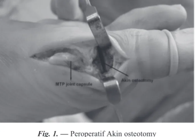
A straight medial incision centered over the base of the proximal phalanx is used. The capsule is elevated and retracted exposing the metaphysis of the proximal phalanx. Six mm distal to the joint, a 2 to 3 mm medial wedge is removed with a microsaggittal saw (Figure 1), preserving the lateral cortex and avoiding the joint. We proposed a new fixation method with suture anchors for Akin osteotomy. Suture anchors are basically used to attach tendons to bone. Mainly, bone to bone suturing is provided with the suture anchor. The author (AO) has techniques for fixing bone to bone, such as fixation of the chevron osteotomy and sternal closure for bypass surgery (3,6). A suture anchor has the strength to resist any force causing it
to be pulledout (pulledout strength : Statak 5.0=364, Statak 5.2=343, Statak 3.5=129, Statak 2.5=91, Statak 1.5=37, Mitek G4=216, Mitek G2=147, Mitek G3=53 pounds...) (2).Wedge closure and deformity correction are followed by osteotomy fixation with a suture anchor (Statak, Mitek anchors...) (Figure 2).
RESULTS
At all of the patients approximately 30 days clinical recovery was seen.
Clinical recovery was seen in all patients in approximately 30 days. Full union was realized radiological between 1.5 and 2.5 months. Full union was seen at 3th month in a bilateral case.
Local wound infection incurred at 2 patients. However it has completely recovered with proper treatment.
Fig. 1. — Peroperatif Akin osteotomy
Postoperative plaster was not apllied. First 3 days elevation and local ice application was performed. After 3 days he/she was permitted for mobilization with slippers. On the 15th day the stitches were
removed. On the 16th days he/she was permitted to
wear wide shoes and to walk with short distances. Preoperative AOFAS values for pain, function and alignment were measured. Pain values were 8 patients 20 points, 27 patients 0 points. Function values were, activity, 14 patients 4 points, 21 patients 0 points ; footwear requirements, 18 patients 5 points, 17 patients 0 points, MTP joint movement, 11 patients 5 points, 24 patients 0 points, IP joint movement, 5 patients 5 points, 30 patients 0 points, MTP-IP stability, 35 patients 0 points, callus related to hallux MTP-IP, 35 patients 5 points. The alignment values were 35 patients 0 points (Table 1).
Post-operative AOFAS values were measured and evaluated as follows. Pain values were 33 patients 40 points, 2 patients 30 points. Function values
were, activity, 30 patients 10 points, 5 patients 7 points ; footwear requirements,32 patients 10 points, 13 patients 5 points, MTP joint movement, 22 patients 10 points, 13 patients 5 points, IP joint movement, 34 patients 5 points, 1 patients 0 points, MTP-IP stability, 35 patients 5 points, callus related to hallux MTP-IP, 35 patients 5 points. The alignment values were 29 patients 15 points, 6 patients 8 points (Table 1). These results were found to be statisically highly significant (p<0.001).
DISCUSSION
Various fixation materials for Akin osteotomy have been described in literature such as Krisschner wire (10,11), small plates (12), screwes fixation (13,14,15), staples (13,16), Herbert screw (17,18), absorbable sture (9). Each fixation technique has
its own advantages and disadvantages. K-wires are generally used for fixation but may cause pin migration or skin irritation (12,15,16). The use of Table I. — Preoperativeandpostoperative AOFAS points (Max. 100 points)
Min. and Max . points Patients
Preoperative Postoperative
Points Patients Points
Pain 0-40 27 8 20 33 40 0 2 30 Function Activity 0-10 14 4 30 10 21 0 5 7 Footwear require-ments 0-10 18 5 32 10 17 0 13 5 MTP joint movement 0-10 1124 50 2213 105 IP joint movement 0-5 530 50 341 50 MTP-IP stability 0-5 35 0 35 5 Callus rela-ted to hallux MTP-IP 0-5 35 0 35 5 Aligment 0-15 35 0 6 29 15 8 p<0.001
The use of screws for fixation may cause cracking of the phalanx, and the screws may then have to be removed later. When small anchors are used for screws for fixation may cause cracking of the
phalanx, and the screws may then have to be removed later (14).
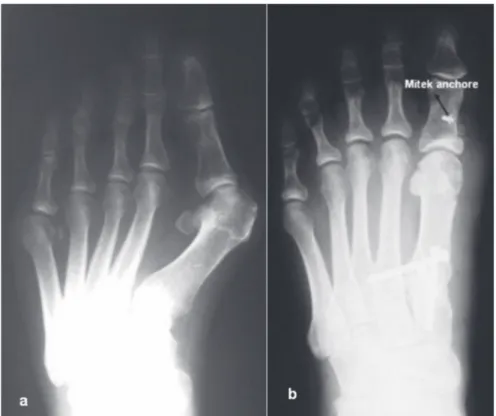
Fig. 4. — 45 year old female left foot; a) preoperative and b) postoperative (Akin procedure) radiography
8. Garrido IM, Rubio ER, Bosch MN et al. Scarf and
Akin osteotomies for moderate and severe hallux valgus : clinical and radiographic results. Foot Ankle Surg 2008 ; 14 : 194-203.
9. Hansen ST. Functional reconstruction of the foot and
ankle. Honsen, (2000) ; p 544.
10. James L. Beskin, MD. Akin’s Phalangeal Osteotomy for
Bunion Repair. In : Myerson M, director. Current Therapy In Foot and Ankle Surgery. St. Louis : CV Mosby ; 1993 : 54-57
11. Jose A.V.Sanhudo. Clinical Tip : Modified Akin
Osteotomy. Foot Ankle Int 2005 ; 26 ; 901-902.
12. Kalman Toth, Péter Kellerman, Károly Wellinger.
Fixation of Akin osteotomy for hallux abductus with absorbable suture Archives of Orthopaedic and Trauma
Surgery 2010 ; 130 : 1257-1261.
13. Kilmartin TE Phalangeal osteotomy versus first metatarsal
decompression osteotomy for the surgical treatment of hallux rigidus : a prospective study of age-matched and condition-matched patients. J Foot Ankle Surg 2005 ; 44 : 2-12.
14. Kitaoka HB, Alexander IH, Adelaar RS et al. Clinical
rating system for the ankle-hindfoot, midfoot, hallux, and lesser toes. Foot Ankle Int. 1994 ; 15 : 349-353.
15. Oznur A. A new technique for fixation of distal chevron
osteotomy. Foot Ankle Int 2002 ; 23 : 954-955.
16. Sabo D, Buchner M. Die Behandlung des Hallux valgus
– Syndroms mit Scarf-Osteotomie, Akin-Osteotomie und Weil- Osteotomie. Fuss Sprung 2004 ; 2 : 76-84.
17. Thomas J Master techniques in podiatric surgery : the foot
and ankle. Lippincott ; 2005 ; 61-72.
18. Thordarson DB, Rudicel SA, Ebramzadeh E, Gill, LH.
Outcomestudy of hallux valgus surgery- an AOFAS multi-center study. Foot Ankle Int. 2001 ; 22 : 956-959. fixation of the phalanx in Akin osteotomy, there
is sufficient strength of fixation and no need for removal. Suture anchor fixation encourages rigid fixation and allows the patient to return to normal activities earlier. Patients are instructed to walk with postoperative shoe (Figure 3, Figure 4). There were no complications due to the use of suture anchors. Osteotomy stability, reduction, and fixation were achieved successfully in all patients. Considering its lesser morbidity, excellent cosmesis and no need for hardware removal, this new technique offers an attractive alternative in Akin osteotomy.
REFERENCES
1. Akin OF. The treatment of hallux valgus : A new operative
approach and its result. Med Sentinel. 1925 ; 33 : 678-679.
2. Barber FA, Herbert MA, Click JN. The ultimate strength
of suture anchors. Arthroscopy 1995 ; 11 : 21-28.
3. Basile A, Battaglia A, Campli A. Comparison of Chevron–
Akin osteotomy and distal soft tissue reconstruction – Akin osteotomy for correction of mild hallux valgus. Foot Ankle
Surg 2000 ; 6 : 155-163.
4. Chang TJ. Master Techniques in podiatric surgery. Foot
Ankle 2004 ; 72 : 560.
5. Cohen MM. The oblique proximal phalangeal osteotomy
in the correction of hallux valgus. J Foot Ankle Surg 2003 ; 42 : 282-289.
6. Dogan OF, Oznur A, Demircin M. A new technical
approach for sternal closure with suture anchors (Dogan technique). Heart Surg Forum. 2004 ; 7 : E328-32.
7. Frey C, Jahss M, Kummer FJ. The Akin procedure : an