Contents lists available atScienceDirect
Food and Chemical Toxicology
journal homepage:www.elsevier.com/locate/foodchemtoxMultidirectional biological investigation and phytochemical profile of Rubus
sanctus and Rubus ibericus
Gokhan Zengin
a,∗, Claudio Ferrante
b, Ismail Senkardes
c, Reneta Gevrenova
d,
Dimitrina Zheleva-Dimitrova
d, Luigi Menghini
b, Giustino Orlando
b,∗∗, Lucia Recinella
b,
Annalisa Chiavaroli
b, Sheila Leone
b, Luigi Brunetti
b, Carene Marie Nancy Picot-Allain
e,
Kannan RR. Rengasamy
f, Mohamad Fawzi Mahomoodally
eaDepartment of Biology, Faculty of Science, Selcuk University, Konya, Turkey
bDepartment of Pharmacy, University “G. d'Annunzio” of Chieti-Pescara, Chieti, 66100, Italy
cDepartment of Pharmaceutical Botany, Faculty of Pharmacy, Marmara University, Istanbul, Turkey
dDepartment of Pharmacognosy, Faculty of Pharmacy, Medical University of Sofia, Bulgaria
eDepartment of Health Sciences, Faculty of Science, University of Mauritius, 230 Réduit, Mauritius
fDepartment of Bio-resources and Food Science, Konkuk University, Seoul, South Korea
A R T I C L E I N F O Keywords: Rubus LC-MS Toxicity Antioxidant Anti-inflammatory Wound healing A B S T R A C T
In the present study, the biological properties, including, the enzyme inhibitory and antioxidant activities, as well as, the phytochemical profile of the ethyl acetate, methanol, and water extracts of Rubus sanctus Schreb. and
Rubus ibericus Juz. leaves were determined using in vitro bioassays. Wide range of phytochemicals, including,
hydroxybenzoic acids, hydroxycinnamic acids, acylquinic acids, ellagitannins, flavonoids, and triterpenoid sa-ponins were determined using UHPLC-ESI/HRMS technique. The ethyl acetate and methanol extracts of the studied Rubus species effectively inhibited acetyl and butyryl cholinesterase. On the other hand, R. sanctus water extract showed low inhibition against α-amylase and prominent inhibitory action against α-glucosidase. Data collected from this study reported the radical scavenging and reducing potential of the studied Rubus species. Investigation of the protective effects of the different extracts of R. sanctus and R. ibericus in experimental model of ulcerative colitis was performed. The extracts were also tested on spontaneous migration of human colon cancer cells (HCT116) in wound healing experimental paradigm. Only R. sanctus methanol extract inhibited spontaneous HCT116 migration in the wound healing test. Our results suggested that R. sanctus and R. ibericus may be potential candidates as sources of biologically-active compounds for the development of nutraceuticals, pharmaceuticals, and/or cosmetics.
1. Introduction
The Rubus genus consists of 900–1000 species distributed world-wide (Ryu et al., 2018). Archaeologists found evidence of the use of Rubus dating around 8000 BCE, postulating that species of the Rubus genus have been long used as food and medicinal source (Hummer, 2010). Besides, ethnobotanical data substantiate the use of Rubus spe-cies for therapeutic applications by several cultures across the globe. For instance, the young shoots of the Rubus species were traditionally used to heal wounds, insect bites, and pimples (Süntar et al., 2011). The aerial part of R. fruticosus was used against cough, the fruit juice was recommended for colitis, the roots were used against diarrhoea,
chewing the leaves of R. fruticosus was recommended to relieve tooth-ache, a tea prepared from the roots was used for labour pain, and a decoction prepared from R. fruticosus roots was used to treat dysentery (Verma et al., 2014). Australian aborigines have long consumed Rubus fruits to induce mild laxative effect (Bakar et al., 2016). Indeed, Rubus fruits have long been consumed worldwide, for their possible health benefits or simply because of their good taste (Lee et al., 2012). A herbal tea prepared from the decoction of R. sanctus roots was used to alleviate pain and against rheumatism (Süntar et al., 2011). The fruits of R. sanctus were used as a diuretic and against diarrhoea, haemor-rhoids, diabetes mellitus, and rheumatism (Akkol et al., 2015). R. dis-color (synonym of R. ibericus) fruits, leaves, and roots were used to treat
https://doi.org/10.1016/j.fct.2019.03.041
Received 23 January 2019; Received in revised form 19 March 2019; Accepted 20 March 2019 ∗Corresponding author.
∗∗Corresponding author.
E-mail addresses:[email protected](G. Zengin),[email protected](G. Orlando).
Food and Chemical Toxicology 127 (2019) 237–250
Available online 23 March 2019
0278-6915/ © 2019 Elsevier Ltd. All rights reserved.
nephritis and prostatitis. Additionally, the leaves were used to heal wounds and treat diarrhoea (Veličković et al., 2016). In Traditional Chinese Medicine, a mixture containing R. chingii was used to manage infertility, impotence, frequent urination, low backache, and poor sight (Bakar et al., 2016). R. parvifolius roots were widely used for the treatment of hepatitis, rheumatism, and abdominal pain caused by postpartum stasis (Xu et al., 2017). Traditionally, teas and alcoholic infusions prepared from the leaves, shoots, and fruits of R. grandifolius were used for the management of diabetes, as depurative, diuretic, and to treat sore throat (Spínola et al., 2019).
Rubus species are rich sources of bioactive compounds, possessing multiple biological properties. Several studies reported the biological activities of Rubus species. For instance, euscaphic acid, isolated from R. rosifolius, has been reported to show significant antioxidant activity. Ellagic acid, quercetin, and kaempferol, identified from R. rosifolius were related to the chemopreventive properties of the plant (Campbell et al., 2017). Phenolic rich extracts of R. rosifolius presented microbial properties with quorum sensing properties and anti-oxidant activity (Oliveira et al., 2016). Three compounds isolated from
R. idaeus rhizome showed neuroprotective effects in vitro (Xu et al.,
2017). R. hirsutus fruits showed high antioxidant activity (Fu et al., 2015). R. grandifolius extracts inhibited glucosidase, β-glucosidase, α-amylase, lipase, and aldose reductase (Spínola et al., 2019). R. fair-holmianus methanol root extract effectively lowered cell viability, ATP proliferation, and increased LDH release from human breast cancer cells (George et al., 2017). Previously, Shin et al. (2014) demonstrated protective effects induced by R. coreanus in experimental model of ul-cerative colitis.Akkol et al. (2015)demonstrated the inhibitory action of extracts of R. sanctus aerial parts on collagenase and elastase.
Based on the multiple biological activities of several Rubus species, this study was designed to assess the possible inhibitory action of R. sanctus and R. ibericus against key enzymes relevant to Alzheimer's disease (acetyl and butyryl cholinesterases), skin hyperpigmentation complications (tyrosinase), and type 2 diabetes (amylase, and α-glucosidase). Besides, as far as our literature review could ascertain, there has not been any study on the inhibitory action of R. sanctus and R. ibericus on the selected enzymes. The antioxidant potential of the selected Rubus species was also evaluated. The phytochemical profiles of the ethyl acetate, methanol, and water extracts of R. sanctus and R. ibericus were determined by UHPLC-ESI/HRMS. The protective effects of R. sanctus and R. ibericus extracts, in an experimental model of ul-cerative colitis constituted of rat colon specimens challenged with li-popolysaccharide (LPS) ex vivo, was assessed. Finally, the chemopre-ventive effects of R. sanctus and R. ibericus extracts on human colon cancer (HCT116) cell migration and invasion capacities (wound healing test) were investigated.
2. Materials and methods 2.1. Plant materials
The Rubus species were collected in Kastamonu-Turkey (R. ibericus: Hanönü village, between Yeniköy and Yılanlı, 619 m; R. sanctus: Hanönü village, centre of Yılanlı, 1015 m) in June 2018. The tax-onomical identification was performed by the botanist Dr. Ismail Senkardes (Marmara University, Faculty of Pharmacy, Pharmaceutical Botany, Istanbul) and a voucher specimen of each species was kept in the herbarium of Marmara University. The leaves were allowed to dry for 10 days at the room temperature. Then, these samples were pul-verised with a laboratory mill.
2.2. Extraction
To prepare ethyl acetate and methanol extracts, the leaves samples (5 g in 100 mL solvent) were stirred overnight (24 h) at room tem-perature and filtered. After filtration, the extracts were concentrated
using a rotary evaporator under vacuum at 40 °C. Water extract was prepared by boiling 5 g of leaves samples in 100 mL water for 20 min. The mixture was then filtered and dried by using a lyophiliser. The extracts were stored at +4 °C until further analysis.
2.3. Quantification of phytochemicals
With reference to our previous studies (Uysal et al., 2017), the total amount of phenolics (TPC) (by standard Folin-Ciocalteu method) and flavonoids (TFC) (by aluminum chloride method) were determined. The final results were expressed as equivalents of standard compounds, i.e., gallic acid (mg GAE/g) and rutin (mg RE/g) for TPC and TFC, respec-tively.
2.4. Metabolite profiling by UHPLC-ESI/HRMS
The UHPLC-ESI/HRMS analyses were achieved on a Q Exactive Plus heated electrospray ionization (HESI-II) – high resolution mass spec-trometer (HRMS) (ThermoFisher Scientific, Inc., Bremen, Germany) equipped with an ultra-high-performance liquid chromatography (UHPLC) system Dionex Ultimate 3000RSLC (ThermoFisher Scientific, Inc.) (Zengin et al., 2017). The analytical details were given in Supplemental material.
2.5. Determination of antioxidant and enzyme inhibitory effects
The enzyme inhibitory activity of R. ibericus and R. sanctus extracts were tested against α-amylase, α-glucosidase, acetyl cholinesterase (AChE), butyryl cholinesterase (BChE), and tyrosinase. The procedures of these assays were reported in our earlier work (Uysal et al., 2017). The enzyme inhibitory effects were expressed as equivalents of acar-bose (for α-amylase and α-glucosidase), galantamine (for AChE and BChE), and kojic acid (for tyrosinase).
Antioxidant capacity of R. ibericus and R. sanctus extracts were spectrophotometrically determined using different methods including phosphomolybdenum, radical scavenging assays (DPPH and ABTS), reducing potentials (FRAP and CUPRAC), and ferrous ion chelating. The results were expressed as trolox (mg TE/g) and ethylenediaminete-traacetic acid equivalents (mg EDTAE/g). The procedures of assays were reported in our earlier work (Uysal et al., 2017).
The results of antioxidant and enzyme inhibitory assays were tistically with one-way ANOVA (by Tukey's test, p < 0.05). The sta-tistical procedures were performed by SPPS v. 17.0. Multivariate ana-lysis (Pearson Correlation, heat map and Sparse Partial Least Squares (sPLS-DA) analysis) were performed by using R software v. 3.5.1. 2.6. Biological assays
2.6.1. Artemia salina lethality bioassay
Artemia salina cysts were hatched in oxygenated artificial sea water (1 g cysts/L). After 24 h, brine shrimp larvae were gently transferred with a pipette in 6 well plate containing 2 mL of Rubus extracts at dif-ferent concentrations (0.1–20 mg/mL) in artificial sea water. Ten larvae per well were incubated at 25–28 °C for 24 h. After 24 h the number of living napulii were counted under light microscope and compared to control untreated group. Results were expressed as percentage of mortality calculated as:
×
T S
T 100,
where, T is the total number of incubated larvae and S is the number of survival napulii. Living nauplii were considered those exhibiting light activating movements during 10 s of observation. For each mental condition two replicates per plate were performed and experi-mental triplicates were performed in separate plates.
2.6.2. In vitro studies
The HCT116 cell lines were cultured in DMEM (Euroclone) supple-mented with 10% (v/v) heat-inactivated fetal bovine serum and 1.2% (v/v) penicillin G/streptomycin in 75 cm2tissue culture flask (n = 5 individual culture flasks for each condition). The cultured cells were maintained in humidified incubator with 5% CO2at 37 °C.
For cell differentiation, HCT116 cell suspensions at a density of 1 × 10⁶cells/mL were treated with various concentrations (10, 50, and 100 ng/mL) of phorbol myristate acetate (PMA, Fluka) for 24 h or 48 h (induction phase). Thereafter, the PMA-treated cells were washed twice with ice-cold pH 7.4 phosphate buffer solution (PBS) to remove PMA and non-adherent cells, whereas the adherent cells were further main-tained for 48 h (recovery phase). Morphology of cells was examined under an inverted phase-contrast microscope.
To assess the basal cytotoxicity of R. sanctus and R. ibericus extracts, a viability test was performed on 96 micro well plates, using 3-(4,5-dimethylthiazol-2-yl)-2,5-diphenyltetrazolium bromide (MTT) test. Cells were incubated with extracts (ranging in the concentration 10–1000 μg/mL) for 24 h. Aliquot of 10 μL of MTT (5 mg/mL) was added to each well and incubated for 3 h. The formazan dye formed was extracted with DMSO and absorbance was recorded as previously de-scribed (Menghini et al., 2018). Effects on cell viability were evaluated in comparison to untreated control group.
Finally, HCT116 cell line was exposed to Rubus extracts, in wound healing experimental paradigm. HCT116 cells (6 × 10³cells/well) were seeded on 6-well plastic plates. Cells monolayers were preliminarily treated with a proliferation inhibitor mitomycin C (Sigma-Aldrich, St. Louis, Missouri, USA) at the non-toxic concentration of 5 μM, in order to exclude the effect of cell proliferation. After 2 h on cells in the con-fluence interval 85–90%, a wound was generated by scratching the cell monolayer using a 0–200 μL pipette tip. The sample was washed twice with PBS to remove detached cells. Cells were incubated in serum free media supplemented with Rubus extracts at the non-toxic concentration of 100 μg/mL. Cell migration was visualised by capturing at least 3 microscope images/well at time 0, 24 and 48 h. An inverted light mi-croscope Leika equipped with Nikon 5100 camera was used to capture image at 4x magnification. The quantification of scratch area with no cells was quantified using Image-J software (NIH). Using GraphPad software (version 6.0), mean data at T0, 24 and 48 h were calculated for untreated control and Rubus extracts and expressed as percentage var-iation with reference to relative 100% of at 0 h.
2.6.3. Ex vivo studies
Male adult Sprague-Dawley rats (200–250 g) were housed in Plexiglass cages (40 cm × 25 cm × 15 cm), two rats per cage, in air-conditioned colony rooms (22 ± 1 °C; 60% humidity), on a 12 h/12 h light/dark cycle (light phase: 07:00–19:00 h), with free access to tap water and food, 24 h/day throughout the study, with no fasting periods. Rats were fed a standard laboratory diet (3.5% fat, 63% carbohydrate, 14% protein, 19.5% other components without caloric value; 3.20 kcal/ g). Housing conditions and experimentation procedures were strictly in accordance with the European Union ethical regulations on the care of animals for scientific research.
The experiments were approved by Local Ethical Committee (University “G. d’Annunzio” of Chieti-Pescara) and Italian Health Ministry (Italian Health Ministry authorization N. F4738.N.XTQ, de-livered on 11th November 2018). Rats were sacrificed by CO2 inhala-tion (100% CO2at a flow rate of 20% of the chamber volume per min) and colon specimens were immediately collected and maintained in humidified incubator with 5% CO2at 37 °C for 4 h, in RPMI buffer with added bacterial LPS (10 μg/mL) (incubation period).
During the incubation period, tissues were treated with scalar sub-toxic concentrations of R. sanctus and R. ibericus extracts (100 μg/mL). The activity of extracts was compared to sulfasalazine (2 mg/mL), an anti-inflammatory reference drug. Tissue supernatants were collected, and nitrite production was determined by mixing 50 μL of the assay
buffer with 50 μL of Griess reagent (1.5% sulfanilamide in 1 M HCl plus 0.15% N-(1-naphthyl) ethylenediamine dihydrochloride in distilled water, [v/v]). After 10 min incubation at room temperature, the ab-sorbance at 540 nm was determined and nitrite concentrations were calculated from a sodium nitrite standard curve.
On the other hand, individual colon specimens were dissected and subjected to extractive procedures to evaluate serotonin 5-hydro-xytrptamine (5-HT) (ng/mg wet tissue) as previously reported (Brunetti et al., 2014;Ferrante et al., 2016). As regards to 5-HT analysis, tissues were homogenized in ice bath for 2 min with Potter-Elvehjem homo-genizer in 1 mL of 0.05 N perchloric acid containing 0.004% sodium EDTA and 0.010% sodium bisulfite. Thereafter, samples were analyzed by HPLC coupled to electrochemical detection consisting of ESA Cou-lochem III detector equipped with ESA 5014B analytical cell.
Additionally, malondialdehyde (MDA) level was determined by the thiobarbituric acid reactive substances (TBARS) method (Mihara et al., 1980). Briefly, tissue specimens were added with 1% H3PO4and 0.6% thiobarbituric acid, and then incubated at 96 °C for 20 min. Absorbance was recorded at 532 nm, and the MDA level was expressed as g/mL.
Besides, LDH activity was measured by evaluating the consumption of NADH in 20 mM HEPES-K+(pH 7.2), 0.05% bovine serum albumin, 20 μM NADH and 2 mM pyruvate using a microplate reader (excitation 340 nm, emission 460 nm) according to manufacturer's protocol (Sigma-Aldrich, St. Louis, Missouri, USA). Data were from triplicate test and expressed as relative variations compared to vehicle-treated cells (Menghini et al., 2018).
2.7. Statistical analysis
Statistical analysis was performed using GraphPad Prism version 5.01 for Windows (GraphPad Software, San Diego, CA). Means ± S.E.M. were determined for each experimental group and analyzed by one-way analysis of variance (ANOVA), followed by Newman-Keuls comparison multiple test. Statistical significance was considered as p < 0.05. Regarding the animals randomized for each experimental group, the number was calculated on the basis of the “Resource Equation” N=(E + T)/T (10 ≤ E ≤ 20) (Charan and Kantharia, 2013), and in accordance with the guidelines suggested by the “National Centre for the Replacement, Refinement and Reduction of Animals in Research” (NC3RS) and reported on the following web site: https://www.nc3rs.org.uk/experimental-designstatistics. N is the number of animals per treated group. E represents the degrees of freedom of the analysis of variance (ANOVA). T is the number of treatments. Considering that E values should be between 10 and 20, the animal number N for ex vivo analysis was chosen in accordance to an E value of 20.
3. Results and discussion
3.1. Quantification of total bioactive components
The Folin-Ciocalteu and aluminum chloride assays are rapid and simple quantitative phytochemical analyses currently used for the de-tection of bioactive secondary metabolites, namely, phenolics and fla-vonoids. Phenolics, consisting of one or more aromatic rings linked hydroxyl groups, are the most abundant class of secondary metabolites, involved in the reproductive, growth, and defence mechanisms of plants (Huot et al., 2014). The total phenolic content of the studied extracts ranged between 17.22 and 152.55 mg GAE/g extract and R. ibericus water extract showed the highest total phenolic content (Table 1). Likewise, Veličković et al. (2016) also reported that the aqueous extracts of R. ibericus leaves contained highest phenolic con-tent. Flavonoids are a subclass of phenolic compounds and are well known for their antioxidant properties (Chen et al., 2018). In the pre-sent study, the total flavonoid content of the studied extracts ranged between 25.70 and 40.52 mg RE/g extract and R. ibericus ethyl acetate
extract showed the highest total flavonoid content. A group of authors studied the flavonoid content of R. ibericus leaves collected from dif-ferent locations and extracted the plant materials using difdif-ferent sol-vents. Flavonoid yield was higher when acetone was used as extraction solvent, the sample collected from the different regions showed distinct difference in flavonoid content (Veličković et al., 2016).
3.2. UHPLC-ESI/HRMS results
Based on the accurate mass measurements, MS/MS fragmentation patterns, relative abundance of the precursor and fragment ions, ele-mental compositions, monoisotopic peak profiles, as well as comparison with reference standards and literature data, a variety of Rubus com-pounds were identified or tentatively elucidated. For the majority of assayed compounds, the mass accuracy of M-H]- in MS/MS analyses was within a level of 5 ppm.
3.2.1. Hydroxybenzoic acids and hydroxycinnamic acids, and their derivatives
The total ion chromatograms for the tested extracts were given in supplementary material (Fig. S1). Hydroxybenzoic acids 2, 4 and 9, and hydroxycinnamic acids 6, 8 and 12 were identified on the basis of the retention times, accurate masses, fragmentation patterns and compar-ison with authentic standards (Table 2). Peaks 1 ([M-H]- at m/z 331.067) and 3 ([M-H]-at m/z 315.072) yielded abundant fragment ions at m/z 169.013 [gallic acid-H]-and 153.018 [protocatechuic acid-H]-, respectively, indicating the loss of 162 Da. Thus 1 and 3 were identified as gallic acid-O-hexoside and protocatechuic acid O-hexoside, respectively. In the same way, 5 and 7 were tentatively identified as two isomers of caffeic acid-O-hexoside. MS/MS spectra of 10 ([M-H]-at
m/z 503.155) and 11 ([M-H]-at m/z 504.123) were acquired (Table 2).
The base peak at m/z 161.023 [caffeic acid-HeH2O]- together with fragment ions at m/z 179.034 [caffeic acid-H]-and 135.044 [caffeic acid-HeCO2]-allowed to identify caffeic acid derivatives. Fragmenta-tion pattern of 10 displayed subsequent losses of hexose units at m/z 341.0902 [M-H-Hex]-and 323.079 [M-H-2Hex]-, indicating caffeoyl-dihexoside. Prominent ions at m/z 282.070 [M-H-caffeoyl-60]- (11), 252.060 ([M-H-caffeoyl-90]- and 222.049 ([M-H-caffeoyl-120]- were consistent with cross ring cleavages of the hexose unit0,4X−,0,3X−and 0,2X−, respectively. Thus, 11 was ascribed to dicaffeoyl-hexoside. 3.2.2. Ellagitannins and ellagic acid derivatives
The abundant peak 17 at m/z 300.999 ([M-H]-) matched the stan-dard reference ellagic acid. Ellagic acid-O-pentoside (15) and ellagic acid-O-deohyhexoside (16) were identified based on the prominent ion at m/z 300.999 and matching MS/MS fingerprint as 17, and data published by (Oszmiański et al., 2015). Concerning compound 14 ([M-H]−at m/z 934.073), the loss of a hexahydroxydiphenoyl (HHDP) moiety (301 Da) at m/z 633.073 and a base peak at m/z 300.999 [el-lagic acid-H]-, along with fragment ions at m/z 257.009 [ellagic
acid-HeCO2]-, 245.009 [ellagic acid-H-2CO]-, 229.014 [ellagic acid-He-COeCO2]-and 217.014 [ellagic acid-H-3CO]-, highlighted the presence of galloyl-bis-HHDP-hexoside. This compound could be related to the ellagitannins casuarictin/potentillin, previously reported in Rubus spe-cies (Donno et al., 2013;Oszmiański et al., 2015). Peak 13 ([M-H]-at m/z 1401.597) afforded prominent ions at m/z 633.0735 and 300.999 corresponding to galloyl-HHDP-hexose and ellagic acid, respectively. This fragmentation pattern could be tentatively assigned to lamber-tianin C (Oszmiański et al., 2015).
3.2.3. Acylquinic acids
Eighteen acylquinic acids were identified in the majority of the tested Rubus extracts (Table 2). Herein, the hydroxycinnamic acids are mainly linked to quinic acid. The assignment of the different acylquinic acids was based on the hierarchical key developed by Clifford and colleagues (Clifford et al., 2003, 2005). Peaks 18, 20 and 21 were identified as 3-Oe, 5-Oe and 4-O-caffeoylquinic acids ([M-H]−at m/z 353.088), respectively, according to the relative abundance of the characteristic fragment ions at m/z 191.055 [quinic acid-H]-, 179.034 [caffeic acid-H]- and 173.045 [quinic acid-HeH
2O]- and 135.044 [caffeic acid-HeCO2]-(Clifford et al., 2003,2005). Based on compar-ison with authentic standards, compounds 18 and 20 were identified as neochlorogenic and chlorogenic acid, respectively. In the same manner, peaks 19, 24 and 25 were ascribed to 3-Oe, 4-Oe and 5-O-p-coumar-oylquinic acids ([M-H]−at m/z 337.093), while peaks 23, 26 and 27 ([M-H]−at m/z 367.103) were assigned as 1-Oe, 4-Oe and 5-O-fer-uloylquinic acid (Table 2). Among the diacylquinic acids, peaks 28, 29 and 30 were related to 3,4-Oe, 3,5-O and 4,5-O-dicaffeoylquinic acids ([M-H]−at m/z 515.120); 28 and 30 yielded indicative fragment ions at 173.045 together with 353.088 ([M-H-caffeoyl]-, while the presence of 29 was evidenced by the relative abundance of the ions at 191, 179 and 135 (Clifford et al., 2003;Clifford et al., 2005;Zheleva-Dimitrova et al., 2017). Compounds 31–34, [M-H]−at m/z 529.136 related to caffeoyl-feruloylquinic acid (Clifford et al., 2003,2005). Furthermore, 31 and 34 were discernible by the base peaks at m/z 193.050 [ferulic acid-H] -and 135.044 [caffeic acid-HeCO2]-, respectively. The formation of the abundant fragment ion at m/z 367.104 [M-H-caffeoyl]-(86.1%) (31) was favored for 3-feruloyl-5-caffeoylquinic acid, while 3-caffeoyl-5-feruloylquinic acid was witnessed by the abundant ions at 179.034 [caffeic acid-H]- (62.8%) (34) together with 161.023 [caffeic acid-HeH2O]-(53.8%). Typical ions of 4-feruloyl-5-caffeoylquinic acid (32) fragmentation were observed at m/z 173.045 (base peak), 367.103 (64.4%) and 193.050 (17.9%). By comparison with 1,5-dicaffeoylquinic acid (Clifford et al., 2005,2007), 33 was assigned to 1-caffeoyl-5-fer-uloylquinic acid, evidenced by the fragment ions at m/z 161.023 (base peak) and 367.103 (20.3%).
3.2.4. Flavonoids
Five flavonoid aglycones 53–57 were tentatively identified in the studied extracts (Table 2). Regarding 53, typical neutral losses of the flavonoid aglycones were observed at m/z 245.082 [M-HeCO2]-, 205.051 [M-H-3CO]-, 203.070 [M-HeC
3O2eH2O]-. Fragment ions at
m/z 163.038 (1,4B−), 137.023 (1,3A−) and 121.028 (1,2B−) were
at-tributed to the Retro-Diels Alder (RDA) cleavages of the flavonoid skeleton (de Rijke et al., 2006). Consequently, 53 was identified as epicatechin. Indeed, the most important fragmentation pattern for 54 (luteolin) is the RDA cleavage which afforded ions at m/z 133.028 (1,3B−), 151.003 (1,3A−) and 107.012 (0,4A−). Fragments at m/z 241.050 [M-HeCO2]- and 201.018 [M-H-3CO]- had low abundance (below 1%) which is consistent with the previous study ( Zheleva-Dimitrova et al., 2018). Regarding 55 (quercetin), the precursor ion at 301.036 gave a series of neutral losses at m/z 273.041 [M-HeCO]-, 255.030 [M-H-2CO]-, 229.050 [M-HeCOeCO
2]-. RDA cleavage gener-ated 1,3A− at m/z 151.003, 0,2A− at m/z 163.003, 1,2A− at m/z 178.998,1,2B−at m/z 121.0287 and0,4A−at m/z 107.012 (Table 2). It is worth noting that 57 (isorhamnetin) yielded fragment ion at m/z Table 1
Total phenolic and flavonoid contents of Rubus sanctus and R. ibericus extracts. Extract Total phenolic content (mg
GAE/g extract) Total flavonoid content (mgRE/g extract)
R. sanctus-EA 17.22 ± 1.21e 25.70 ± 0.62d R. sanctus -MeOH 83.31 ± 2.75d 29.12 ± 0.59c R. sanctus -Water 134.53 ± 2.45b 35.40 ± 3.99b R. ibericus-EA 21.57 ± 0.50e 40.52 ± 0.50a R. ibericus -MeOH 115.53 ± 3.88c 27.88 ± 0.20cd R. ibericus -Water 152.55 ± 1.45a 38.08 ± 0.23ab
Values expressed are means ± S.D. of three parallel measurements. GAE: Gallic acid equivalent; RE: Rutin equivalent; EA: Ethyl acetate; MeOH: Methanol. Different superscripts indicate differences among the extracts (p < 0.05).
Table 2 Metabolites detected in the extracts from Rubus ibericus and R. sanctus . Peak № Accurate mass [M-H] −m/z Molecular formula [M-H] -MS/MS data m/z tR 1(4) min Exact mass [M-H] -m/z Delta ppm Tentative assignment Reference Standard (RS)/Reference Hydroxybenzoic and hydroxycinnamic acids and derivatives 1 331.0672 C13 H15 O10 331.0672 (100), 169.0132 (53.31), 151.0023 (16.51), 125.0230 (20.67) 0.99 331.0659 0.303 Gallic acid-O-hexoside a , b , c , d , e , f 2 169.0132 C7 H5 O5 169.0132 (31.85), 125.0230 (100) 1.18 169.0131 −6.133 Gallic acid a , b , c , d , e , f RS 3 315.0724 C13 H15 O9 315.0727 (36.24), 153.0183 (100), 123.0439 (2.15), 109.0281 (44.28) 1.91 315.0710 0.935 Protocatechuic acid-O-hexoside a , b , c , d , e , f 4 153.0182 C7 H5 O4 153.0188 (11.57), 109.0281 (100), 123.0439 (85.33) 2.24 153.0182 −7.659 Protocatechuic acid a , b , c , d , e , f RS 5 341.0881 C15 H17 O9 341.0881 (21.0), 323.0789 (21.0), 281.0667 (100), 251.0561(55.1), 221.0452 (48.8), 179.0341 (95.6), 161.0234 (68.63), 135.0439 (68.9) 2.91 341.0867 0.813 Caffeic acid-O-hexoside a , b , c , d , e , f Clifford et al. (2007) 6 163.0390 C9 H7 O3 163.0390 (23.64), 135.0438 (9.15), 119.0489 (100) 3.10 163.0389 −6.731 p-Coumaric acid a , b , c , d , e , f RS 7 341.0887 C15 H17 O9 341.0887 (21.9), 281.0667 (3.0), 251.0560 (62.1), 221.0453 (50.4), 179.0341 (100), 161.0234 (65.9), 135.0439 (72.3) 3.26 341.0867 2.770 Caffeic acid-O-hexoside isomer a , b , c , d , e , f Clifford et al. (2007) 8 179.0341 C9 H7 O4 179.0341 (17.47), 135.0439 (100) 3.72 179.0338 −5.150 Caffeic acid a , b , d , e RS 9 153.0182 C7 H5 O4 153.0182 (65.10), 135.0074 (28.61), 122.0362 (1.56), 109.0281 (100) 4.03 153.0182 −7.463 Gentisic acid a , b , c , d , e , f RS 10 503.1440 C21 H27 O14 503.1548 (48.8), 341.0902 (3.6), 323.0787 (21.6), 179.0344 (34.3), 161.0234 (100), 135.0437 (39.4) 6.29 503.1406 6.701 Caffeoyldihexoside d , e Oszmiański et al. (2015) 11 504.1273 C24 H24 O12 504.1234 (96.7), 342.0916 (18.0), 282.0702 (29.2), 252.0595 (20.7), 222.0487 (10.8), 179.0342 (46.9), 161.0234 (100), 135.0439 (45.7) 6.29 504.1234 −6.677 Dicaffeoyl-hexoside a , b 12 163.0391 C9 H7 O3 163.0390 (21.22), 135.0438 (4.12), 119.0489 (100) 7.04 163.0389 −6.056 m -Coumaric acid d , e RS Ellagitannins and ellagic acid derivatives 13 1401.5967 C66 H97 O32 1401.5967 (72.0), 897.0446 (7.7), 633.0735 (12.2), 300.9989 (100), 229.0138 (8.6) 4.34 1401.5968 −0.117 Ellagitannins (Lambertianin C) d Oszmiański et al. (2015) 14 934.0726 C41 H26 O26 934.0726 (39.8), 663.0733 (6.6), 300.9989 (100), 257.0090 (3.9), 245.0092 (2.8), 229.0137 (6.1), 217.0136 (1.5) 4.55 934.0718 0.847 Galloyl-bis-hexahydroxyphenoyl- hexoside a , b , c , d , e , f Donno et al. (2013) 15 433.0415 C19 H13 O12 433.0415 (100), 300.9990 (82.7), 257.0091 (1.3), 229.0142 (1.9) 4.76 433.0412 0.510 Ellagic acid-pentoside a , b , c , d , e , f Oszmiański et al. (2015) 16 447.0571 C20 H15 O12 447.0571 (82.7), 300.994 (47.7), 299.9912 (100) 4.95 477.0569 0.382 Ellagic acid-deoxyhexoside d Oszmiański et al. (2015) 17 300.9991 C14 H6 O8 300.9991 (100), 257.0092 (0.4), 245.0091 (2.3), 229.0140 (3.2), 217.0131 (0.6), 145.0281 (3.4), 117.0317 (1.3) 5.10 300.9990 0.396 Ellagic acid a , b , c , d , e , f RS Acylquinic acids 18 353.0880 C16 H17 O9 353.0880 (46.2), 191.0553 (100), 179.0342 (67.3), 173.0447 (2.0), 161.0235 (4.6), 135.0439 (50.5), 93.0331 (2.0) 2.48 353.0867 0.580 Neochlorogenic (3-caffeoylquinic) acid a , b , c , d , e , f RS 19 337.0932 C16 H17 O8 337.0932 (7.7), 191.0552 (7.6), 173.0443 (4.1), 163.0390 (100), 119.0489 (28.0), 93.0329 (2.7) 3.12 337.0929 0.829 3-coumaroyl-quinic acid a , b , c , d , e , f Clifford et al. (2005) 20 353.0881 C16 H17 O9 353.0881 (6.0), 191.0554 (100), 179.0345 (3.1), 173.0450 (2.5), 161.0234 (2.3), 111.0433 (1.1), 97.4881 (0.7), 93.0331 (3.1), 127.0389 (1.7), 135.0439 (2.6) 3.29 353.0867 0.920 Chlorogenic (5-caffeoylquinic) acid a , b , c , d , e , f RS 21 353.0881 C16 H17 O9 353.0881 (28.3)191.0554 (56.1), 179.0341 (64.6), 173.0446 (100), 161.0229 (3.8), 135.0439 (58.8), 111.0438 (3.2), 93.0331 (21.8) 3.47 353.0867 0.920 4-caffeoylquinic acid a , b , c , d , d , e , f Clifford et al. (2005) 22 367.1035 C17 H19 O9 367.1035 (14.7), 193.0499 (100), 173.0448 (5.3), 149.0593 (2.4), 134.0361 (58.6), 93.0331 (2.6) 3.53 367.1035 0.094 3-feruloylquinic acid a , b , c , d , e , f Clifford et al. (2005) 23 367.1038 C17 H19 O9 367.1038 (41.0), 193.0499 (16.1), 173.0444 (2.6), 161.0233 (100), 134.0362 (9.1), 127.0388 (1.5), 85.0280 (12.4) 3.94 367.1035 0.830 1-feruloylquinic acid a , d Clifford et al. (2005) 24 337.0929 C16 H17 O8 337.0929 (7.3), 191.0555 (0.5), 173.0446 (100), 163.0390 (20.0), 119.0400 (7.3), 93.0331 (19.1), 97.4970 (0.7) 4.12 337.0929 0.473 4-coumaroyl-quinic acid a , b , c , d , e , f Clifford et al. (2005) 25 337.0939 C16 H17 O8 337.0939 (8.6), 191.0554 (100), 173.0444 (15.9), 163.0393 (6.4), 119.0489 (6.2), 93.0330 (20.1) 4.14 337.0929 4.532 5-coumaroyl-quinic acid d , e Clifford et al. (2005) 26 367.1038 C17 H19 O9 367.1038 (13.7), 193.0500 (18.0), 191.0552 (5.7), 173.0446 (100), 161.0230 (3.83), 134.0361 (14.8), 111.0439 (2.4), 93.0331 (24.2) 4.50 367.1034 −1.840 4-feruloylquinic acid a , b , c , d , e , f Clifford et al. (2005) 27 367.1034 C17 H19 O9 367.1034 (62.8), 193.0501 (0.7), 173.0445 (3.80), 161.0.234 (100), 134.0327 (1.0), 127.0388 (2.5), 111.0438 (0.4), 93.0332 (2.5), 85.0280 (17.0) 4.65 367.1034 −0.260 5-feruloylquinic acid a , d Clifford et al. (2005) (continued on next page )
Table 2 (continued ) Peak № Accurate mass [M-H] −m/z Molecular formula [M-H] -MS/MS data m/z tR 1(4) min Exact mass [M-H] -m/z Delta ppm Tentative assignment Reference Standard (RS)/Reference 28 515.1198 C25 H23 O12 515.1198 (100), 353.0883 (17.8), 335.0774 (8.2), 203.0335 (0.5), 191.0554 (41.55), 179.0341 (67.0), 173.0447 (78.8), 161.0234 (24.8), 135.0439 (67.8), 111.0436 (1.7), 93.0331 (19.8) 5.78 515.1184 0.487 3,4-dicaffeoylquinic acid a , b , c , d , e , f Clifford et al. (2005) 29 515.1199 C25 H23 O12 515.1199 (13.8), 353.0879 (93.8), 335.0776 (0.8), 191.0553 (100), 179.0341 (49.9), 173.0477 (4.66), 161.0284 (5.50), 135.0439 (50.3), 111.0438 (1.64), 93.0331 (3.7), 85.0279 (8.0) 5.94 515.1184 0.720 3,5-dicaffeoylquinic acid a , b , c , d , e , f Clifford et al. (2005) 30 515.1196 C25 H23 O12 515.1196 (6.3), 353.0880 (48.9), 335.0781 (1.1), 191.0554 (38.2), 179.0341 (68.5), 173.0446 (100), 161.0235 (6.0), 135.0439 (65.6), 11.0437 (1.2), 93.0331 (23.7), 85.0279 (6.2) 6.32 515.1184 0.254 4,5-dicaffeoylquinic acid a , b , c , d , e , f Clifford et al. (2005) 31 529.1354 C26 H25 O12 529.1354 (7.2), 367.1035 (86.1), 193.0500 (100), 161.0236 (77.7), 134.0361 (63.3), 6.92 529.1351 0.549 3-feruloyl-5-caffeoylquinic acid a , b Clifford et al. (2005) 32 529.1350 C26 H25 O12 529.1350 (74.0), 367.1030 (64.4), 193.0503(17.9), 173.0448(100), 134.0362 (14.8), 111.0439 (4.2), 93.0332 (24.5) 7.26 529.1351 −0.263 4-feruloyl-5-caffeoylquinic acid a , b Clifford et al. (2005) 33 529.1357 C26 H25 O12 529.1357 (7.2), 367.1032 (20.3), 349.0930 (2.8), 191.1402 (1.8), 161.0234 (100), 134.0355 (2.0), 93.0329 (1.7) 7.32 529.1351 1.003 1-caffeoyl-5-feruloylquinic acid a , d Clifford et al. (2005) 34 529.1357 C26 H25 O12 529.1357 (77.1), 367.1054 (18.6), 191.0249 (2.2), 179.0339 (62.8), 161.0233 (53.8), 134.0366 (7.1), 135.0439 (100) 7.69 529.1351 1.116 3-caffeoyl-5-feruloylquinic acid a , d Clifford et al. (2005) 35 677.1513 C34 H29 O15 677.1513 (97.6), 515.1188 (44.4), 353.0874 (52.6), 335.0780 (15.4), 229.5099 (4.0), 191.0552 (51.5), 179.0341 (75.6), 173.0447 (93.7), 161.0234 (28.5), 135.0440 (100), 111.0435 (5.0), 93.0330 (24.5) 7.86 677.1512 0.172 3,4,5-tricaffeoylquinic acid a , b Clifford et al. (2007) Flavonoids 36 609.1476 C27 H29 O16 609.1476 (2.5), 447.0934 (28.7), 285.0409 (65.8), 255.0296 (56.5) 3.89 609.1461 2.515 Luteolin-O-dihexoside a , d Ferreres et al. (2007) 37 6091467 C27 H29 O16 609.1467 (97.6), 429.0856 (1.0), 285.0400 (51.9), 284.0327 (100), 255.0302 (58.7), 227.0346 (34.2), 163.0029 (1.4), 151.0027 (2.0), 107.0122 (1.5) 4.87 609.1461 0.923 Kaempferol-O-dihexoside a , c , d , e , f Ferreres et al. (2007) 38 609.1462 C27 H29 O16 609.1462 (100), 301.0349 (49.5), 300.0276 (87.9), 271.0247 (51.8), 255.0296 (22.1), 243.0293 (12.6), 227.0347 (1.6), 163.0028 (1.7), 151.0026 (8.6), 107.0125 (1.9) 5.14 609.1461 0.118 Quercetin-3-O-rutinoside (rutin) a , b , c , d , e , f RS 39 463.0885 C21 H19 O12 463.0885 (100), 301.0349 (53.2), 300.0277 (98.2), 271.0249 (56.4), 255.0297 (21.5), 243.0298 (11.2), 227.0344 (4.4), 151.0025 (9.9), 135.0070 (0.7), 107.0119 (2.0) 5.27 – 0.585 Quercetin-3-O-galactoside (Hyperoside) a , b , c , d , e , f RS 40 579.1359 C26 H27 O15 579.1359 (84.6), 429.0829 (1.4), 327.0502 (0.3), 309.0408 (0.5), 285.0398 (42.9), 284.0327 (100), 255.0296 (61.3), 227.0346 (40.3), 229.0503 (4.0), 211.0398 (2.2), 178.9976 (2.0), 163.0024 (1.4), 151.0024 (2.6), 135.0073 (1.4), 107.0124 (2.5) 5.28 579.1355 0.547 Kaempferol-2″ e O-pentosylhexoside a , b , c , d , e , f Ferreres et al. (2007) 41 477.0669 C21 H17 O13 477.0669 (54.6), 301.0356 (100), 271.0251 (1.0), 255.0300 (4.2), 243.0296 (0.9), 227.0341 (2.3), 211.0392 (2.6), 178.9978 (9.2), 163.0026 (4.3), 151.0023 (25.4), 135.0072 (0.3), 121.0281 (7.0), 107.0124 (9.9) 5.33 477.0675 −1.203 Quercetin-O-hexuronide a , b , c , d , e , f Oszmiański et al. (2015) 42 463.0882 C21 H19 O12 463.0882 (97.6), 301.0348 (42.3), 300.0277 (100), 271.0249 (54.0), 255.0298 (21.4), 243.0297 (13.5), 227.0344 (3.1), 151.0025 (7.2), 147.0078 (0.2), 135.0073 (1.2), 107.0123 (3.0) 5.39 463.0882 0.002 Quercetin-3-O-glucoside (isoquercitrin) a , b , c , d , e , f RS 43 461.0728 C21 H17 O12 461.0728 (48.8), 357.0616 (0.8), 327.0498 (1.4), 297.0393 (0.3), 285.0406 (100), 241.0504 (1.0), 229.0515 (0.3), 217.0507 (1.3), 151.0025 (5.4), 133.0282 (11.1), 107.0124 (3.0) 5.48 461.0725 0.501 Luteolin-O-hexuronide d , e , f de Rijke et al. (2006) 44 433.0805 C20 H17 O11 433.0805 (100), 301.0356 (58.7), 300.0272 (98.4), 271.0248 (56.5), 255.0301 (18.9), 243.0303 (13.0), 151.0027 (4.4) 5.60 433.0776 Quercetin-O-pentoside a , b , c Oszmiański et al. (2015) 45 593.1511 C27 H29 O15 593.1511 (91.5), 285.0403 (100), 255.0300 (53.6), 229.0504 (8.4), 227.0346 (37.5), 161.0226 (1.0), 151.0023 (3.3), 135.0072 (1.0), 107.0126 (3.1) 5.74 593.1512 0.415 Kaempferol-3-O-rutinoside d , e , f RS 46 447.0564 C20 H15 O12 447.0564 (95.2), 315.0150 (100), 299.9913 (89.9), 270.9887 (34.7), 255.0299 (2.5), 227.0356 (1.1) 5.87 477.0569 0.110 Isorhamnetin-O-pentoside a , b , c , d , e , f de Rijke et al. (2006) (continued on next page )
Table 2 (continued ) Peak № Accurate mass [M-H] −m/z Molecular formula [M-H] -MS/MS data m/z tR 1(4) min Exact mass [M-H] -m/z Delta ppm Tentative assignment Reference Standard (RS)/Reference 47 477.1020 C22 H21 O12 477.1020 (100), 315.0497 (19.4), 314.0434 (60.7), 300.0272 (11.6), 299.0197 (14.4), 271.0249 (37.5), 243.0297 (24.6), 179.0340 (35.3), 161.0233 (24.4), 151.0023 (5.8), 135.0440 (29.5) 5.89 477.1038 −3.897 Isorhamnetin-3-O-glucoside a , b , c , d , e , f RS 48 461.0732 C21 H17 O12 461.0732 (9.5), 285.0406 (100), 257.0457 (4.1), 229.0502 (10.2), 211.0398 (2.2), 201.0548 (1.1), 151.0022 (1.1), 163.0029 (1.4), 135.0073 (1.5), 131.0492 (0.3), 107.0123 (2.8) 5.95 461.0725 1.368 Kaempferol-O-hexuronide a , b , c , d , e , f Oszmiański et al. (2015) 49 447.0938 C21 H19 O11 447.0938 (100), 285.0327 (28.2), 284.0397 (75.1), 255.0298 (57.6), 227.0346 (58.9), 151.0025 (2.8), 135.0073 (0.57), 107.0124 (1.1) 5.96 447.0933 1.041 Kaempferol-3-O-glucoside a , b , c , d , e , f RS 50 447.0875 C21 H19 O11 447.0938 (100), 285.0405 (77.8), 271.0249 (14.6), 243.0304 (16.7), 227.0337 (4.1), 151.0026 (3.7), 133.0281 (9.4), 107.0121 (2.6) 6.39 447.0933 −12.894 Luteolin-7-O-glucoside a , b , c , d , f RS 51 609.1250 C30 H25 O14 609.1250 (100), 447.0944 (5.3), 285.0406 (84.3), 255.0299 (30.0), 229.0506 (7.4), 227.0349 (22.9), 211.0394 (1.4), 179.0340 (13.0), 161.0234 (30.3), 151.0024 (2.2), 135.0439 (18.7), 107.0123 (2.3) 7.07 609.1250 0.035 Kaempferol-O-caffeoylhexoside a , b , d de Rijke et al. (2006) 52 593.1306 C30 H25 O13 593.1306 (100), 447.0936 (3.1), 285.0403 (82.8), 257.0457 (2.9), 255.0300 (56.4), 239.0346 (1.6), 229.0502 (6.9), 227.0347 (39.4), 211.0395 (3.30), 151.0026 (3.7), 119.0489 (3.8), 117.0332 (3.8), 107.0120 (2.9) 7.77 593.1301 0.937 Kaempferol-3- O-p-coumaroyl-glucoside (tiliroside) a , b , c , d , e , f RS 53 289.0720 C15 H13 O6 289.0720 (100), 245.0822 (41.2), 205.0505 (15.7), 203.0702 (18.9), 163.0383 (2.0), 137.0230 (18.0), 121.0279 (8.5) 3.99 289.0718 0.687 Epicatechin RS 54 285.0406 C15 H9 O6 285.0406 (100), 241.0498 (0.78), 201.0180 (0.7), 151.0025 (6.5), 133.0282 (26.5), 107.0124 (4.8) 7.69 285.0405 0.452 Luteolin a , b , c , d , e , f RS 55 301.0360 C15 H9 O7 301.0360 (100), 273.0412 (3.9), 255.0297 (3.5), 229.0499 (1.8), 178.9978 (25.4), 163.0027 (0.7), 151.0025 (49.5), 11.60 (121.0281 (15.2), 107.0124 (14.5) 7.70 301.0354 2.007 Quercetin a , b , c , d , f RS 56 285.0404 C15 H9 O6 285.0404 (−0.320), 239.0354 (1.0), 229.0512 (1.4), 107.0122 (1.2) 8.97 285.0405 Kaempferol a , b , c , d , e , f RS 57 315.0515 315.0515 (81.4), 300.0277 (100), 301.0305 (10.5), 255.0302 (2.4), 243.0299 (1.7), 227.0351 (3.0) 8.98 315.0510 1.632 Isorhamnetin a , b , c , d , e , f RS Triterpenoid saponins 58 711.3967 C36 H57 O11 711.3967 (99.0), 665.3905 (13.6), 503.3378 (100), 485.3265 (2.0) 7.26 711.3961 0.865 Ilexosapogenin A-hexoside a , b , c , d , e , f Jung et al. (2001) 59 679.3703 C36 H55 O12 679.3703 (100), 517.3170 (32.9), 499.3042 (0.4), 473.3264 (0.4), 455.3171 (6.7), 437.3052 (2.5), 393.3796 (0.4)( 8.26 679.3699 0.588 Barrinic acid-hexoside a , b , c , d , e , f Jung et al. (2001) 60 709.3809 C37 H57 O13 709.3809 (77.8), 663.3756 (12.1), 501.3226 (100), 483.3106 (1.3), 455.3175 (0.6), 439.3161 (0.4), 437.3079 (0.5), 421.3124 (3.4) 8.36 709.3805 0.557 Hydroxygypsogenic acid-hexoside a , b , c , d , e , f Jung et al. (2001) 61 679.3704 C36 H55 O12 679.3704 (100), 517.3185 (31.6), 455.3193 (3.3), 437.3068 (2.5) 8.39 679.3699 0.677 Barrinic acid-hexoside isomer a , b , c , d , e , f 62 709.3809 C37 H57 O13 709.3808 (67.7), 663.3767 (10.4), 501.3225 (100), 483.3120 (4.4) 8.59 709.3805 0.472 Hydroxygypsogenic acid-hexoside isomer a , b , c , d , e , f 63 695.4022 C37 H59 O12 695.4022 (91.9), 649.3929 (16.5), 487.3429 (100), 423.3273 (3.4) 9.10 695.4012 1.380 Arjunolic acid-hexoside a , b , c , d , e , f Jung et al. (2001) 64 695.4012 C37 H59 O12 695.4022 (76.3), 649.3929 (10.5), 487.3429 (100) 9.31 695.4012 1.380 Arjunolic acid-hexoside isomer a , b , c , d , e , f 65 503.3361 C30 H47 O6 503.3361 (100), 485.3277 (6.7), 473.3252 (1.1), 459.3103 (12.2), 457.3341 (3.3), 441.3391 (9.1), 439.3219 (1.2), 421.3129 (3.1), 407.2959 (0.9) 11.49 503.3378 −3.482 Ilexosapogenin A c , f Jung et al. (2001) 66 517.3173 C30 H45 O7 517.3173 (100), 499.3062 (2.6), 473.3303 (3.9), 455.3168 (11.0), 439.2848 (3.5), 421.2749 (0.6) 12.12 517.3171 0.354 Barrinic acid c , f Jung et al. (2001) 67 487.3430 C30 H47 O5 487.3430 (100), 469.2954 (0.6), 441.2969 (2.4), 423.2913 (0.5), 407.2961 (17.2), 405.2784 (1.9), 389.2856 (.5) 13.21 487.3430 0.251 Arjunolic acid c , f Jung et al. (2001) 68 501.3220 C30 H45 O6 501.3230 (100), 483.3107 (2.8), 471.3132 (0.5), 455.3171 (1.5), 437.3069 (1.2), 421.3119 (8.0), 419.2961 (1.9), 401.2869 (1.6), 393.3166 (0.6) 13.84 501.3222 1.731 Hydroxygypsogenic acid c , f Jung et al. (2001) aRubus sanctus -MeOH. bR. sanctus -Water. cR. sanctus -EA). dR. ibericus -MeOH. eR. ibericus -Water. fR. ibericus -EA. tR 1(4) retention times are referred to Rubus sanctus methanolic extract ( 1), R. ibericus methanolic extract ( 4)and R. ibericus ethylacetate extract ( 6).
301.031 [M-HeCH3]-together with radical aglycone [M-HeCH3]-•at
m/z 300.028 (base peak) as has been seen previously (Cuyckens and
Claeys, 2004). The identification of aforementioned flavonoid agly-cones was confirmed by comparison with authentic standards.
Three isobaric flavonoids 36–38 shared the same [M-H]- at m/z 609.146 (exact mass). The MS/MS spectra of 36 and 37 showed losses of two hexose units yielding aglycone at m/z 285.041. Concerning 37, RDA cleavage 0,2A− at m/z 163.003 and1,3A−at m/z 151.003 sug-gested flavonol kaempferol, supported by abundant fragment ions at m/
z 255. 030 (eCH2O) (51.8%) and 227.035 (eCH2OeCO) (34.2%). The
aglycone of 36 was consistent with luteolin, witnessed by1,3B−at 133. 028. Consequently, 36 and 37 were tentatively identified as luteolin-Oe and kaempferol-O-dihexoside, respectively. Compounds 38, 39, 41, 42 and 44 afforded the same abundant ion at m/z 301.035 (6) [quer-cetin-H]-supported by the radical aglycone at m/z 300.027 (8) as was seen previously for the quercetin-3-O-glycosides (Cuyckens and Claeys, 2004). Based on fragmentation pattern and comparison with authentic standards, 38, 39 and 42 were identified as rutin, hyperoside and iso-quercitrin, respectively, while 41 and 44 were assigned to quercetin-O-glucuronide and quercetin-O-pentoside, respectively.
The fragmentation fingerprints of 40, 45, 48, 49, 51 and 52 were associated with kaempferol derivatives, witnessed by the abundant fragment ion at m/z 285.040 supported by the ions at m/z 255, 227, 211, 179, 163, 151, 135 and 107 (40). The fragmentation of [M-H]-at m/z 579.136 (40) yielded low abundant ions at m/z 429.083 [M-H-132-H2O]-, 327.050 [M-H-132-120]-and 309.041 [M-H-120-132-H2O]- in-dicating the presence of hexose and pentose moieties. The loss of 150 Da (132 + 120) suggested that latter could be linked to the hy-droxyl group of the primary hexose. Moreover, the ion at m/z 309 in-dicated that the pentose unit is linked at position 2″, since the fragment 120 Da (0,2X
0-) implies 3″, 4″, 5″and 6" (Ferreres et al., 2007). The presence of the radical aglycone at m/z 284.033 (base peak) was in agreement with 3-O-glycosidic bond of the primary hexose (Cuyckens and Claeys, 2004). Thus, 40 was tentatively identified as kaempferol-2″eO-pentosyl-3-O-hexoside. The fragmentation pathway of 51 and 52 involved consequent losses of caffeoyl and p-coumaroyl residue, re-spectively, at m/z 447.094, and hexose at m/z 285.041.
The caffeoyl residue (51) was evidenced by the prominent ions at
m/z 179, 161 and 135, as was seen in caffeoylquinic acids (Table 2).
Accordingly, 51 was assigned to kaempfero-O-caffeoylhexoside. Based on comparison with authentic standard, 52 was identified as kaemp-ferol-3-O-(6-p-coumaroyl)-glucoside (tiliroside), commonly found in
Rubus sp. (Gevrenova et al., 2013;Han et al., 2012). In the same way,
retention times, fragmentation patterns and monoisotopic profiles of 45, 47, 49 and 50 were in good agreement with those of the reference standards kaempferol-3-O-rutinoside, isorhamnetin-3-O-glucoside, kaempferol-3-O-glucoside and luteolin-7-O-glucoside.
3.2.5. Triterpenoid saponins
MS/MS spectra of three isobaric pairs 59/61, 60/62 and 63/64 were acquired (Table 2). Concerning 59/61, the loss of a hexose unit afforded a fragment ion at m/z 517.317 corresponding to the [sapo-genin-H]- which was consistent with a molecular formula C
30H45O7 (0.558 ppm). Its fragmentation pathway involved losses of 18 Da (H2O) and 44 Da (CO2), together with the concomitant losses of (H2O + CO2) at m/z 455.317, (2H2O + CO2) at m/z 437.305 and (2H2O+2CO2) at m/z 393.380, suggesting triterpenoid acid with at least two tertiary hydroxyl groups (Sandjo et al., 2017). This conclusion was consistent with barrinic acid-hexoside previously identified in the roots of Rubus
ctaraegifolia (Jung et al., 2001). Formate adducts [M + HCO2]-were
observed at m/z 709.381 (60/62) and 695.402 (63/64). Their sapo-genins were ascribed to the hydroxygypsogenin and arjunolic acid, respectively (Jung et al., 2001). In addition, ilexosapogenin A-hexoside
was evidenced as main triterpenoid saponins in all studied extracts (Table 2) (Jung et al., 2001). Barrinic acid (66) was the dominant compound in both ethyl acetate extracts together with the abundant sapogenins ilexosapogenin A (65), arjunolic acid (67) and hydro-xygypsogenic acid (68) (Table 2).
3.3. Antioxidant capacity
To determine the antioxidant properties of the different extracts of R. sanctus and R. ibericus, several antioxidant assays were employed. The total antioxidant capacity of the extracts, measured using the phosphomolybdenum assay, followed this order water > methanol > ethyl acetate extracts. It is worth mentioning that the total antioxidant capacity corresponded with total phenolic determi-nations. Indeed, several reports have demonstrated the relationship between phenolic content and antioxidant activity (Amzad Hossain and Shah, 2015;Encarnação et al., 2016). The anti-radical activity of the extracts was assessed using the DPPH and ABTS assays. In general, the methanol and water extracts of the studied Rubus species showed good anti-radical activities. Free radicals, being unstable and highly reactive, are capable of damaging biological molecules leading to cell damage and homeostatic disruption (Lobo et al., 2010). Multiple lines of evi-dence support the free radical scavenging properties of several species of the Rubus genus (Bhandary et al., 2012; Ding, 2011;Venskutonis et al., 2007). Upon evaluation of the reducing activity of the different extracts of R. sanctus and R. ibericus, the water extracts of both Rubus species showed highest reducing potential against FRAP and CUPRAC (Table 3). It is worth mentioning that R. ibericus showed higher activity compared to R. sanctus. This finding is in line withVeličković et al. (2016)who reported that higher reducing activity of R. ibericus aqueous leaves extract in the FRAP assay. Besides, rutin, previously identified from the aqueous extract of R. ibericus leaves (Keser et al., 2015) and known to possess potent reducing action (Apak et al., 2008) might be responsible for the observed activity. The chelating potential of the different extracts of the studied Rubus species was also investigated. According to data presented inTable 3, the water extracts (52.20 and 59.83 mg EDTAE/g extract, for R. sanctus and R. ibericus, respectively) of both Rubus species showed higher chelating activity.
3.4. Enzyme inhibitory properties
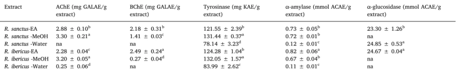
We investigated the inhibitory activities of the ethyl acetate, me-thanol, and water extracts of R. sanctus and R. ibericus against enzymes related to Alzheimer's disease, skin hyperpigmentation complications, and diabetes type 2. As shown inTable 4, the ethyl acetate and me-thanol extracts inhibited both acetyl and butyryl cholinesterase. The development of cholinesterase inhibitors is still the most popular clin-ical strategy targeted for the management of Alzheimer's disease. In fact, in the brain of Alzheimer's disease patients, the abnormal low level of acetylcholine has been related to pathological features of Alzheimer's disease, particularly cognitive decline (Li et al., 2018). R. coreanus ethanol extract showed inhibitory activity against acetylcholinesterase
in vitro and exerted memory ameliorating effects in vivo (Kim et al.,
2013).
The ability of the extracts to inhibit tyrosinase was also established. In general, the ethyl acetate (121.55 and 124.28 mg KAE/g extract, for R. sanctus and R. ibericus, respectively) and methanol (131.44 and 132.05 mg KAE/g extract, for R. sanctus and R. ibericus, respectively) extracts of the studied Rubus species showed potent inhibitory action against tyrosinase. Plant extracts showing inhibitory activity towards skin-regulating enzymes, such as tyrosinase, are considered as pro-mising candidates for the development of dermatological treatments and cosmetics as skin-whitening agents (Papaioanou et al., 2018).
In the present study, we assessed the ability of R. sanctus and R. ibericus extracts to inhibit the activity of α-amylase and α-glucosidase. The inhibition of carbohydrate hydrolysing enzymes is considered as an interesting therapeutic strategy to control glycaemic level (Zengin et al., 2018). However, it is worth mentioning that the excessive in-hibition of α-amylase has been associated to a number of gastro-intestinal complications caused by undigested food (Uysal et al., 2019). Thus, developing hypoglycaemic agents showing mild or no α-amylase inhibition and potent α-glucosidase inhibitory action is considered as the ideal therapeutic approach to the management of diabetes type 2. In the present study, R. sanctus water extract showed low inhibition against α-amylase (0.12 mmol ACAE/g extract) and prominent in-hibitory action against α-glucosidase (24.85 mmol ACAE/g extract). Data collected from this study support the traditional use of R. sanctus leaves for the treatment of diabetes type 2 (Süntar et al., 2011).
We performed further statistical analysis to understand any re-lationship between total bioactive components and biological activities. As presented inFig. 1, we observed strong correlation between total phenolic and antioxidant properties. It might be suggested that phe-nolics in the tested extracts were responsible for the observed anti-oxidant activities. However, weak correlation was noted between phenolics and enzyme inhibitory effects. In this sense, non-phenolic inhibitors could be attributed to observed enzyme inhibitory effects. Apparently, the extracts were divided depending on species in sPLS-DA analysis. Also, VIP values are higher than 1 for total flavonoid content, phosphomolybdenum and metal chelating assays, which are main parameters to divide the extracts as well as species.
3.5. Biological assays
As a preliminary approach to evaluate potential toxicity, the ethyl acetate, methanol, and water of the selected Rubus species, (0.1–100 mg/mL) were tested on brine shrimp lethality assay. Artemia salina Leach is commonly used to investigate toxicological activities of plant extracts (Ohikhena et al., 2016). The evaluation of Rubus extract toxicity revealed LC50values in the range 2.52–5.12 mg/mL.
Based on LC50values recorded, a concentration of 500 μg/mL was chosen for subsequent assessment on human colon cancer-derived HCT116 cell using the MTT test. The tested extracts (10–500 μg/mL) confirmed a good biocompatibility, as revealed by the null effect on cell line viability in the range (10–100 μg/mL). On the other hand, at the highest tested concentration (500 μg/mL) cell viability decreased under the limit of biocompatibility (viability ≥ 70%).
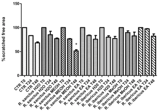
Furthermore, the effect of Rubus extracts on spontaneous HCT116 cell migration, up to 48 h after scratching stimulus, was as-sessed. Results revealed that most of the extracts were ineffective in modulating spontaneous HCT116 cell migration. By contrast, R. sanctus methanol extract significantly inhibited spontaneous cell migration thus suggesting a potential protective effect against migration and in-vasion capacities of HCT116 human colon cancer cells (Fig. 2).
A subsequent panel of experiments was performed on isolated rat colon specimens challenged with LPS, a validated ex vivo experimental paradigm to evaluate the efficacy of drugs and extracts on oxidative and inflammatory pathways involved in ulcerative colitis (Locatelli et al., 2017; Menghini et al., 2016, 2018). Overproduction of reactive oxygen/nitrogen species (ROS/RNS) has long been considered to play a key in tissue damage through disruptive peroxidation reactions on macromolecules, including proteins, lipids, and nucleic acids (Uttara et al., 2009). Particularly, lipid peroxidation has been long involved in tissue chronic inflammatory diseases (Achitei et al., 2013). The role of ROS/RNS, mainly synthesized by activated macrophages and neu-trophils, include neutrophils recruitment at the inflamed tissues (Fialkow et al., 2007;Kruidenier and Verspaget, 2002). To this regard, the assessment of tissue nitrite level is a useful marker of nitric oxide (NO) synthesis, which is an indicator of disease activity in ulcerative colitis (Goggins et al., 2001). NO is a free radical which can react with Table
3 Antioxidant activities of Rubus sanctus and R. ibericus extracts. Extract DPPH (mg TE/g extract) ABTS (mg TE/g extract) CUPRAC (mg TE/g extract) FRAP (mg TE/g extract) Phosphomolybdenum (mmol TE/g) Metal chelating ability (mg EDTAE/g) R. sanctus -EA 24.12 ± 0.81 f 28.19 ± 2.64 e 71.08 ± 6.40 f 26.81 ± 2.33 e 1.65 ± 0.03 f 46.91 ± 2.46 c R. sanctus -MeOH 347.61 ± 13.21 d 279.95 ± 11.13 d 456.23 ± 5.56 d 245.93 ± 5.44 d 2.50 ± 0.12 d 39.68 ± 2.46 d R. sanctus -Water 386.39 ± 10.97 c 543.68 ± 14.28 b 762.96 ± 2.95 b 486.85 ± 3.24 b 3.05 ± 0.04 c 52.20 ± 0.19 b R. ibericus -EA 37.37 ± 0.66 e 34.15 ± 4.53 e 91.42 ± 1.45 e 32.89 ± 2.41 e 1.97 ± 0.04 e 47.30 ± 1.32 c R. ibericus -MeOH 487.60 ± 0.93 a 483.51 ± 7.25 c 681.88 ± 5.44 c 416.13 ± 14.69 c 3.92 ± 0.10 b 53.27 ± 0.14 b R. ibericus -Water 453.74 ± 11.99 b 663.40 ± 12.58 a 921.92 ± 6.85 a 616.63 ± 7.12 a 4.52 ± 0.06 a 59.83 ± 0.52 a Values expressed are means ± S.D. of three parallel measurements. TE: Trolox equivalent; EDTAE: EDTA equivalent; EA: Ethyl acetate; MeOH: Methanol. Different superscripts indicate differences among the extracts (p < 0.05).
multiple tissue biomolecules, thus giving oxidation products including nitrite, nitrate, nitrosyl (NO-heme) species, and Se and N-nitroso pro-ducts. The level of these NO-related products reflects the nitrosative stress due to inflammation-induced upregulation of the inducible NO synthase (iNOS) (Saijo et al., 2010).
R. sanctus methanol and ethyl acetate extracts were equally able to reduce LPS-induced nitrite level in isolated colon (Fig. 3), while sulfa-salazine resulted ineffective in downregulating nitrite levels. The null effect of sulfasalazine on nitrite level corroborates with the recent findings byCetin et al. (2017)which observed a null effect displayed by sulfasalazine on nitrosative stress pathway, evaluated as nitrite level. R. sanctus methanol and R. ibericus ethyl acetate extracts were also able to reduce colon MDA levels, upregulated by LPS challenging (Fig. 4). The extracts were as effective as sulfasalazine, which was able to restore basal MDA level in isolated rat colon challenged with LPS, according to the recent findings bySoliman et al. (2019). Consistent with the effect
on MDA level, the extracts reduced LDH level on rat colon, showing activity as effective as sulfasalazine (Fig. 5). LDH could be considered a predictive marker of tissue damage, especially in the gut, and reduced LDH activity following extracts treatment was related to protective ef-fects in IBDs (Kannan and Guruvayoorappan, 2013;Nagarjun et al., 2017). Actually, the downregulation of nitrite, MDA and LDH level induced by the extracts is consistent with their total phenol and fla-vonoid content (Raihan et al., 2009). The relative abundance in kaempferol could explain, albeit partially, the major blunting effect exerted by R. sanctus methanol extract on LPS-induced nitrite, MDA and LDH level, in isolated rat colon..
5-HT pro-inflammatory role in ulcerative colitis has been previously suggested (Regmi et al., 2014), possibly involving the activation of 5-HT3 receptors (Mousavizadeh et al., 2009). Previously, it was observed that antioxidant and anti-inflammatory chamomile and devil's claw extracts reduced 5-HT steady state level, in rat colon challenged with Table 4
Enzyme inhibitory properties of Rubus sanctus and R. ibericus extracts.
Extract AChE (mg GALAE/g
extract) BChE (mg GALAE/gextract) Tyrosinase (mg KAE/gextract) α-amylase (mmol ACAE/gextract) α-glucosidase (mmol ACAE/gextract)
R. sanctus-EA 2.88 ± 0.10b 2.18 ± 0.31b 121.55 ± 2.39b 0.73 ± 0.05b 23.30 ± 1.26b R. sanctus -MeOH 3.30 ± 0.21a 1.41 ± 0.03c 131.44 ± 0.37a 0.72 ± 0.01b na R. sanctus -Water na na 78.14 ± 3.23d 0.12 ± 0.01c 24.85 ± 0.53a R. ibericus-EA 2.28 ± 0.04c 2.49 ± 0.24a 124.28 ± 1.04b 0.82 ± 0.06a 24.67 ± 0.04a R. ibericus -MeOH 3.20 ± 0.05a 0.27 ± 0.04d 132.05 ± 1.57a 0.67 ± 0.04b na R. ibericus -Water 0.25 ± 0.06d na 83.99 ± 2.62c 0.11 ± 0.01c na
Values expressed are means ± S.D. of three parallel measurements. GALAE: Galatamine equivalent; KAE: Kojic acid equivalent; ACAE: Acarbose equivalent; na: not active; EA: Ethyl acetate; MeOH: Methanol. Different superscripts indicate differences among the extracts (p < 0.05).
Fig. 1. Statistical Evaluation (A: Relationship between total bioactive compounds and biological activities; B: Clustering of extracts in according to biological activities and total bioactive components based on Heatmap; C: sPLS-DA results obtained from biologicals activities of the tested extracts; D: Influence of 13 variables (total bioactive components and biologicals activities) for the total map (VIP variable importance in the prediction).
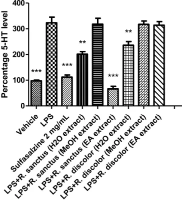
LPS (Menghini et al., 2016). Several studies confirmed that steady state tissue 5-HT concentration also proved to be a valuable index of neu-rotransmitter activity, including synthesis and release (Bungo et al., 2009;Clark et al., 2006). Aqueous extracts from each species revealed equally effective in blunting LPS-induced 5-HT steady state levels, in rat colon (Fig. 6). Additionally, R. sanctus ethyl acetate extract revealed to be more effective than sulfasalazine (Menghini et al., 2016). The in-hibitory effects exerted by R. sanctus and R. ibericus extracts could be
related to multiple concomitant mechanisms. On one side, the phenol and flavonoid content could reduce 5-HT level as a result of the anti-oxidant activity. On the other side, the inhibitory effect on 5-HT activity could be induced by multiple components of flavonoid fraction which could reduce 5-HT release and antagonize pro-inflammatory 5-HT3 Fig. 2. Effect of MeOH, water and EA extracts (100 μg/mL) of R. ibericus and R. sanctus on spontaneous HCT116 cell migration (wound healing test). Data are means ± SD of three experiments performed in triplicate. ANOVA, P < 0.05; post-hoc, *P < 0.05 vs. CTR48.
Fig. 3. Effect of MeOH, water and EA extracts (100 μg/mL) of R. ibericus and R.
sanctus on LPS-induced nitrite level (mmoL/g wet tissue) in rat colon specimens
(N = 5 per group). ANOVA, P < 0.0001; post-hoc, ***P < 0.001 vs. LPS. Fig. 4. Effect of MeOH, water and EA extracts (100 μg/mL) of R. ibericus and R.
sanctus on LPS-induced malondialdehyde (MDA) production in rat colon tissues
challenged with LPS (N = 5 per group). ANOVA, P < 0.001; post-hoc, **P < 0.01 vs. LPS.
mediated-pathway. (Chen et al., 2002;Herbrechter et al., 2015). 4. Conclusion
Results of the present investigation revealed the potential of the selected Rubus species as effective enzyme inhibitors and antioxidant agents. Besides, findings of this study highlight the importance of sol-vent choice in the extraction of bioactive compounds from plants. The water extracts showed high phenolic content and antioxidant activity while the ethyl acetate and methanol extracts of R. sanctus and R. ibericus showed potent enzyme inhibitory activity. In the quest for safer hypoglycaemic agents, R. sanctus water extract revealed to be a pro-mising candidate, showing low amylase inhibition and prominent α-glucosidase inhibitory activity.
On the other hand, R. sanctus methanol extract showed anti-in-flammatory activity in colon cells, showing significant blunting effects on LPS-induced levels of well-established markers of oxidative stress and tissue damage such as nitrites, MDA, and LDH. Besides, R. sanctus methanol extract displayed a significant inhibition of spontaneous mi-gration of HCT116 cell line, thus suggesting a potential protective effect against migration and invasion capacities of human colon cancer cells. Further studies are warranted to isolate and characterize bioactive compounds present in the studied Rubus extracts for the development of novel nutraceuticals, pharmaceuticals and/or cosmetics.
Conflicts of interest
The authors declare that there are no conflicts of interest.
Acknowledgements
The study was supported by Italian Ministry of University (FAR grants): FAR 2018 granted to Prof. Claudio Ferrante; FAR 2017 granted to Prof. Giustino Orlando.
Appendix A. Supplementary data
Supplementary data to this article can be found online athttps:// doi.org/10.1016/j.fct.2019.03.041.
References
Achitei, D., Ciobica, A., Balan, G., Gologan, E., Stanciu, C., Stefanescu, G., 2013. Different profile of peripheral antioxidant enzymes and lipid peroxidation in active and
non-active inflammatory bowel disease patients. Dig. Dis. Sci. 58, 1244–1249.
Akkol, E.K., Süntar, I., Ilhan, M., Aras, E., 2015. In vitro enzyme inhibitory effects of
Rubus sanctus Schreber and its active metabolite as a function of wound healing
ac-tivity. J. Herb. Med. 5, 207–210.
Amzad Hossain, M., Shah, M.D., 2015. A study on the total phenols content and anti-oxidant activity of essential oil and different solvent extracts of endemic plant
Merremia borneensis. Arab. J. Chem. 8, 66–71.
Apak, R., Güclü, K., Özyürek, M., Celik, S.E., 2008. Mechanism of antioxidant capacity assays and the CUPRAC (cupric ion reducing antioxidant capacity) assay. Microchim.
Acta 160, 413–419.
Bakar, A., Fadzelly, M., Ismail, N.A., Isha, A., Ling, M., Lee, A., 2016. Phytochemical composition and biological activities of selected wild berries (Rubus moluccanus L., R.
fraxinifolius Poir., and R. alpestris Blume). J. Evid. Based Complementary Altern. Med.
2016, 1–10.
Bhandary, B., Lee, H.Y., Back, H.I., Park, S.H., Kim, M.G., Kwon, J.W., Song, J.Y., Lee, H.K., Kim, H.R., Chae, S.W., Chae, H.J., 2012. Immature Rubus coreanus shows a free radical-scavenging effect and inhibits cholesterol synthesis and secretion in liver
cells. Ind. J. Pharm. Sci. 74, 211–216.
Brunetti, L., Leone, S., Orlando, G., Ferrante, C., Recinella, L., Chiavaroli, A., Di Nisio, C., Fig. 5. Effect of MeOH, water and EA extracts (100 μg/mL) of R. ibericus and R.
sanctus on LPS-induced lactate dehydrogenase (LDH) activity in rat colon
spe-cimens (N = 5 per group). ANOVA, P < 0.001; post-hoc, **P < 0.01, ***P < 0.001 vs. LPS.
Fig. 6. Effect of MeOH, water and EA extracts (100 μg/mL) of R. ibericus and R.
sanctus on serotonin (5-HT) level (ng/mg wet tissue) in rat colon specimens
challenged with LPS (N = 5 per group). ANOVA, P < 0.001; post-hoc, **P < 0.01, ***P < 0.001 vs. LPS.