A study on the
of gasteroid fungi
collected from Trabzon
Ertuğrul SESLİ *, Gabriel MORENO , Alberto ALTÉS
Department of Biology Education, Karadeniz Technical University, Trabzon, TURKEY
Dpto. Biología Vegetal, Universidad de Alcalá, 28871 Alcalá de Henares (Madrid), SPAIN 1 2 2 1 2 Abstract Key Words Özet Anahtar Kelimeler
Fruiting bodies were collected in Trabzon province in 2009 and 2010. Macroscopic and studies were performed on the dried specimens. Surface structures of the spores of
Pers. ( Corda), Pers. and Pers.
( Chevall.) were illuminated under . Color photos were taken in the field and new localities were determined. New descriptions of the Turkish collections were given and the spore structures of them were ilustrated for the first time.
: Gasteroid fungi, Scanning electron microscope, taxonomy, Trabzon, Turkish Mycota
Fruktifikasyon organları 2009 ve 2010 Yıllarında Trabzon il sınırları içerisinden toplandı. Makroskopik ve mikroskobik çalışmalar kurutulmuş örnekler üzerinde gerçekleştirildi.
Pers. ( Corda), Pers. ve Pers. (
Chevall.) sporlarının yüzey yapıları taramalı elektron mikroskobu altında aydınlatıldı. Renkli resimler arazide çekildi ve yeni lokaliteler saptandı. Türkiye'den toplanan örneklerin yeni tanımları verildi ve spor yapıları ilk kez aydınlatıldı.
: Gasteromycetes, Taramalı elektron mikroskobu, taksonomi, Trabzon, Türkiye Mikotası
Geastrum rufescens Geastraceae Lycoperdon molle L. umbrinum Agaricaceae
Geastrum rufescens Geastraceae Lycoperdon molle L. umbrinum Agaricaceae
Trabzon'dan toplanan gasteroid mantarların spor morfolojisi
üzerinde bir çalışma
spore morphology
microscopic
scanning electron microscope
Introduction
Nowadays it is known that gasteromycetes, those fungi better known as puffballs, earthstars, stinkhorns, bird's nest fungi, false truffles, and gastroid agarics, is really an artificial group included in Basidiomycota. The most known fungi in this group are the puffballs and earthstars which they bear spores in a case. The fruiting body of earthstar is roundish to oval when young and its outer peridium splits into starlike segments. The inner peridium is a sac which sometimes is borne on a stalk. The sterile tissue inside the inner peridium is called columella (Sunhede, 1989).
Scanning electron microscopic studies of spores are very important for the identification of gasteromycetes, as we can confirm in the genus
(Moreno et al., 1997).
Tulostoma
The aim of the present study was to illuminate the spore morphology of
Pers., Pers. and
Pers. under Scanning electron microscope. Although, these fungi have been reported for Turkish mycota, this is the first detailed study including short descriptions, photos, localities and surface structure of the spores.
Geastrum rufescens Lycoperdon molle L. umbrinum
Materials and methods
Descriptions
Material studied
The materials were collected in Trabzon province of Turkey in 2009 and 2010. The collections were deposited in a personal fungarium in the Karadeniz Technical University and some duplicates in the Herbarium of the Universidad de Alcalá, Spain (AH). Author names and fungal names were given according to Index Fungorum (www.indexfungorum.org) a n d M y c o b a n k ( w w w. m y c o b a n k . o r g ) . Microscopical studies and preparation of the specimens for light microscopy were made according to Clémençon (2009). Microscopic characters (i.e. spore size, which includes ornamentation) were observed with a Nikon Eclipse 80i on material mounted in Hoyer's medium. Scanning electron microscope studies of spores were made with a Zeiss DSM-950 (Universidad de Alcalá). The spores of dried specimens were rehydrated with 50% ammonium hydroxide for 24 h, fixed in 3% glutardialdehyde in water, dehydrated in a series of aqueous ethanol solutions of increasing concentration (70%, 80%, 90% and 100%) for 15 min in each, and thereafter immersed in acetone for at least 2 h. The spores were then critical point dried, deposited onto an aluminium stub, and coated with gold-palladium in a Polaron E-5000 sputter coater for 120 sec at 1.4 kV and 18 mA in an argon atmosphere creating an approximately 500 Å thick metal coating.
Identifications were made according to Breitenbach & Kränzlin (1986), Sunhede
(1997), Sarasini (2005) and Calonge (1998).
Pers. 1801
(Syn; Vittadini 1842 =
Vittadini 1842)
:Sevinç village (Maçka
–Trabzon), decayed log,
26.07.2010 ( 2788); ibidem, 15.08.2009
( 2676).
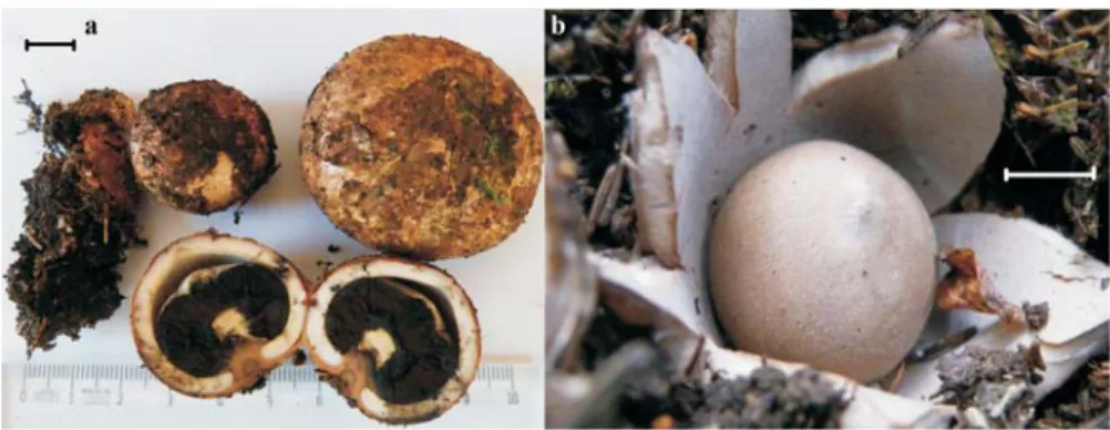
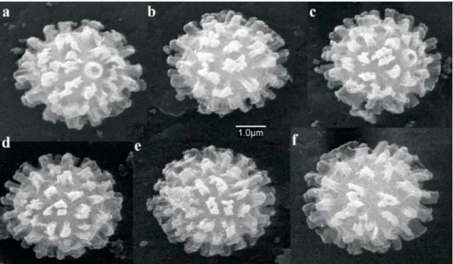
Fruiting bodies 3–8 cm diam (Figure 1), consist of a well developed outer and inner peridium, even in the immature collection. The thick fleshed outer peridium is pale flesh colored when young and it turns pink brown or reddish in mature. The inner peridium which contains the gleba is spherical, 3 cm diam, short stalked and shows hirsute surface (Sunhede, 1997; Breitenbach and Kränzlin, 1986). We found out that the material shows a well developed columella around which are radially arranged the capillitium and the powdery mass of spores. Spores verrucose, 3-6 µm diam. Under scanning electron microscope the spore ornamentation appears formed by numerous columnar processes, some anastomosed (Figure 2), very similar of those observed by Sunhede (1997).
is common in Turkey. The total number of species of
recorded in Turkey to date is 17 (Sesli & Denchev, 2008). Geastrum rufescens G. schaefferi G. vulgatum Picea orientalis Sesli Sesli Geastrum rufescens Geastrum
Figure 1. Fruiting bodies of , showing different degrees of maturation: a. 2788; b. 2676 (bars: 1 cm)
Geastrum rufescens Sesli Sesli
Figure 2. Spore ornamentation of under Scanning electron microscope
( 2788)
G. rufescens Sesli
Lycoperdon molle Pers. 1801
: Karaçam (Akçaabat –
Trabzon), under , 17.09. 2010
( 2911).
Gregarious, fruiting bodies spherical to prolonged, 3–8 cm high, and spore sacs 3–6 cm diam. Exoperidium granulose and persistent; bluish colored in the young basidiocarps, changing to brown color when mature (the material was collected immature in the field, and matured during the drying process in the lab)
(Figure 3).
scanning electron microscope the ornamentation is formed by rather cylindrical warts, with flattened apex (Figure
.
The genus is common in
Turkey. The total number of species recorded in Turkey to date is about 18 (Sesli & Denchev, 2008).
Material studied
Picea orientalis Sesli
Lycoperdon
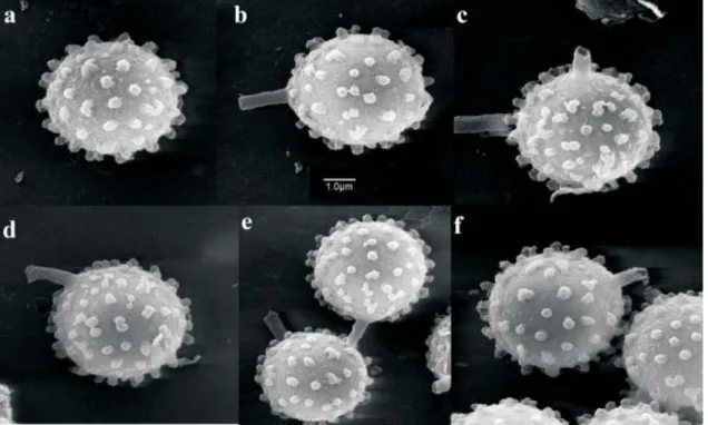
Subgleba well developed, chambered, olivaceous. Spores verrucose, 5-6,5 μm diam. Under
4). Capillitium with pits of aprox. 0,5 μm diam; abundant sterigmata debris present
Figure 4. Spore ornamentation of under Scanning electron microscope ( 2911)
Lycoperdon molle Sesli
Lycoperdon umbrinum Pers.:Pers.
1801
: Hıdırnebi (Akçaabat
–Trabzon), under , 15.10.2010
( 3018).
Fruiting bodies spherical, tuberous or pyriform (Figure 5), 8 cm high, and spore sacs 7 cm diam. Exoperidium formed by connivent spines, regularly arranged. Stoma well delimited, circular. Subgleba well developed, chambered. Gleba olivaceous. Spores 4,5-5,5
µm diam. Under Scanning electron microscope the spore ornamentation is formed by less developed and more separated verrucae than
those of 2911 (Figure 6).
Capillitium scantily branched and apparently not septate, with pits of about 0,5 µm diam; some sterigmata debris in the gleba.
belongs to the morphological species complex, together with , but they are well distinguished by molecular analysis (Larsson & Jeppson, 2008).
Material studied Picea orientalis Sesli L. molle Sesli Lycoperdon umbrinum L. molle L. lambinonii
Figure 5. Fruiting bodies ofL. umbrinum(a.Sesli3018, b.Sesli3038; bars: 2 cm)
Figure 6. Spore ornamentation of under Scanning electron microscope ( 3018)
Lycoperdon umbrinum Sesli
Acknowledgements
This research was financially supported by the Karadeniz Technical University (Scientific Research Projects: 2009.116.002.2 and 2009.116.002.6). We thank Prof.Dr. Orhan Aydın and Nuran Ertuğrul for their assist, and Antonio Priego of the Universidad de Alcalá Electron Microscopy facility for his collaboration.
References
Breitenbach J., Kränzlin F. (eds.), , vols.2., Verlag Mykologia, Switzerland (1986). Calonge F.D.,
. Vol. 3. J.Cramer, 271 p., Stuttgart (1998). Clémençon, H.,
. Eching: IHW Verlag, Berchtesgaden (2009). Larsson, E., Jeppson, M.,
. Mycol. Res. 112: 4-22 (2008). Moreno, G., Altés, A., Ochoa, C., Wright, J.E.,
Mycol. Res. 101: 957-965 (1997).
Sarasini M., . Associazione Micologica Bresadola, 406p., Trento (2005). Sesli E., Denchev C.M.,
Mycotaxon 106: 65-67 (2008) + [complete version, 1-145, new version uploaded in January 2013]. Sunhede S.,
. Synopsis Fungorum 1. Fungiflora . 535 pp., Oslo (1989).
Fungi of Switzerland
Flora Micológica Ibérica. Gasteromycetes I. Lycoperdales, Nidulariales, Phallales, Sclerodermatales, Tulostomatales
Methods for working with macrofungi: Laboratory cultivation and preparation of larger fungi for light microscopy
Phylogenetic relationships among species and genera of Lycoperdaceae based on ITS and LSU sequence data from north European taxa
Notes on type materials of Tulostoma. Some species with mixed holotypes.
Gastermiceti Epigei
Checklists of the myxomycetes, larger ascomycetes, and larger basidiomycetes in Turkey. Geastraceae (Basidiomycotina). Morphology, ecology and systematics with special emphasis on the North European species