Journal of Cukurova University Faculty of Medicine
Radiographical Variations of the Normal Female Urethra:
Classification with Voiding Cystourethrography
Normal Kadın Üretrasının Radyografik Varyasyonları: Voiding Sistoüretrografi
ile Klasifikasyon
Bahattin Ulu
1, Elif Karadeli
2, Orhan
Özbek
3, Ahmet
Özturk
2, Dilek Kösehan
4,
Şükriye
Yavuzer
2, Emin Alp Niron
21 Samsun Gazi State Hospital, Department of Urology, SAMSUN 2 Baskent Unıversity, Dapartment of Radiology, ANKARA 3 Selçuk University, Deparment of Radiology, KONYA 4 Fatih University, Department of Radiology, İSTANBUL
Çukurova Üniversitesi Tıp Fakültesi Dergisi (Journal of Cukurova University Faculty of Medicine) 2012; 37(2):98-102
ABSTRACT
Objective: Female urethra can be visualized in different anatomical shapes during voiding cystourethrography.We presented the
different types of urethral shapes which we encountered and to correlate our larger series with literature.
Materials and methods: The VCU examinations of pediatric patients for the investigation of mainly vesicoureteral reflux or other urinary
system pathologies mostly referred with the prediagnosis of urinary tract infections and enuresis nocturna were retrospectively evaluated. 381 patients without any micturation symptoms and with normal urethral meatus underwent 426 vesicoureteral reflux examinations. The shapes of urethra were described and then classified according to the sort of shapes appeared during vesicoureteral reflux examination.
Results: We identified six different types of urethral morphology. The most common configuration in our study was cylindirical shaped
urethra (n=170) (44%). These were subclassified as thick (n=38) (9.9%), thin (n=25) (6.5%), long (n=65) (17%) and short (n=42) (11%). Others were goblet (n=69) (19%), fusiform (n=66) (17%), spinning top (n=44) (12%), wedge (n=27) (7%), and beaded (n=5) (1%) shaped urethras.
Conclusion: During the vesicoureteral reflux procedure female urethra can be visualised in diverse shapes. Radiologist and
pediatricians must become familiar to the normal anatomical shapes of female urethra for not to interpret it as pathological.
Keywords: female urethra,voiding cystourethrography, radiographical variations
ÖZET
Amaç: Kadın üretrası voiding sistoüretrografisi boyunca farklı anatomik şekiller de görülebilir. Biz, literatürle bizim serimizi korele etmek
ve bizim karşılaştığımız üretra şekillerinin farklı tiplerini sunduk.
Yöntem: Enüresis nokturna ve üriner sistem enfeksiyonları nedeniyle esas olarak vesikoüreteral reflü veya diğer üriner sistem
patolojilerini araştırmak için çoçuk hastaların voiding sistoüretrografileri retrospektif olarak değerlendirildi. Normal üretral meatuslu, herhangi bir miksiyon semptomu olmaksızın 381 hasta 426 VSU tetkiki ile değerlendirildi. Üretra şekillleri tanımlandı, Voiding sistoüretrografisi tetkiki boyunca görünen üretra şekillerinin tiplerine göre sınıflandırıldı.
Bulgular: Biz 6 farklı üretra tipi tanımladık. En sık konfigürasyon silindirik şekilli üretraydı (n=170) (%44). Bunlar kalın (n=38) (%9.9), ince (n=25) (% 6.5, uzun (n=65)(%17) ve kısa (n=42) (%11) olarak alt gruplara ayrıldı. Diğerleri kadeh (n=69) (%19), iğsi (n=66) (%17), fırıldak (n=44) (%12), kama (n=27) (%7), ve boncuk (n=5) (%1) şeklinde üretralardır .
Sonuç: Kadın üretrasını voiding sistoüretrografisi incelemesi boyunca farklı şekillerde görebiliriz. Radyologlar ve çoçuk doktorları kadın
üretrasının normal anatomik şekillerine patolojik durumları ayırt edebilmek için alışık olmalıdırlar.
INTRODUCTION
Female urethra can be visualized in different
anatomical shapes during voiding
cystourethrography (VCU), most likely because of its anatomical properties. Different shapes of female urethra can cause some difficulty of interpretations by the unexperienced radiologist and particularly by pediatricians. Whitaker firstly described the different shapes of urethra in girls as cylindirical, fusiform, arrowhead, wine glass, conic and strand shaped1. The aim of this study is to present the different types of urethral shapes which we encountered and to correlate our larger series with literature.
MATERIALS and METHODS
Our series comprises the pediatric patients who underwent VCU examination in our hospital between May 1996 and May 2006 for the investigation of mainly vesicourethral reflux (VUR) or other urinary system pathologies mostly referred with the prediagnosis of urinary tract infections (UTI) and enuresis nocturna. Some of children in our study had renal failure and had VCU examination in pretransplant evaluation. Those patients whom the urethral meatal pathologies detected during clinical examination or urethral
catheterization were excluded from the study. 381 patients without any micturation symptoms and with normal urethral meatus underwent 426 VCU examinations. 31 patients underwent more than one examination. The age of patients ranged between 3 months to 17 years. Mean age was 6 years. The shapes of urethra were described and then classified according to the sort of shapes appeared during VCU examination. VUR was graded from I to V.
RESULTS
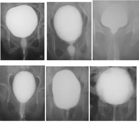
Radiographically we identified six different types of urethral morphology and classified them as cylindirical (long, short, thick or thin), fusiform, spinning top, wedge, goblet and beaded shaped in our series. 31 patients had more than one examination and showed the same type of uretheral configuration in each examination. The most common configuration in our study was cylindirical shaped urethra (n=170) (44%). These were subclassified as thick (n=38) (9.9%), thin (n=25) (6.5%), long (n=65) (17%) and short (n=42) (11%). Others were goblet (n=69) (19%), fusiform (n=66) (17%), spinning top (n=44) (12%), wedge (n=27) (7%), and beaded (n=5) (1%) shaped urethras (Figure 1).
Figure legends
Figure 1 (a-i). The diverse shapes of female urethra seen during micturation in VCU examination. a. Beaded, b.
Goblet, c. Wedge, d. Fusiform, e. Spining top, f. Cylindirical (thin), g. Cylindirical (thick), h. Cylindirical (short), i. Cylindirical (long) shaped.
DISCUSSION
Female urethra is not surrounded by dense resisting structures, as in the male, which admits of considerable dilatation2. Most likely due to this anatomical properties the caliber and configuration of normal female urethra can be influenced by the intramural pressure and flow volume of running urine. These factors may result in a wide range of normal urethral shape. Due to this there is no general accepted classification concerning the configurations of female urethra during VCU2,3. Whitaker and coworkers classified the shapes of urethra as cylindirical, fusiform, arrowhead, wine glass, conic and strand shaped in his study with 60
the urethral flow resistance and the shapes of urethra and found that 7 patients with goblet and strand shaped urethras had urine flow with high resistance1. Pompino and coworkers examined the correlation between meatal anomalies (stenosis, short urethra, anterior prolongation, asymmetry) and urethral configuration. They found that anomalies of the meatus cannot be related to variations of the urethral shapes as seen in the micturating cystourethrogram. They can however be the cause of enuresis, chronic urinary infection, urethrovaginal reflux, and disturbances in micturation. The most common configurations of urethra seen during VCU in their study was strand,
bladder trabeculation, and urethral notching (beading) were accepted as suspicious signs. Although these radiological signs have been detected in half the patients with detrusor instability, they also occurred in three of eight children with a stable bladder3. The most striking shape of urethra is the spinning top urethra in literature5. This term is used to describe the widened posterior urethra seen mainly in girls. In literature the association of spinning top urethra and bladder instability was investigated in many studies. It was found that if no bladder instability can be found, spinning top urethra should be considered to be a normal variant3,4,5. In our series we encountered 44 ‘spinning top’ urethra. In addition Batista found that beaded shape of urethra (urethral notching) was also one of the suspicious sign of bladder instability besides spinning top urethra6. In our series clinical findings were mainly related to UTI and purpose of doing VCU was to rule out VUR and other urinary tract abnormalities. None of the patient in our series having any sign or symptoms of detrusor instability. Due to the absence of clinical indications no urodynamic study was done to any of our patients. In 298 out of 381 patients VCU’s were within normal limits. 83 patients showed VUR in different grades. According to the literature and our series female urethras may be seen in different shapes normally during VCU’s. Urethral shapes that we encountered in our series were more or less similar to those of uretheral configuration desribed in literature. Our classification of the urethral shapes were as cylindirical (long, short, thick or thin), fusiform, spinning top, wedge, goblet and beaded. Normal male urethra is seen its classical anatomical shape during VCU. But this is not the
case for normal female urethra. This is most likely due to absence of rigid supporting structures around it like male urethra. This may cause different types of normal dilatation while micturations.
VCU is still most commonly employed radiological examination for the detection of UTI and VUR in children. During the procedure urethra is usually visualised and evaluated. Radiologist and pediatricians must become familiar to the shapes of female urethra for not to interpret it as pathological.
Conflict of interesting and funding; We
evaluated radiographical variations of female urethra with VCU and presented the different types of urethral shapes which we encountered and to correlate our larger series with literature. We did not use funding.
REFERENCES
1. Whitaker J, Johnston GS. Correlation of urethral resistance and shape in girls. Radiology. 1968; 91:457-61.
2. John A. Corocco. Gray’s Anatomy, Bounty Books, 15th edition, Newyork 1977, female urethra, 1007. 3. Hausegger KA, Fotter R, Sorantin E, Schmidt P.
Urtehral morphology and bladder instability. Pediatr Radiol. 1991; 21:278-280.
4. Pompino HJ, Hoffmann D.Anomalies of the external urethral orifice in girls. Prog Pediatr Surg. 1984; 17:49-56.
5. Dogra PN, Ansari MS. Spining top urethra and lower urinary tract dysfunction in a young female. Scientific World Journal. 2004; 7(4): 108-10.
6. Batista JE, Caffaratti J, Arano P, Regalado R, Garat JM. The reliability of cysto-urethrographic signs in the diagnosis of detrusor instability in children. Br J Urol. 1998; 81(6): 900-4.
Yazışma Adresi / Address for Correspondence:
Doç. Dr. Elif Karadeli Başkent University Deparment of Radiology Fevzi Çakmak Cad. No: 45 Bahçelievler/ANKARA Phone number: 0 90 312 2126868 Fax number: 0 90 312 2237333 e-mail: [email protected] geliş tarihi/received :27.04.2012 kabul tarihi/accepted:23.05.2012