INVITED AUTHOR
Yumuşak Dokunun Estetik Tedavi Planındaki Yeri Korkud DEMİREL
ORIGINAL ARTICLES
Effect of Tooth Whitening (Bleaching) Agent on Dentin Microhardness Sema YILDIRIM, H. Miray UYAN
Evaluation of Microtensile Bond Strenght of Conventional and New Generation Flowable Composite Resins to Dentin With Different Adhesive Systems
Buket AYNA, Emrah AYNA, Sema ÇELENK, Behiye BOLGÜL, Ersin UYSAL CASE REPORTS
Minimally Invasive Restoration of Restoration of Erosive Lesions With Direct composıte laminate veneers: A Case Report
Mağrur KAZAK
Rubber Dam Clamp Ingestion During Root Canal Treatment: A Case Report Ece ÇALIŞKAN, Hafize Hande GÜRBÜZ, Rüştü DAĞLAROĞLU, Raif ERIŞEN A Multidisciplinary Approach to Localized Gingival Recession: A Case Report
Burcu KARADUMAN, Cenker KOYUNCUOĞLU, Sevda ATALAY, Ece ÇALIŞKAN, Nurcan TEZCİ, Sabri Hasan MERİÇ Resin Nano Ceramic Endocrown: An Alternative to Post-Core Supported Crowns
Işıl Kaya BÜYÜKBAYRAM, Gülce ALP REVIEWS
Endodontic Systems Working With Reciprocal Movements for Instrumentation Of Root Canals Selçuk GÖKYAY, Ece ÇALIŞKAN
DİŞ HEKİMLİĞİ FAKÜLTESİ DERGİSİ
JOURNAL OF THE FACULTY OF DENTISTRY
JOURNAL OF THE FACULTY OF DENTISTRY
AYDIN DENTAL
ISTANBUL AYDIN UNIVERSITY
JOURNAL OF THE FACULTY OF DENTISTRY
AYDIN DENTAL
Ahu URAZ, Gazi University, Turkey Arzu ATAY, Gulhane Military Medical Academy, Turkey Aylin BAYSAN, The London School of Medicine and Dentistry, London, U.K.
Behçet EROL, Istanbul Aydın University, Turkey Bora ÖZDEN, Ondokuz Mayıs University, Turkey
Can DÖRTER, Istanbul University, Turkey Cansu ALPASLAN, Gazi University, Turkey Cem TANYEL, Istanbul University, Turkey Cemal ERONAT, Ege University, Izmir, Turkey Didem ÖNER ÖZDAŞ, Istanbul Aydın University, Turkey
ECE ÇALIŞKAN, Istanbul Aydın University, Turkey Elif KALYONCUOĞLU, Ondokuz Mayıs University, Turkey
Esra SOMTÜRK, Istanbul Aydın University, Turkey Feyza OTAN ÖZDEN, Ondokuz Mayıs University, Turkey Fulya TOKSOY TOPÇU, Gulhane Military Medical Academy, Turkey Günseli GÜVEN POLAT, Gulhane Military Medical Academy, Turkey
Hakan ÖZBAŞ, Istanbul University, Turkey Handan ERSEV, Istanbul University, Turkey
Hasan Sabri MERİÇ, Istanbul Aydın University, Turkey Kadriye DEMİRKAYA,Gulhane Military Medical Academy, Turkey
Kemal SÜBAY, Istanbul University, Turkey Korkud DEMİREL, Istanbul University, Turkey Leyla KURU, Marmara University, Istanbul, Turkey
Raif ERİŞEN, Istanbul University, Turkey Rezzan ÖZER, Dicle University, Turkey Rüdiger JUNKER, Danube Private University, Austria
Sema BELLİ, Selçuk University, Turkey Sema ÇELENK, Dicle University, Turkey Sercan KÜÇÜKKURT, Istanbul Aydın University, Turkey
Serdar CINTAN, Istanbul University, Turkey Simel AYYILDIZ, Gulhane Military Medical Academy, Turkey Şeniz KARAÇAY, Gulhane Military Medical Academy, Turkey
Şölen GÜNAL, Istanbul Aydın University, Turkey Ümit KARAÇAYLI, Gulhane Military Medical Academy, Turkey
Vesela STEFANOVA, Medical University of Plovdiv, Bulgaria Sahibi - Proprietor
Mustafa Aydın
Yazı İşleri Müdürü - Editor-in-Chief Nigar Çelik
Editör - Editor
Jülide Özen Yayın Kurulu - Associate Editors Seda YILMAZ
Esra YÜCE Sercan KÜÇÜKKURT
Academic Studies Coordination Office (ASCO)
İdari Koordinatör - Administrative Coordinator Nazan Özgür
Teknik Editör - Technical Editor Hakan Terzi
Dili - Language English - Türkçe
Yayın Periyodu - Publication Period Published twice a year
April and October
Yazışma Adresi - Correspondence Address Beşyol Mahallesi, İnönü Caddesi, No: 38 Sefaköy, 34295 Küçükçekmece/İstanbul Tel: 0212 4441428 - Fax: 0212 425 57 97 web: www.aydin.edu.tr - e-mail: [email protected] Baskı - Printed by
Matsis Matbaacılık, Teyfikbey Mahallesi, Dr.Ali Demir Caddesi No: 51 34290 Sefaköy/İSTANBUL Tel: 0212 624 21 11 - Fax: 0212 624 21 17
e-mail: [email protected]
ISSN: 2149-5572
EDITORIAL BOARD
İstanbul Aydın Üniversitesi, Diş Hekimliği Fakültesi, Aydın Dental Dergisi özgün bilimsel araştırmalar ile uygulama çalışmalarına yer veren ve bu niteliği ile hem araştırmacılara hem de uygulamadaki akademisyenlere seslen-meyi amaçlayan hakem sistemini kullanan bir dergidir.
Istanbul Aydın University, Journal of The Faculty of Dentistry, Aydın Dental is A Double-Blind Peer-Reviewed Journal Which Provides A Platform For Publication Of Original Scientific Research And Applied Practice Studies. Positioned As A Vehicle For Academics And Practitioners To Share Field Research, The Journal Aims To Appeal To Both Researchers And Academicians.
INVITED AUTHOR Yumuşak Dokunun Estetik Tedavi Planındaki Yeri
Korkud DEMİREL... 1 ORIGINAL ARTICLES
Effect of Tooth Whitening (Bleaching) Agent on Dentin Microhardness
Sema YILDIRIM, H. Miray UYAN... 7 Evaluation of Microtensile Bond Strenght of Conventional and New Generation Flowable Composite
Resins to Dentin With Different Adhesive Systems
Buket AYNA, Emrah AYNA, Sema ÇELENK, Behiye BOLGÜL, Ersin UYSAL... 15 CASE REPORTS
Minimally Invasive Restoration of Restoration of Erosive Lesions With Direct Composite Laminate Veneers: A Case Report
Mağrur KAZAK... 25 Rubber Dam Clamp Ingestion During Root Canal Treatment: A Case Report
Ece ÇALIŞKAN, Hafize Hande GÜRBÜZ, Rüştü DAĞLAROĞLU, Raif ERIŞEN... 31 A Multidisciplinary Approach to Localized Gingival Recession: A Case Report
Burcu KARADUMAN, Cenker KOYUNCUOĞLU, Sevda ATALAY, Ece ÇALIŞKAN, Nurcan TEZCİ, Sabri Hasan MERİÇ... 35 Resin Nano Ceramic Endocrown: An Alternative to Post-Core Supported Crowns
Işıl Kaya BÜYÜKBAYRAM, Gülce ALP... 43 REVIEWS
Endodontic Systems Working With Reciprocal Movements for Instrumentation Of Root Canals
Sayın Aydın Dental Journal okurları,
2015 yılında Mütevelli Heyeti Başkanımız Sn. Dr. Mustafa AYDIN, Rektörümüz Sn. Prof. Dr. Yadigar İZMİRLİ ve Dekanımız Sn. Prof. Dr. Sabri Hasan MERİÇ himayesinde bilimsel araştırma ve yayın ilkelerine bağlı olarak yayınlanacak olan Aydın Dental Journal dergisi yılda iki kez Türkçe ve İngilizce dilinde olmak üzere kesintisiz olarak yayın hayatına başlamıştır.
Günümüzde diş hekimliği, son teknolojiye ve gelişen bilime dayalı, devamlılık arz eden bir bilim dalı haline gelmiştir. Bu gelişmeler doğrultusunda, dergimiz diş hekimliği biliminde yurt dışından ve yurt içinden akademisyenlerin ve diş hekimlerinin kendi araştırmalarını ve deneyimlerini paylaşabilecekleri bir ortam yaratacaktır. Hedefimiz kısa süre içinde geniş katılımlarla yüksek bilimsel seviyeye ulaşıp, dergimizin uluslararası indekslerde yer almasını sağlamak ve üniversitemizin adını bu alanda da duyurmaktır.
Gelecek sayılarımızda davetli yazar, derleme, özgün araştırma ve olgu raporlarının yayımlanmasını sağlayarak geniş katılımlarla yüksek bilimsel seviyeye ulaşmayı amaçlamaktayız.
Derginin yayım aşamasında bilim kurulunda yer alarak engin mesleki bilgi ve tecrübeleri ile yüksek bilimsel kalitede araştırmaların yayımlanabilmesinde destek veren çok değerli hocalarımıza, yayım aşamasında desteklerini esirgemeyen Sn. Prof. Dr. Ahmet Metin GER ve ekibine ve aynı zamanda tüm akademisyen arkadaşlarıma teşekkür eder, dergimizin yayın hayatının başarılı olmasını dilerim.
Saygılarımla, Prof. Dr. Jülide ÖZEN Aydın Dental Journal Editörü
Dear Aydin Dental Journal Readers,
The Journal of Aydin Dental, as being sticked to scientific and inquisitive publication principles, has just started to be published bi-annually under the auspices of our honorable Head of Trustees Dr. Mustafa Aydın, President Prof. Dr. Yadigar Izmirli and Dean Prof.Dr. Sabri Hasan Meric. The dentistry has become a scientific field of study which complies with current technological developments and requires sustainment. In the light of these recent developments, our journal will create an opportunity to the academicians, either from Turkey or abroad, to share their studies and outcome of researches about the dentistry. Our goal is to reach the highest scientific level by achieving wide-ranging participation as soon as possible which will enable our journal to be listed among international indices journals and also contribute to the recognition of the Istanbul Aydin University worldwide.
We aim to publish the articles of various guest authors, compilations, peculiar researches and case reports which will lead us to highest scientific levels through a widespread participation in our future issues.
I appreciate the contributions of our valuable teachers who strived vigorously for the publication of high level scientific quality Journal of Aydin Dental by being part of the scientific committee during the publication period through their immense knowledge and experience. Also, special thanks to our honorable teacher Prof. Dr. Ahmet Metin Ger and his team members including whole academician colleagues who contributed to the journal. I wish a successful, long lasting publication lifespan to our journal.
Best regards, Prof. Dr. Jülide ÖZEN Editor of Aydin Dental Journal
Yumuşak Dokunun Estetik Tedavi Planındaki Yeri
Korkud DEMİREL
1Estetik açıdan periodontal değerlendirme:
“Sağlam kafa sağlam vücutta bulunur’’1 deyişi gibi estetik değerlendirme yapabilmek için önce
dokuların sağlıklı olması gerekmektedir. Diş eti kanamaları, mikrobiyal dental plak varlığı ve Diş etinde iltihabi süreçten kaynaklanan renk ve yapı değişiklikleri bir taraftan estetik beklentilerin karşılanmasını olanaksız kılmakta, diğer taraftan elde edilecek olası başarının zaman içerisinde korunabilmesini imkansız hale getirmektedir. Estetik değerlendirmelere geçilmeden önce söz konusu bölgede sondalanabilir cep derinliklerinin vestibül ve palatinal yüzlerde 3mm yi geçmiyor olması, ara yüzlerde ise en fazla 4mm olması gerekmektedir. Diş eti sağlığının değerlendirilmesinde ikinci aşamada sondalama sonrası cep içerisinden kanama olmaması ve Diş eti kenarında renk değişikliği ve kanama bulunmaması gerekmektedir. Periodontal hastalığın olduğu durumlarda estetik değerlendirmelere geçilmeden önce hastalığın ortadan kaldırılması gerektiği unutulmamalıdır.
Dudakların sınırları ve gülme hattının seviyesi:
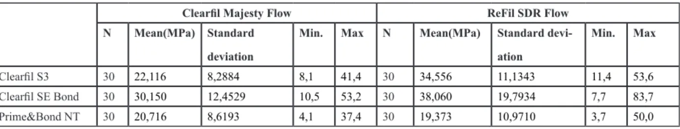
Normal gülme hattı kavramı gülümseme sırasında üst dudağın konumu belirler. Haliyle Diş eti kenarının konumu gülümseme sırasında görülecek Diş eti miktarını etkileyecektir. Gülümseme sırasında orta keser ve köpek dişlerinde diş eti kenarının dudak tarafından örtülmesi ve yan keserlerde 1-2mm Diş eti kenarının açığa çıkması normal olarak kabul edilir (Resim 1a). Gülme hattının zaman içerisinde apikale doğru konum değiştirdiği ve normal olan bir gülme hattının zaman içerisinde düşük gülme hattına (Resim1 b) dönüşeceği unutulmamalıdır. Gülümseme sırasında orta keser ve köpek dişlerinde diş etlerinin ortaya çıkması ise yüksek gülme hattı olarak isimlendirilir (Resim 1c).
Diş etinin sınırları, simetrisi ve biyotipi:
Klinik kuronun en tepe noktasına Zenith noktası denilmekte ve bu nokta her zaman klinik kuronun mesiyal-distal yönde orta noktasına denk gelmemektedir 2 (Resim 1). Diş eti kenarının oluşturduğu
çizgi orta hat rehber alındığında simetri oluşturmaktadır ve simetri orta hatta yaklaşıldığında
1 (Prof.Dr.), İstanbul Üniversitesi, Dişhekimliği Fakültesi, Periodontoloji [email protected]
Resim 1. a,b,c: Gülme hattının belirlenmesinde Vermillon hattı ile üst ön bölgede serbest diş eti kenarının ilişkisi
Yumuşak Dokunun Estetik Tedavi Planındaki Yeri
2
daha fazla önem kazanmaktadır. Diş eti kenarı biyotipine bağlı olarak derin kavisler çizebilir veya daha ziyade düz bir çizgiyi andırabilir. Diş etinin biyotipi doku kalınlığının ölçülmesi ile belirlenir ve ince, normal, kalın olmak üzere alt gruplara ayrılır (Resim 1). Diş etinin biyotipi estetik uygulamalarda önemli olduğu kadar diş etinin sert fırçalama, subgingival kuron kenarı sonlanması veya kötü ağız bakımı gibi kronik bir uyarana vereceği yanıtın tahmin edilmesinde de önem taşımaktadır. İnce biyotipe sahip bölgelerde doku kronik irritasyona her zaman diş eti çekilmesi ile yanıt verir.
İnterdental papil:
İnterdental papil alveol kemiği, dişlerin ara yüz duvarları ve birbirleri ile temas noktaları tarafından oluşturulan boşluğu dolduran diş eti dokusudur. Ön dişlerde papilin varlığı dişler arasındaki temas noktası ve alveol kemiği arasındaki mesafe ile yakından ilişkilidir 3 (Şekil 2). Özellikle protetik
uygulamalarda biyolojik sınırları zorlamadan papilin eksik olduğu bölgelerde interdental alanın daraltılmasıyla papilin boşluğu doldurması sağlanabilir.
Resim 1. Klinik kuronun en tepe noktasına Zenith noktası denilmektedir. Şekilde siyah çizgi ile işaretlenen bu
noktalar orta keser dişlerde ve kaninlerde klinik kuronun orta noktasına denk gelmemekte, sadece yan keser dişlerde her iki nokta çakışmaktadır.
Diş etlerinde sık karşılaşılan estetik sorunlar:
Estetik sorunların ortaya çıkmaması için restoratif işlemlerde öncelikle biyolojik prensiplere dikkat edilmeli ve bireyin ağız bakımı işlemlerini gereğince yapması sağlanmalıdır. Ancak kötü alışkanlıklar, aksamış ağız bakımı, iatrojenik nedenler ve gelişimsel nedenlerle de dişetlerinin ve dolayısı ile ağızın görüntüsü istenmeyen değişimler göstermiş olabilir. Bu estetik sorunların tedavisinde etyolojinin doğru saptanması tedaviden elde edilecek yararı arttıracaktır. Diş etlerinin fazla görünmesi gelişimsel nedenlere iyi bir örnek oluştururken diş eti ve papil kayıpları zararlı alışkanlıklar ve kötü ağız bakımı sınıfına girmektedir.
Diş etlerinin fazla görünmesi:
Dişetlerinin gülümseme sırasında fazlasıyla görünmesinin nedenleri incelendiğinde dört ana başlık dikkat çekmektedir.
Üst dudak yetersizliği, fazla gelişmiş üst çene, gecikmiş pasif sürme, dikey boyut kompensasyon sürmesi. Bunların arasında gecikmiş pasif sürme ve dikey boyut kompensasyon sürmesi kuron boyu uzatma işlemleri ile değişen miktarlarda ortadan kaldırılabilir. İskeletsel bir sorun olan maksillanın fazla büyük olması dikey yüz oranlarını etkilemekte ve orta yüz yüksekliğinin artmış olması ile karakterizedir. Bu bireylerde sadece gülme sırasında dişetleri görünmekle kalmayıp dinlenme halinde ve konuşurken de dişetleri dikkat çekmektedir.
İskeletsel bir sorun olması nedeni ile tedavisinde ortodontik ve ortognatik cerrahi yaklaşımlar önem kazanmaktadır. Kuron boyu uzatma işlemleri ve protetik işlemler ile düzeltilmesi olanaksızdır. Dişlerdeki malpozisyonlar dişetlerinin fazla görünmesinde diğer bir nedendir ve yaygın olduğu olgularda ortodontik tedavi doğru yaklaşım olarak kabul edilmektedir. Ancak bazı olgularda özellikle tek dişi ilgilendiren minör malpozisyonlarda kuron boyu uzatma işlemi ve protetik uygulamalarla sınırlı yarar elde edilebilir.
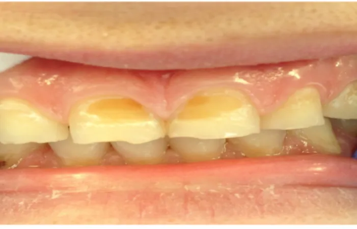
Yüksek gülme hattının en sık karşılaşılan nedenleri arasında gecikmiş pasif sürme değerlendirilmelidir. Aktif sürme tamamlandıktan sonra diş eti kenarı apikale doğru yer değiştirir ve mine sement sınırından 1 mm kuronalde veya tam sınır üzerinde nihai konumuna yerleşir 4. Bazı
bireylerde aktif sürmenin tamamlanmasına karşın diş eti kenarının bu apikale doğru migrasyonu gerçekleşmez. Sonuç olarak bu bireylerde dişetlerinin fazla görünmesi estetik soruna yol açar (Resim 2).
Resim 2. Tip1 gecikmiş pasif sürme olgusu. Dişlerde kare görüntü, iltihap olmamasına karşın diş eti kenarında
Yumuşak Dokunun Estetik Tedavi Planındaki Yeri
4
Gecikmiş pasif sürme sonucu gerçekleşen bu estetik sorunun tedavisinde kuron boyu uzatma cerrahisi uygulanmaktadır. Gecikmiş pasif sürme iki klinik tipe ayrılmaktadır 5. Her iki tipte de
diş eti kenarı mine sement sınırının kuronalinde yer almakla birlikte Tip 1 gecikmiş pasif sürme olgularında geniş bir yapışık diş eti şeridi olmasına karşın Tip 2 olgularında mukogingival sınırın kuronalde yer alması nedeniyle yapışık Diş eti alanı daralmakta ve cerrahi işlem sırasında yapışık diş eti miktarının korunmasına özen göstermek gerekmektedir.
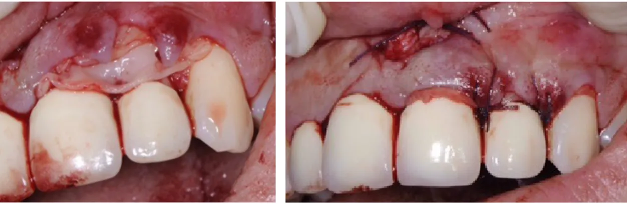
Diş eti çekilmeleri:
Diş eti çekilmelerinin etyolojisinde kronik mekanik travma olabildiği gibi periodontal hastalık sonucunda da diş eti çekilmeleri ile karşı karşıya kalınmaktadır. Etyolojisinden bağımsız olarak diş eti çekilmelerinin sınıflamasında kullanılan bir sistem6 hem çekilmenin niteliğini belirlemekte,
hem de tedaviden sonra çekilmenin ne kadar kapatılacağı konusunda bilgi vermektedir (Resim 3).
Diş eti çekilmelerinin tedavisinde kullanılan yöntemler arasında saplı ve sapsız greft operasyonları, yönlendirilmiş doku rejenerasyonu ve rezorbe olan biyomateryaller yaygın şekilde kullanılmaktadır. Kullanılan yöntemler arasında subepitelyal bağ dokusu greftinin başarısı en yüksek olarak belirlenmekte ve doku renk uyumunun en iyi olduğu bilinmektedir 7-8 (Resim 4).
Resim 3. Diş eti çekilmelerinin sınıflaması:
a) Tip1. Diş eti çekilmesi mukogingival sınıra ulaşmamıştır ve interdental alanda yumuşak doku ve alveol kemiği kaybı
görülmemektedir. Bu tip çekilmelerde kök yüzeyinin cerrahi yöntemlerle tamamen kapatılması beklenir.
b) Tip 2. Diş eti çekilmesi mukogingival sınıra kadar ulaşmış veya geçmiştir ancak interdental alanda yumuşak doku ve alveol
kemiği kaybı görülmemektedir. Bu tip çekilmelerde de kök yüzeyinin cerrahi yöntemlerle tamamen kapatılması beklenir.
c) Tip 3. Diş eti çekilmesi mukogingival sınıra kadar ulaşmış veya geçmiştir. İnterdental alanda yumuşak doku veya alveol kemiği
kaybı mevcuttur veya kök yüzeyinin örtülmesini engelleyebilecek kadar konum bozukluğu vardır. Bu tip çekilmelerde de kök yüzeyinin cerrahi yöntemlerle tamamen kapatılması beklenmez ve kısmi örtülme elde edilebilir.
d) Diş eti çekilmesi mukogingival sınıra kadar ulaşmış veya geçmiştir. İnterdental alanda yumuşak doku veya alveol kemiği
kaybı görülmekte ve/veya kök yüzeyinin örtülmesine engel miktarda dişte konum bozukluğu vardır. Bu tip çekilmelerde de kök yüzeyinin cerrahi yöntemlerle kapatılması beklenmez.
İnterdental papil kayıpları:
Estetik periodontal zorluklar arasında en önemli yeri tutan ve aynı zamanda da tedavisi en güç olan sorunların başında papil kayıpları gelmektedir. Papil kayıplarının etyolojisinde periodontal hastalıklar başı çekmekte ardından hatalı protetik uygulamalar ve hastanın kürdan kullanmak gibi kötü alışkanlıkları gelmektedir. Papil kayıplarının ortadan kaldırılmasında periodontal veya protetik yöntemlerden biri veya her ikisinin beraber kullanılması tercih edilebilir (Resim 5).
Periodontal teknikler içerisinde papilin hacmini arttırmaya yönelik sapsız doku greftleri kullanılırken alveol kemiğin hacmini arttırmaya yönelik kemik dokusu greftleri nadir de olsa kullanılmaktadır.
Resim 4. Tip 2 Diş eti çekilmesi. Her ne kadar çekilme mukogingival sınırı aşmış olsa da interdental alanda
yumuşak ve sert doku kaybı olmaması tedavide tam başarıyı mümkün kılmıştır. Subepitelyal bağ dokusu grefti ile örtülen kök yüzeyi tedaviden 7 yıl sonra da, 15 yıl sonra da diş eti ile örtülü kalmış ve diş eti konumu hiç
değişmemiştir.
Resim 5. Papil kayıplarının tedavisinde sıklıkla subepitelyal bağ dokusu greftleri kullanılmasına karşın başarı
6
KAYNAKÇA
[1] Atatürk MK, https://www.tbmm.gov.tr/kultursanat/me_ata_soz.htm 27 Aüustos 2015. [2] Allen EP. Surgical crown lengthening for function and esthetics. Dent. Clin. North Am.
1993; 37: 163-179.
[3] Tarnow DP, Magner AW, Fletcher P. The effect of the distance from the contact point to the crest of bone on the prescence and asbsence of the interproximal dental papilla. J Periodontol 1992;63: 995-996.
[4] Itoiz ME, Carranza FA. The gingiva. In: Carranza FA, Newman MG. Clinical periodontology 8th ed. Philadelphia: WB Saunders, 1996:12-29.
[5] Coslet JG, Vanarsdall RL, Weisgold A.. Diagnosis and classification of delayed passive eruption of the dentogingival junction in the adult. Alpha Omegan 1977; 70: 24-28 [6] Miller PD. A classification of marginal tissue recession. Int J Periodont. Rest Dent.
1985 ; 5: 8-13.
[7] Efeoğlu A, Demirel K, Okan E: Diş eti çekilmelerinin tedavisinde kullanılan cerrahi yöntemlerin değerlendirilmesi Academic Dental 2001; 3: 20-26.
[8] Yaman D, Demirel K, Aksu S, Basegmez C. Treatment of Multiple Adjacent Miller Class III Gingival Recessions with a Modified Tunnel Technique: A Case Series. Int J Periodontics Restorative Dent. 2015 Jul-Aug;35(4):489-97. doi: 10.11607/prd.2049.
Effect of Tooth Whitening (Bleaching) Agent on Dentin
Microhardness
H. Miray UYAN
1Sema YILDIRIM
2Abstract
Objectives: The aim of this study is to investigate the effect of whitening agents on dentin micro hardness.
Materials and methods: 60-incisor maxillary (20 per group) tooth were applied root chanel treatment: one is control group and the others are applied to whitening products namely, whiteness super endo (Dentscare LTDA;37% Carbamide peroxide) and opalescence endo(Ultradent,USA;35% Hydrogen peroxide). Whitening agents were applied in every four days for 12 days. Hardness assessed by using Vickers test, after the end of treatment. Scanning electron microscopy (SEM) analyses performed after the end of bleaching treatment.
Results: The results show that the micro hardness decreased for both agents; where as, the micro hardness of whiteness superendo is less than opalescence endo.
Keywords: dentin micro hardness, intracoronal bleaching, whitening agent
Diş Beyazlatma Ürünlerinin Dentin Mikrosertliğine Etkileri Özet
Amaç: Bu çalışmanın amacı diş beyazlatıcı ürünlerin dentin mikrosertleşmeleri üzerine etkilerini incelemektir.
Materyel ve Yöntemler: 60-kesici maksiller (grup başına 20 adet) dişe kanal tedavisi uygulanmıştır. Bir tanesi kontrol olmak üzere, diğerlerine sırasıyla şu beyazlatıcı ürünler uygulanmıştır. whiteness super endo (Dentscare LTDA;37% Karbamid peroksit) ve opalescence endo (Ultradent,USA;35% Hidrojen peroksit). Beyazlatıcı ürünler 12 gün süreyle, her 4 günde bir kez uygulanmıştır. Tedavinin sonunda sertleşme Vickers testi ile değerlendirilmiştir. Beyazlatıcı tedavinin sonunda Taramalı elektron mikroskobu ile (TEM) değerlendirmeler yapılmıştır.
Sonuçlar: Sonuçlar, her iki ürünün de mikro sertleşmeleri azalttığını göstermekle birlikte; whiteness superendo’ nun mikro sertleşmeleri opalescence endoya göre daha azdır.
Anahtar Kelimeler: Dentin mikro sertleşmeler, intrakoronal beyazlatma, beyazlatıcı ürün
1 (DDS, PhD.) İstanbul Medipol Üniversitesi, Diş Hekimliği Fakültesi Endodonti Ana Bilim Dalı,
Effect of Tooth Whitening (Bleaching) Agent on Dentin Microhardness
8
1. Introduction
The whiteness of the teeth is always an essential element of health and esthetics (1). Since the importance of esthetic considerations increasing nowadays, giving the natural appearance of the tooth to the patient by restoration has been one of the most important issues of modern dentistry (2).
Various factors can be the cause of coloration of the teeth. This situation occur direct discoloration of enamel dentin or reflection of discoloration of dentin from semi-transparent enamel (3). There are wide variety of chemical agent were used for many years to treat the discoloration of enamel dentin (4).
The bleaching agents, a more conservative method, has become a good alternative for restoration the natural color of stained teeth, instead of prosthetic applications, such as; crown and veneer. Since often usage of tooth whitening applications, there are many clinical studies has made in order to evaluate the efficacy, reliability and potential effect this method (5).
Whitening treatment is a method that aimed to reaction of the free oxygen, which emerge from whitening agents with colored molecules in order to return the natural color of the teeth(6). The most common materials used for bleaching; hydrogen peroxide, sodium perborate, and carbamide peroxide which is usually used for extra coronary bleaching where as; sodium perborate is also used for the intracoronary bleaching.
It is believed that the bleaching also causes increased brittleness of the coronal tooth structure because of desiccation of the dentin and enamel(7). However, this has not been proven conclusively because little is known about the effect of bleaching on the biomechanical properties of teeth. Since dentin constitutes a major part of the tooth structure, any change in the biomechanical properties of dentin after bleaching is likely to have an impact on the overall strength of the tooth (8).
Many studies have looked at the various biomechanical properties of dentin and some of more commonly studied properties include micro hardness and strength properties, such as tensile strength and shear strength(9). A number of recent studies have compared the biomechanical properties of dentin from vital teeth. The results of these studies indicated that endodontically treated teeth were not weaker than vital teeth. In contrast, the number of studies that examined the biomechanical properties of bleached dentin is very limited. Lewinstein et al. examined the effect of hydrogen peroxide and sodium perborate on the micro hardness of human dentin. Intact teeth were sectioned longitudinally and bleaching agents were applied to the polished dentin surfaces for up to 30 min. It was found that 30% hydrogen peroxide reduced the micro hardness of dentin after 5 min, but treatment with sodium perborate mixed with hydrogen peroxide did not alter the micro hardness of dentin at the of the observation period(10).
This study examined the effects of different bleaching agents on dentin and it is aimed to compare the results with each other.
2. Materials and Methods
Sixty intact human maxillary incisors extracted for orthodontic reasons were used. The teeth were placed in distilled water and the soft tissue attached to the root surface was removed with a scalpel. Endodontic access cavities were prepared using a diamond bur in a high-speed hand piece. The pulp tissue was removed ana a no.15 h type file was inserted into the root canal until the tip of the file was seen at the apical foramen. The working length was determined by subtract 1 mm from the length of the file. Cleaning and shaping was carried out using 1% sodium hypochlorite as the irrigant. The root canal was dried with paper points and filling with AH Plus and 15-40 gutha percha points. Cavit was packed into the root canal.
The teeth were randomly distributed into 3 groups, and applied different whitening agents, (Table1) Group 1, opalescense endo, 35% hydrogen peroxide -containing; Group 2, Whiteness super endo %37 carbamide peroxide-containing; Group 3, cotton pellet soaked with distilled water. Each tooth was stored in an individually labeled, capped plastic vial containing distilled water. The plastic vials were stored at 37 C for 12 days. After 12 days, the teeth were sectioned and dentine from the teeth was subjected to microhardness testing.
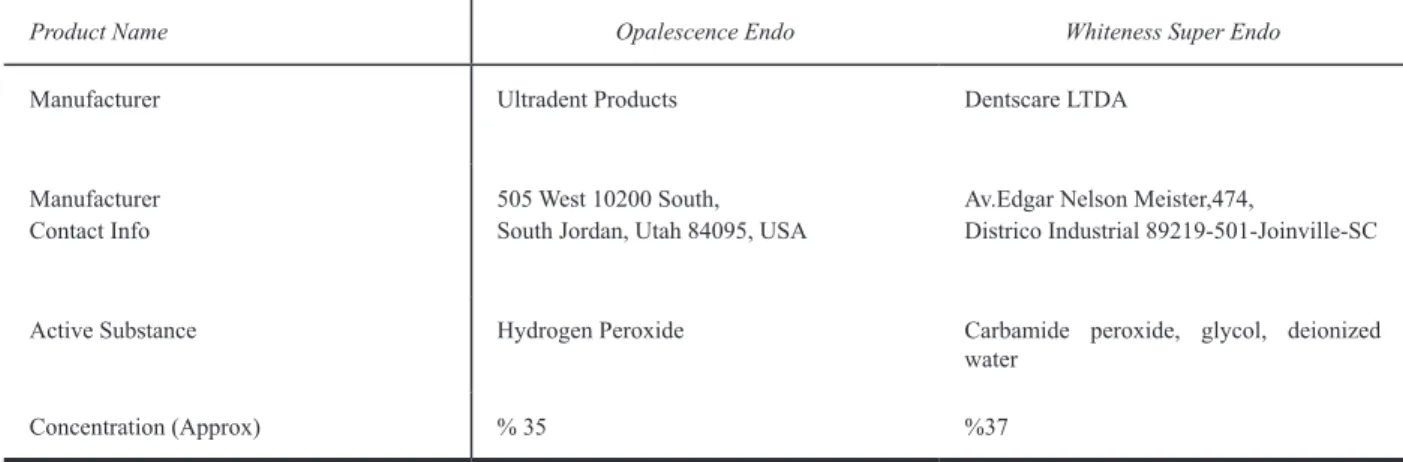
Table 1. Bleaching Agents
Product Name Opalescence Endo Whiteness Super Endo
Manufacturer Ultradent Products Dentscare LTDA
Manufacturer
Contact Info 505 West 10200 South, South Jordan, Utah 84095, USA Av.Edgar Nelson Meister,474,Districo Industrial 89219-501-Joinville-SC
Active Substance Hydrogen Peroxide Carbamide peroxide, glycol, deionized
water
Concentration (Approx) % 35 %37
The root of each was embedded in a block of acrylic resin, 2mm apical to the CEJ to facilitate subsequent sectioning and testing of the specimens. Each specimen was blotted dry and a vickers hardness test was performed using a digital variance was conducted on the data obtained. Data comparisons were conducted using Mann Whitney U tests. All statistical analyses were performed at the 0,05 level of significance.
During the experimental procedures, bleaching products were removed by suction and washed thoroughly with distilled, de-ionized water. After the bleaching procedures, samples were fixed with 2% glutaraldehyde, washed in distilled, de-ionized water, critical point dried and sputter coated with gold (120 s, 70 nm) for SEM analysis. Photomicrographs (3500-x magnification, 15 kV) were obtained from the samples and compared with group 3 by a single assessor, evaluating qualitatively the dentin surface for alterations.
Effect of Tooth Whitening (Bleaching) Agent on Dentin Microhardness
10
3. Results
According to the statistical analysis, the microhardness of the control group was higher than both Opalescence Endo and Whiteness Super endo, where as; the maximum reduction in microhardness has noticed in the Whiteness Super Endo group.
The result, which is obtained by evaluation of dentin microhardness, is the proof of mineral loss or earnings on the hard tissue of tooth. There is a positive correlation between microhardness and mineral structure of teeth. Although reduction in the microhardness could decrease the fragility, when it comes to fragility of teeth there are many other factors, which should take in to consideration. Reduction in microhardness due to mineral loss in dentin, can cause weaken teeth and increase the tendency to tooth decay which is the major cause of increased the fragility of the teeth.
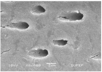
Figure 1. Control Group
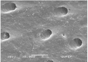
Figure 3. Whiteness Super endo Group
It is observed that, a deep erosion area formed on the dentine tubule, due to the effect of bleaching agents, which could be the main reason for mineral loss of dentin and also reduction of microhardness which cause increase of the fragility and tendency of tooth decay.
4. Discussion
Various studies have shown that hydrogen peroxide, especially in high concentration, affects the biomechanical properties of dentine even after exposure for a short period. Saleh & Ettman found that irrigation with 3% hydrogen peroxide and 5% sodium hypochlorite used alternatively and left for 60 s significantly reduced the microhardness of dentine(11).
Lewinstein et al. showed that the reduction in microhardness of dentine after treatment with 30% hydrogen peroxide was time-related. In their study, the specimens were assessed at 5, 15 and 30 min after application of hydrogen peroxide and significant, progressive reduction in microhardness was observed (12).
Hydrogen peroxide and Carbamide peroxide can cause a decrease in dentin microhardness during intracoronal bleaching. In this case, the degradation of morphological structure of the dentin and the deterioration of inorganic structure is an expected result.
Although the effect of whitening agents on the dentin are not fully understood, some studies reveal that, hydrogen peroxide cause the dissolution of the inorganic material of dentin, thus affect decrease in the calcium phosphor ratio, and loss of mineral on the organic matrix of dentin due to protein denaturalization. The hardness of dentin affected from the Ph of the whitening agents, buffer capacity of the dentin, and also the density difference of dentin tubules. Some researchers investigate the effect of whitening agents and conjunction use of these agents with materials that known to be effect on dentin microhardness.
Effect of Tooth Whitening (Bleaching) Agent on Dentin Microhardness
12
Daniel pinto de Oliveira et al. studied on the differentiation of dentin microhardness when applied whitening agents alone and conjunction with chlorhexidine gel and find out that chlorhexidine jel has no effect on dentin microhardness. However, they find out that chlorhexidine gel can be used for an antimicrobial agent in cavity during intracoronal bleaching(13). Chng et al studied the effect of the different whitening agents with different concentration on the dentin microhardness, hydrogen peroxide, sodium perborate and carbamide peroxide with different concentration was used in this study, and results show that the highest rate of reduction on dentin microhardness observed with hydrogen peroxide 35 %, carbamide peroxide 35 %(14). Zalkind et al. had reported that there is a close relationship between the enamel dentin change and bleaching effect. Studies show that, whitening agent can change the mineral composition and also micro morphology of the enamel dentin thus reduction on dentin microhardness.
The linear relationship between the calcium and phosphor loss and reduction on dentin microhardness show that hardness measurements can be used as an indicator for the relationship between mineralization degree of enamel dentin and caries enamel. Mineral loss induced by bleaching agents, occurs under the enamel surface similar to initial caries lesions. This loss of mineral content is seen as an increase in range of enamel prisms, increase in surface roughness and increase of the adhesion of Streptococcus mutants. Hosaya et al reported that streptococcus mutants colonies increased on the whitened enamel dentin, after repeated bleaching sessions bacterial adhesion growth occurred, and the maximum number of bacterial colonies formed after five bleaching sessions followed by etching(15). Caries lesions affecting dentine enamel easily move toward tissues and can cause the deeper cavities. In addition to this, caries lesions formed on the whitened enamel tissue, due to extra coronal bleaching method, can also occur on the dentin tissue during intracoronal bleaching. Intracoronal bleaching application for the teeth, which were root canal treatment, progression of caries lesions is expected without causing pain. This situation would adversely affect restorations made after.
In order to achieve optimal results in the bleaching treatment, the structure and the concentration and also preventable side effect profile of whitening agent is also important. Although has no clinical symptoms, bleaching agents can cause a chemical and micro structural changes which affect the surface properties of teeth, the degree of mineralization, and probably the development of caries-like lesions should not be forgotten.
It is observed that different bleaching agents used in this study decrease the microhardness of dentin tissue. On the contrary, the hardness has been observed higher for none bleached teeth. Although decrease in the microhardness mainly reduces the fragility, in the case of teeth, other factors must be take in to consideration.
Teeth whitening procedure damage the tooth structure, which is irreversible. The fragility of the tooth, is related with the amount of water contained in dentin, mineralization, and change of the collagen and non-collagen proteins. Especially the bleaching agents, that contain peroxide, due to dissolution and decay in the structure of dentin collagen, that weakens the tooth structure, increase the tendency to caries, and thus increases the fragility.
REFERENCES
[1] Hasson H,Ismail AI,Neıva G.Home -based chemically induced whitening of teeth in adults The Cochrane Collaboration 2007.
[2] Goldstein RE,Garber DA.Complete Dental Bleaching Quintessence Pub Co,Chicago,1995. [3] Rotstein I,Friedman S.pH variation among materials used for ıntracoronal bleaching.J Endod 17,376,1991.
[4] Costas FL,Wong M,Intracoronal ısolatin barriers:Effect of location on root leakage and effective ness of bleaching agents.J Endod 17,8,1991.
[5] Cherry DV, Bowers DE,Thomas Jr L,Redmond AF.Acute toxicological effects of ingested tooth whitens in female rats.J Dent Res 1993,72:1298-1303
[6] Freccia WF,et al.An ın vıtro comparison of non vital bleaching techniques in the discolored tooth.J Endod 8,70,1982.
[7] Rotstein I,et al. Histochemical analysis of dental hard tissues following bleaching .J Endod 22,23,1996.
[8] Martin dunitz,greenwall linda.Bleaching technıques in restorative dentistry.2001.20-24 [9] Goldstein RE, Garber DA. Complete dental bleaching.Chıcago: quıntessence
Publishing;1995.165
[10] Lewinstein I, Hirschfeld Z, Stabholz A, Rotstein I. Effect of hidrogen peroxide and sodium perborate on the microhardness of human enamel and dentin. J Endod 20:61-3,1994. [11] Kwong K, Mohammed S, McMillan MD, Stokes AN. Evaluation of a 10 percent carbamide peroxide gel vital bleaching agent. N Z Dent J 1993;89:18-22.
[12] Lewinstein I, Hirschfeld Z, Stabholz A, Rotstein I. Effect of hidrogen peroxide and sodium perborate on the microhardness of human enamel and dentin. J Endod 20:61-3,1994. [13] In vitro assessment of a gel base containing 2% chlorhexidine as a sodium perborate ‘s vehicle for ıntracoronal bleaching of discolored teeth. 2006,32(7),672-674.
[14] Lım MY et all.An in vitro comparison of bleaching efficiacy
[15] Singleton LS; Wagner MJ. Peroxide tooth whitener concentration. Versus composite resin etching. J Dent Res, 71: .281-286, 1992.
Evaluation of Microtensile Bond Strength of Conventional
and New Generation Flowable Composite Resins to Dentın
Wıth Different Adhesive Systems
Buket AYNA
1Emrah AYNA
2Sema ÇELENK
3Behiye BOLGÜL
4Ersin UYSAL
5 AbstractObjective: The purpose of this study was to evaluate the effect of conventional and new generation
two flowable composite resins with three different adhesive systems on bond strenght to dentin by using the microtensile bond test.
Material and Method: Fourty two non-carious human third molars were sectioned parallel to
the occlusal plane to expose occlusal dentin. The dentin surfaces were ground with 600-grid silicon carbide (SIC) paper. Teeth were randomly divided in to six groups (n=7). Group 1-Clearfil S3Bond+ Clearfil Majesty Flow, Group 2-Clearfil SE Bond+Clearfil Majesty Flow, Group
3-Prime&Bond NT +Clearfil Majesty Flow, Group 4-Clearfil S3Bond+ReFil SDR Flow, Gorup
5-Clearfil SE Bond+ReFil SDR Flow, Group 6-Prime&Bond NT+ReFil SDR Flow. The restored teeth were serially sectioned to obtain 1mm2 sticks. Each stick was submitted to the microtensile
test performed at a crosshead speed of 1mm/minute. One-way ANOVA, and Tamhane’s tests were used to compare the data.
Results: The results indicated that Clearfil SE Bond showed higher microtensile bond strength when compared to the other adhesives in Clearfil Majesty Flow group (p< 0.05). Clearfil SE Bond
and Clearfil S3Bond showed similar microtensile bond strength (p>0.05) whereas microtensile bond strength of Prime&Bond NT was significantly decreased (p<0.05). However, in the comparision of the microtensile bond strength values of Clearfil Majesty Flow and ReFil SDR Flow groups adhesive, it was determined a statistically significant difference between two groups for the only Clearfil S3Bond (p<0.05).
Conclusion: In the experimental conditions of this study it was seen that adhesive systems may
have different effects on the bond strenght to dentine tissue.
1 (DDS, PhD, Associate Professor) Department of Pedodontics, Faculty of Dentistry, Dicle University, Corresponding Author:[email protected] 2 (DDS, PhD, Professor) Department of Prosthodontics, Faculty of Dentistry, Dicle University
3 (DDS, PhD, Professor) Department of Pedodontics, Faculty of Dentistry, Dicle University
4 (DDS, PhD, Associate Professor) Department of Pedodontics, Faculty of Dentistry, Mustafa Kemal University 5 (PhD) Vocational High Scholl, Department of Technique, Dicle University
Evaluation of Microtensile Bond Strength of Conventional and New Generation Flowable Composite Resins to Dentın Wıth Different Adhesive Systems
16
Keywords: Flowable composites, adhesive systems, microtensile bond strength, dentin.
Geleneksel ve Yeni Nesil Akışkan Kompozit Rezinlerin Farklı Adeziv Sistemler ile Dentine Mikrogerilim Bağlanma Dayanımlarının Değerlendirilmesi
Özet
Amaç: Bu çalışmanın amacı, geleneksel ve yeni nesil iki akışkan kompozit rezinin üç farklı
adeziv sistem ile dentine bağlanma dayanımı üzerine etkisini mikrogerilim bağlanma dayanımı test yöntemi ile değerlendirmektir.
Gereç ve Yöntem: Kırk iki adet çürüksüz insan üçüncü büyük azı dişin okluzal minesi kesilerek
okluzal dentin açığa çıkarıldı. Dentin yüzeyleri, 600-grid silikon karbid kâğıtla (SIC) zımparalandı. Dişlerden rastgele altı grup oluşturuldu (n=7). Grup 1-Clearfil S3Bond+ Clearfil Majesty Flow,
Grup 2-Clearfil SE Bond+Clearfil Majesty Flow, Grup 3-Prime&Bond NT +Clearfil Majesty Flow, Grup 4-Clearfil S3Bond+ReFil SDR Flow, Grup 5-Clearfil SE Bond+ReFil SDR Flow,
Grup 6-Prime&Bond NT+ReFil SDR Flow. Restore edilen dişler, 1mm2’lik çubuklar elde edilecek
şekilde kesildi. Her bir çubuk, dakikada 1 mm hızla hareket eden cihazla mikrogerilme testine tabi tutuldu. Sonuçların karşılaştırılmasında tek-yönlü ANOVA, t ve Tamhane testleri kullanıldı.
Bulgular: Clearfil Majesty Flow gruplarında Clearfil SE Bond daha yüksek mikrogerilim
bağlanma dayanımı göstermiştir (p< 0.05). ReFil SDR Flow grubunda ise, Clearfil SE Bond ve Clearfil S3Bond’un mikrogerilim bağlanma dayanım değerlerinin, Prime&Bond NT’ye göre
yüksek olduğu bulunurken (p<0.05), kendi aralarında bir fark bulunamadığı belirlenmiştir (p>0.05). Bununla birlikte, Clearfil Majesty Flow ve ReFil SDR Flow gruplarındaki adezivlerin mikrogerilim bağlanma dayanım değerlerinin karşılaştırılmasında yalnızca Clearfil S3Bond için iki
grup arasında istatistiksel olarak anlamlı bir farklılık gösterdiği tespit edilmiştir (p<0.05).
Sonuç: Bu in vitro çalışma koşullarında, adezivlerin dentine bağlanma dayanımı üzerinde farklı etkilere neden olabileceği görüldü.
Anahtar Kelimeler: Akışkan kompozitler, adeziv sistemler, mikrogerilim bağlanma dayanımı,
dentin.
Introduction
In today’s dentistry; increasing emphasis on aesthetic has brought a lot of research which is based on composite resins and adhesives systems.
The importance of the bonding between hard tissue of teeth with dental materials is great at the success of composite resins which has a wide application area in the posterior and anterior region. As opposed to the bonding to the enamel the bonding to the dentine continues to cause problem for dentists due to its tubular structure and the formation of smear layer during cavity preparation.1,2
Composite resins are being bonded to the dental tissues micromechanically by using new generation of adhesive systems with two different techniques; the total-etch and self-etch.3,4 Producers tended
to simplify the three-step total-etch adhesives, and have developed a two-step total-etch system due to difficulty of moisture control and surplus of application steps.5,6 Self-etch adhesives have been
developed to eliminate operator errors which occur during the usage of the total-etch adhesives by reducing the number of phases of the adhesive aplication and eliminating technique sensitivity. The mixing step is eliminated with the self-etch adhesives which have been developed in recent years, resin monomer, photoinitiator, tertiaryamine accelerator was collected in a singe bottle.7,8
The edge compliance has a critical importance for composite resin restoration to continue its performance for a long time. Polymerization shrinkage of the resin restorations can cause accumulation of stress in the bonding surfaces and separation between the tooth surfaces and adhesives. Gaps that are formed between the cavity walls and restoration material, can cause postoperative problems such as sensitivity, pulp damage and recurrent caries.9,10 The application
of flowable composite resins as a thin layer to cavity flour, is one of the proposed method to provide a full sealing between the cavity wall and composite restorations during the polymerization shrinkage.11 In addition, the usage of flowable composite resin under the composite resin, has been
determined to significantly increase the bonding strength.12,13
Researchers have used different test methods to measure the bonding strength of restoration materials and adhesive systems. Sano et al.11 have introduced the microtensile bonding strength test for the
first time in 1994. Stress can be measured in the 0.25 and 1mm² sample size with microtensile bonding strength test. In addition, higher bonding strength and a very low variation coeffient are measured with conventional shear and tensile tests. Thus the more reliable results are expected to occur. Many studies indicated that the bond strength could be measured in different regions and depths of the tooth at and also multiple samples could be tested from the same tooth.11,14-16
The purpose of this study was to evaluate the effect of conventional and new generation two flowable composite resins with three different adhesive systems on bond strenght to dentin by using the microtensile bond test.
Materials and Methods
Fourty two non-carious human third molars extracted with periodontal reasons were used in this study. Teeth were stored in distilled water until to be tested after cleaning tissue debris on the teeth. Occlusal enamel was cut to be perpendicular to the long axis of tooth under water cooling with diamond bur by operating at low speed. 600 grid silicon carbide paper (SIC) was applied to the surface for one minute to obtain homogeneous smear layer on dentin surfaces that was uncovered. Teeth were randomly divided in to six groups (n=7). The adhesive systems and flowable composites which is used in the research are shown in Table 1.
Evaluation of Microtensile Bond Strength of Conventional and New Generation Flowable Composite Resins to Dentın Wıth Different Adhesive Systems
18
Group 1: After Clearfil S3Bond which is one-step self-etch adhesive is applied in accordance with
manufacturer recommendations to dentin surfaces, it was polymerized with LED (HS LED 1500, Henry Schein Inc, USA) light-curing unit.
Table1. The adhesive systems and flowable composites which is used in the research
Materials Type Manufacturer
Clearfil S3 one-step self-etch adhesive Kuraray Medical Inc., Tokyo, Japan
Clearfil SE Bond two-step self-etch adhesive Kuraray Medical Inc., Tokyo, Japan Prime&Bond NT two-step total-etch adhesive Dentsply, Konstanz,Germany
Clearfil Majesty Flow flowable composite Kuraray Medical Inc., Tokyo, Japan
ReFil SDR Flow flowable composite Dentsply, Konstanz,Germany
Group 2: After Clearfil SE Bond which is two-step self-etch adhesive is applied in accordance with manufacturer recommendations to dentin surfaces, it was polymerized with LED light-curing unit. Group 3: After 34% phosphoric acid gel applying to the occlusal dentinal surface, Prime & Bond NT which is the total-etch adhesive is applied in accordance with manufacturer recommendations to dentin surfaces, it was polymerized with LED light-curing unit.
Clearfil Majesty Flow as flowable composite was light cured after being placed with a thickness of 2 mm to the dentin surface which was applied adhesive in all three in the group.
Group 4: After Clearfil S3Bond which is one-step self-etch adhesive is applied in accordance with
manufacturer recommendations to dentin surfaces, it was polymerized with LED light-curing unit. Group 5: After Clearfil SE Bond which is two-step self-etch adhesive is applied in accordance with manufacturer recommendations to dentin surfaces, it was polymerized with LED light-curing unit. Group 6: After 34% phosphoric acid gel applying to the occlusal dentinal surface, Prime & Bond NT which is the total-etch adhesive is applied in accordance with manufacturer recommendations to dentin surfaces, it was polymerized with LED light-curing unit.
ReFil SDR Flow as flowable composite was light cured after being placed with a thickness of 2 mm to the dentin surface which was applied adhesive in all three in the group.
Then the teeth were kept in the incubator for 24 hours in the saline solution. After each storage period, the bonded teeth were vertically sectioned into serial slabs and further into beams with cross-sectional areas of approximately 1 mm2.Totally 30 rod was obtained for each group.
Specimens were attached to microtensile testing apparatus (Micro Tensile Tester, Bisco, USA) with the cyanoacrylate adhesive (Zapit, Dental Ventures of America, Corona, CA, USA) and stressed to failure in tension at a crosshead speed of 1 mm/min. The load recorded in Newtons was retrieved in MPa.
Normal distribution assumption of conformity of the data were analyzed with the Kolmogorov-Smirnow test, it’s homogeneity was examined by Levene test. Statistical differences were examined using ANOVA, Independent Samples test and Tamhane test at a significance level of 5% with SPSS 11.0 for Windows (SPSS Inc., USA)
Results
Microtensile bond strength values of the experimental group are shown in Table 2.
As a result of evaluation of the data that is obtained, a statistically significant difference was observed in the microtensile bond strength values of adhesives that is applied Clearfil Majesty Flow (F=7.825; p=0,001) (Figure 1). In multiple comparison of microtensile bond strength values, bond strength values of Clearfil SE Bond were determined to be statistically significantly higher (p˂0.05). 0 5 10 15 20 25 30 35
Clearfil Majesty Flow
Clearfil S3Bond Clearfil SE Bond Prime&Bond NT
Figure 1. Mean microtensile bond strength values of adhesives that is applied Clearfil Majesty
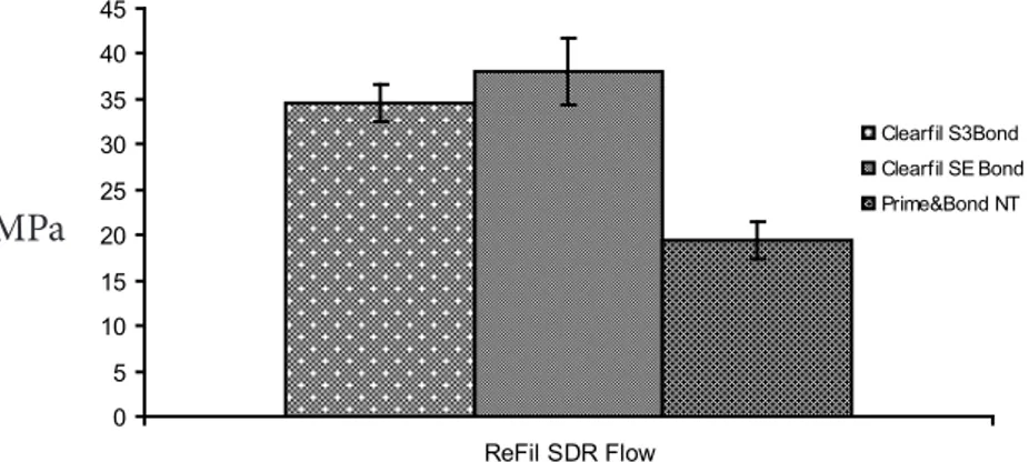
When microtensile bond strength values of adhesive that is applied ReFil SDR Flow are examined to of adhesive it was observed a statistically significant difference. (F=13,960; p=0,000) (Figure 2). In multiple comparison of microtensile bond strength values, bond strength values of Clearfil SE Bond and Clearfil S3Bond was observed statistically significant higher than Prime & Bond NT
(p <0.05). However, no statistically significant difference was shown in the bond strength values between the Clearfil SE Bond and Clearfil S3Bond (p>0.05).
Figure 2. Mean microtensile bond strength values of adhesives that is applied ReFil SDR Flow 0 5 10 15 20 25 30 35 40 45 ReFil SDR Flow Clearfil S3Bond Clearfil SE Bond Prime&Bond NT MPa MPa
Evaluation of Microtensile Bond Strength of Conventional and New Generation Flowable Composite Resins to Dentın Wıth Different Adhesive Systems
20
In the comparison microtensile bond strength values of the adhesives that is applied Clearfil SE Bond and ReFil SDR Flow was determined a statistically significant difference between two groups for Clearfil S3Bond (t=-4,909; p=0,000); there is no statistically significant difference for
Clearfil SE Bond and Prime&Bond NT (p˃0.05).
Table 2. Microtensile bond strength values of the experimental group
Clearfil Majesty Flow ReFil SDR Flow
N Mean(MPa) Standard deviation
Min. Max N Mean(MPa) Standard devi-ation Min. Max Clearfil S3 30 22,116 8,2884 8,1 41,4 30 34,556 11,1343 11,4 53,6 Clearfil SE Bond 30 30,150 12,4529 10,5 53,2 30 38,060 19,7934 7,7 83,7 Prime&Bond NT 30 20,716 8,6193 4,1 37,4 30 19,373 10,9710 3,7 50,0 Discussion
Clinical trials are the most appropriate way to evaluate the effectiveness of restorative materials. However, clinical research of the developing adhesive systems or resins is very difficult technically and ethically. Therefore, laboratory studies are often preferred in dentistry.17 One of the commonly
used methods are microtensile bond strength tests for the evaluation of clinical performance of resin-adhesive systems. In addition to conventional tensile testing methods, the micro test methods which is using 1 mm2 surface area are also used to determine the bond strength between the dental
tissues and restorative materials. It can be stated that the non-uniform stress distribution at the interface of the dental tissue and an adhesive system can be eliminated through the use of samples which have smaller surface areas in test methods. 11,17,18 In this study, microtensile bond strength
test method was used by obtaining bars that have an average of 1 mm2 bonding surface from
samples, as in the study Sano et al11 and Phrukkanon et al19.
Clearfil SE Bond which we used as a two-step self-etch adhesive, has acidic primer in middle strength (pH=1.9). This adhesive system showed high bond strength to normal dentin in many studies.20-22 Clearfil SE Bond contains filler particles that are thought to increase the adhesive’s
tensile capacity against shrinkage stress (silicon dioxide).23 It was declared that theoretically,
simultaneously of the emerging of collagen fibrils and the occurring of the monomer infiltration were sufficient for micromechanical bonding. Also, carboxyl and phosphate groups of Clearfil SE Bond monomers may be chemically bonded to the residual hydroxyapatite. In this way, it is claimed to exhibit high bond strengths. 20,24,25
Clearfil S3Bond that is used as one-step self-etch adhesive, has acidic primer in low strength (pH=
2.7). 26 This type of adhesive systems allow to remain hydroxyapatite around collagen fibrils by
demineralizing dentin fairly shallow, and it creates a superficial hybrid layer. These adhesives behave like a permeable membrane and absorb a significantly water after polymerization due to its hydrophilic nature. Therefore, it is claimed to showed lower bond strength values from two-step self-etch adhesives. 25,27,28
In the two-step total-etch system is the first step in creating the acid application, the second step primer and adhesive Prime & Bond NT, which constitutes one bottle united version. Although the bonding mechanism of this type of adhesive systems are same as three-step total-etch systems, in many studies, it is claimed that the application of the adhesive and the primer in one-step may reduce the hybridization. The fact that two-step “etch-and-rinse” adhesive systems are more sensitive to water and oxygen contamination, may lead to the incomplete polymerization for adhesive resin and lower bond strengths. 6,29
This type of adhesive before depolymerization, nano-filler particles can form clusters which are large enough to prevent the infiltration come together to inter-fibrillar space of the hybrid layer of adhesive. In addition, the aqueous monomer which is the main component of the adhesive can prevent the infiltration of these particles as previously infiltrated to demineralized intertubuler matrix . This situation affects bonding negatively. 30 It is reported that may have reduced the bond
strength values as a result of lack of access to the adhesive to these regions and having the greater demineralization depth which is occured in dentin.5
We believe that all this obtained data in our study explains the different bond strength results. It has been reported in many studies to reduce microleakage and seen in the restoration edge spacing formation by preventing the polymerization shrinkage, as a linear usage under composite restorations of flowable composite resins having a low elasticity coefficient. 31,32Not only the usage
of flowable composite under composite resins play a role in compensating stresses caused by polymerization shrinkage, but also ıt has been reported that stress absorber is doing against the accumulated tension and compressive stress in this region task during chewing forces. 33,34 A new
generation of flowable composite which has more filling rate, is claimed that find wide application due to the increased mechanical properties. 22,35 Although higher bond was observed in the new
generation of flowable composite group, a statistically significant difference was detected only for Clearfil S3Bond, when bond strength of the flowable composite resins is compared.
Nowadays, while many new developments occurs at the system of adhesive dentistry; following these developments on a regular basis and selecting suitable materials of dentist will improve the clinical success of restorative treatment. In this context, we believe that our study results will contribute to the different studies on the subject.
REFERENCES
[1] Can Say E, Nakajima M, Senawongse P, Soyman M, Tagami J. Microtensile bond strength of a filled vs unfilled adhesive to dentin using self-etch and total-etch technique. J Dent 2006; 34: 283-29.
[2] Frankenberger R, Kramer N, Petschelt A. Fatigue behavior of different dentin adhesives. Clin Oral Invest 1999; 3: 11-17.
Evaluation of Microtensile Bond Strength of Conventional and New Generation Flowable Composite Resins to Dentın Wıth Different Adhesive Systems
22
[3] Pashley DH, Carvalho RM Dentine permeability and dentin adhesion. J Dent
1997; 25: 355-372.
[4] Perdigao J. Dentin bonding as a function of detin structure. Dent Clin North Am
2002; 46: 277-301.
[5] Swift EJ. Dentin/enamel adhesives: Review of the literature. Pediatr Dent
2002; 24: 456-61.
[6] Bouillagued S, Gysi P, Wataha JC, Ciucchi B, Cattani M, Godin C, Meyer JM. Bond
strength of composite to dentin using conventional, one-step, and self-etching adhesive systems. J Dent 2001; 29: 55-61.
[7] Chigira H, Manabe T, Hasegawa T, Yukitani W, Fujimitsu T, Itoh K, Hisamitsu H, Wakumoto
S. Efficiacy of various commercial dentin bonding systems. Dent Mater 1994; 10: 363-368.
[8] Finger WJ, Balkenhol M. Practitioner variability effects on dentin bonding with an acetone
based one bottle adhesive. J Adhes Dent 1999; 1: 311-314.
[9] Bauer JG, Henson JL. Microleakage: A measure of the performance of direct filling
materials. Oper Dent 1984; 9: 2- 9.
[10] Davidson CL, Gee AJ, Feilzer A. The competition between the composite-dentin bond
strength and the polimerization contraction stress. J Dent Res 1984; 63: 1396- 1399.
[11] Sano H, Shono T, Sonoda H, Takatsu T, Ciucchi B, Carvalho R, Pashley DH. Relationship
between surface area for adhesion and tensile bond strength-Evaluation of a micro-tensile bond test. Dent Mater 1994; 10: 236-240.
[12] Bek G, Eligüzeloğlu E, Arısu HD, Üçtaşlı MB, Ömürlü H, Türköz E. Akışkan kompozit
rezinlerin dentine mikrogerilim bağlanma dayanımı üzerine etkileri. GÜ Diş Hek Fak Derg 2008; 25(2) : 1-6.
[13] Knezevic A, Tarle Z, Meniga A, Sutalo J, Pichler G, Ristic M. Degree of conversion and
temperature rise during polymerization of composite resin samples with blue diodes. J Oral Rehab 2001; 28: 586-591.
[14] Fernandes CAO, CarvalhoRM, ShonoY, Pashley DH. Bond strengths of adhesives to dentin
as an array. J Dent Res 1998; 77:205 (Abstr. No.798).
[15] Terada RSS, Carvalho RM, Fernandes CAO, Pashley DH. Mapping bond strength to flat
[16] Craig RG, Powers JM, Sakaguchi RL. Craigs restorative dental materials, 12th edition, Mosby- Year Book Inc, 11830 Western Industrial Drive, St. Louis, Missouri, 2006, 52-96.
[17] Tekçe N. İn Vitro Bağlanma Dayanım Testleri ile Klinik Çalışmalar Arasındaki İlişki. EÜ
Dişhek Fak Derg 2013; 34 (2): 57-65.
[18] Armstrong S, Geraldeli S, Maia R, Raposo LH, Soares CJ, Yamagawa J. Adhesion to tooth
structure: a critical review of “micro” bond strength test methods. Dent Mater 2010;26:50-62.
[19] Phrukkanon S, Burrow MF, Tyas MJ. The influence of crosssectional shape and surface
area on the microtensile bond test. Dent Mater 1998; 14: 212-221.
[20] Eren D, Bektaş ÖÖ, Siso SH. Three different adhesive systems; three different bond
strength test methods. Acta Odontol Scand 2013; 71(3-4): 978-83.
[21] Harnirattisai C, Roengrungreang P, Rangsisiripaiboon U, Senawongse P. Shear and
micro-shear bond strengths of four self-etching adhesives measured immediately and 24 hours after application. Dent Mater J 2012; 31(5): 779-87.
[22] Arslan S, Demirbuğa S, Çayabatmaz M, Üstün Y. Geleneksel ve Yeni Nesil Akışkan
Kompozit Rezinlerin Mikromakaslama Bağlanma Dayanımlarının Değerlendirilmesi. Turkiye Klinikleri J Dental Sci 2013; 19(3): 185-92.
[23] Goracci C, Sadek FT, Monticelli F, Cardoso PE, Ferrari M. Microtensile bond strength of
self-etching adhesives to enamel and dentin. J Adhes Dent 2004; 6(4): 313-8.
[24] Fuentes MV, Monticelli F, Goracci C, Toledano M, and Ferrari M. Microtensile bond
strength of different self-etch adhesives to sound human dentin.. 81st General Session of the International Association for Dental Research. Svenska Massan Exhibition 2003; abstract no:0351.
[25] Van Meerbek B, Munck JD, Yoshida Y, Inoue S, Vargas MA, Vijay P, Landuyt KV,
Lambrechts P, Vanherle G. Adhesion to enamel and dentin: Current status and future challenges. Oper Dent 2003; 28; 215-35.
[26] Ostby AW, Bishara SE, Denehy GE, Laffoon JF, Warren JJ. Effect of self-etchant pH on
the shear bond strenght of orthodontic brackets. Am J Orthod Dentofacial Orthop 2008; 134(2): 203-208.
[27] Tanaka J, Ishikawa K, Yatani H, Yamashita A, Suzuki K. Correlation of dentin bond
24
[28] Tay FR, Pashley DH, Yoshiyama M. Two modes of nanoleakage expression in single-step
adhesives. J Dent Res 2002; 81(7): 472-6.
[29] Urabe I, Nakajima S, Sano H, Tagami J. Physical properties of the dentin-enamel junction
region. Am J Dent 2000; 13(3): 129-35.
[30] Gagliardi RM, Avelar RP. Evaluation of micro -leakage using different bonding agents.
Oper Dent 2002; 27(6): 582-6.
[31] Gueders AM, Charpentier JF, Albert AI, Geerts SO. Microleakage after termocycling of 4
etch and rinse and 3 self-etch adhesives with and without a flowable composite lining. Oper Dent 2006; 31: 450-455.
[32] Migues PA, Pereira P, Foxton RM, Walter R, Nunes MF, Swift EJ. Effects of flowable resin
on bond strength and gap formation in Class I restorations. Dent Mater 204; 20: 839-845.
[33] Tredvin CJ, Stokes A, Moles DR. Influnce of flowable liner and marginal location on
microleakage of conventional and packable Class II resin composites. Oper Dent 2005; 30: 32-38.
[34] Van Meerbeek B, Williems G, Cecils JP, Roos JR, Bracm M, Lambrechts P, Vanherle G.
Assesment by nano-indentation of the hardness an elasticity of the resin dentin bonding area. J Dent Res 1993; 72: 1434- 1442.
[35] Altun C. Kompozit dolgu materyallerinde son gelişmeler. Gülhane tıp dergisi
Minimally Invasive Restoration of Erosive Lesions With Direct
Composite Laminate Veneers: A Case Report
Mağrur KAZAK
1Abstract
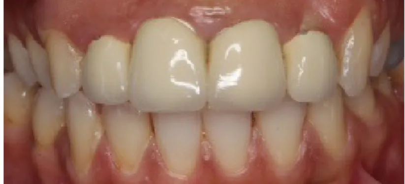
Background: Non-carious cervical lesions (NCCLs) which may be caused by erosion, abrasion, abfraction and attrition are commonly observed in clinic practice. Dental erosion is defined as loss of dental hard tissue due to a chemical irritant that does not involve the influence of bacteria. In case of such lesions progress over time, teeth hypersensitivity and aesthetical problems arise.
Objective: The purpose of this case report was to restore erosive lesions on maxillary central teeth with direct composite laminate veneers.
Case Description: A 34 years old female patient presented to Istanbul Aydın University Dental Faculty Restorative Dentistry Department because of aesthetic reasons.After clinical examination; erosive defects mainly at the maxillary central teeth and worn incisal edges were diagnosed. As a result of the patient’s anamnesis, it was understood that, the patient consumes lemon regularly every week. The erosive lesions were restored with an adhesive system and a nanofill resin composite. Finishing and polishing procedures were performed immediately. After aesthetical restorations, information is given to the patient about the elimination of the factors which cause erosive lesions.
Practical Implications: When clinically evaluated, direct composite laminate veneers are based on the principles of minimally invasive dentistry and reversibility that can be used for the rehabilitation of NCCLs aesthetically.
Keywords: Dental erosion, non-carious cervical lesions, direct composite restoration, dental
laminate veneer, minimally invasive restoration
Eroziv Lezyonlarin Direkt Kompozit Lamina Venerler ile Minimal İnvaziv Restorasyonu: Olgu
Özet
Giriş: Erozyon, abrazyon, abfraksiyon ve atrizyon nedeni ile ortaya çıkan çürüksüz servikal lezyonlar klinikte yaygın olarak görülmektedir. Diş erozyonu, bakteri etkisi içermeyen bir kimyasal iritan sebebi ile diş sert dokusunun kaybı olarak tanımlanmaktadır. Bu tip lezyonların zaman içerisinde ilerlemesi durumunda, dişlerde aşırı duyarlılık ve estetik problemler ortaya çıkmaktadır.
Minimally Invasive Restoration of Erosive Lesions With Direct Composite Laminate Veneers: A Case Report
26
Amaç: Bu olgu sunumunun amacı; üst çene santral dişlerde oluşan eroziv lezyonları direkt
kompozit laminate venerler ile restore etmektir.
Olgu Raporu: 34 yaşındaki kadın hasta estetik sebeplerden ötürü İstanbul Aydın Üniversitesi
Diş Hekimliği Fakültesi Restoratif Diş Tedavisi Bölümü’ne başvurmuştur. Hastanın yapılan klinik muayenesi sonucunda; en fazla üst çene santal dişlerde olmak üzere eroziv defetler ve kesici kenarlarda da aşınmalar tespit edilmiştir. Hastadan alınan anamnez sonucunda, hastanın her hafta düzenli olarak limon tükettiği öğrenilmiştir. Üst çene santral dişlerin eroziv lezyonları, bağlayıcı ajan ve nano dolduruculu kompozit bir rezin materyali kullanılarak restore edilmiştir. Bitirme ve cila işlemleri yapılmıştır. Estetik restorasyonlardan sonra hastaya erozyona sebep olan faktörlerin elimine edilmesi konusunda bilgi verilmiştir.
Pratik Uygulamalar: Klinik açıdan değerlendirildiğinde, minimal invaziv diş hekimliği ve
reverzibilite ilkelerine dayanan direkt kompozit lamina venerler ile çürüksüz servikal lezyonlar estetik olarak restore edilebilirler.
Anahtar Kelimeler: Dental erozyon, çürüksüz servikal lezyonlar, direkt kompozit restorasyon,
dental lamina vener, minimal invaziv restorasyon
Introduction
Non-carious cervical lesions (NCCLs) which may be caused by erosion, abrasion, abfraction and attrition are commonly observed in clinic practice.1 Dental erosion is defined as loss of dental hard
tissue due to a chemical irritant that does not involve the influence of bacteria.2 A chemical irritant
involves acids that reach the mouth and have extrinsic or intrinsic origin.3 Extrinsic acids from the
diet are becoming the most important source of erosive attacks due to the increasing consumption of acidic drinks.4
Progressive erosive lesions can be clinically difficult to diagnose at an early stage and patients often unaware of the tooth loss because of no pain or esthetic problem. More pronounced changes in macromorphology occur when the erosive damage is more severe. Once the restorative treatment is indicated, dental practitioner has to know that esthetic restorations of erosive teeth present challenges. One of the challenge is the presence of sclerotic dentin. Sclerotic dentin is a common substrate that occurs in response to tooth wear caused by attrition, abrasion, abfraction or erosion. This substrate has demonstrated to be a challenge for bonding procedures.5 Currently, there are
several options for the treatment of erosive lesions, which can range from a conservative (adhesive and composite resins restorations) to more invasive procedures (crowns, bridges, or even full-mouth reconstructions) according to the severity of the lesions.6,7 When a restorative treatment is
required, it is important to choose a material resistant to erosion that allows the preservation of the tooth as much as possible.In terms of conservative treatment options, composite resin restorations seem to the best materials for the restoration of NCCLs.8,9
Direct composite laminate veneers (DCLVs) are minimally invasive restorations and the teeth are prepared in a conservative manner.These restorations can be applied on a minimally prepared tooth