11β-hydroxysteroid dehydrogenase type 1
gene expression is increased in ascending
aorta tissue of metabolic syndrome patients
with coronary artery disease
F. Atalar1, B. Vural2, C. Ciftci3, A. Demirkan2,4, G. Akan5, B. Susleyici-Duman6,
D. Gunay7, B. Akpinar8, E. Sagbas8, U. Ozbek2 and A.S. Buyukdevrim9 1Department of Growth-Development and Pediatric Endocrinology, Child Health Institute, Istanbul University, Istanbul, Turkey 2Department of Genetics, Institute for Experimental Medicine, Istanbul University, Istanbul, Turkey
3Department of Cardiology, Medical Faculty, Istanbul Bilim University, Istanbul, Turkey 4Department of Epidemiology and Biostatistics,
Division of Genetic Epidemiology, Erasmus University Medical Center, Rotterdam, The Netherlands
5Department of Medical Biology and Genetics, Medical Faculty, Istanbul Bilim University, Istanbul, Turkey
6Biology Department, Science and Art Faculty, Marmara University, Goztepe, Istanbul, Turkey
7Biochemistry Laboratory, Florence Nightingale Hospital, Istanbul, Turkey 8Department of Cardiovascular Surgery and Florence Nightingale Hospital, Istanbul Bilim University Medical Faculty, Istanbul, Turkey
9Emeritus, Turkish Diabetes Consortium, Istanbul, Turkey Corresponding author: F. Atalar
E-mail: [email protected]
Genet. Mol. Res. 11 (3): 3122-3132 (2012) Received July 10, 2012
Accepted July 30, 2012 Published August 31, 2012
DOI http://dx.doi.org/10.4238/2012.August.31.10
activity and mRNA levels are increased in visceral and subcutaneous adipose tissues of metabolic syndrome subjects. We analyzed 11β-HSD-1 expression in human epicardial adipose (EA) and ascending aorta (AA) tissues of metabolic syndrome patients and examined their contribution to the development of coronary atherosclerosis. The 11β-HSD-1 expression was evaluated by qRT-PCR in EA and AA tissues of 20 metabolic syndrome patients with coronary artery disease (metabolic syndrome group) and 10 non-metabolic syndrome patients without coronary artery disease (controls). 11β-HSD-1 expression was increased in EA and AA tissues of the metabolic syndrome group (4.1- and 5.5-fold, respectively). A significant positive correlation was found between 11β-HSD-1 expression in EA tissue and waist hip ratio and 11β-HSD-1 expression in AA tissue and body mass index, while a negative correlation was found between 11β-HSD-1 expression in EA tissue and HDL. Expression of CD68, a macrophage marker, was significantly increased in both tissues of the metabolic syndrome group; it was 2-fold higher in AA tissue compared to EA tissue in the metabolic syndrome group. Our findings of increased expression of 11β-HSD-1 and CD68 in AA tissue of the metabolic syndrome group lead us to suggest that they contribute to coronary atherosclerosis in metabolic syndrome. This positive correlation between obesity markers and 11β-HSD-1 in AA and EA tissues strengthens the evidence that 11β-HSD-1 has a role in metabolic syndrome. To the best of our knowledge, this is the first report showing 11β-HSD-1 and CD68 expression in AA tissue of metabolic syndrome patients. We suggest that there is tissue-specific expression of 11β-HSD-1 in metabolic syndrome and associated cardiovascular disorders.
Key words: 11β-hydroxysteroid dehydrogenase type 1; Ascending aorta;
Glucocorticoids; Epicardial adipose tissue; Metabolic syndrome; Coronary artery disease
INTRODUCTION
Metabolic syndrome (MS) is a complex disease, which causes a wide range of disor-ders in carbohydrate metabolism, including visceral adiposity, insulin resistance, and impaired glucose tolerance or overt diabetes mellitus, and other disorders as well, such as dyslipidemia and hyperuricemia, arterial hypertension and atherothrombotic events (Masuzaki et al., 2003; Stewart, 2003). The presence of MS predicts a 2- to 4-fold increase in the risk of cardiovas-cular disease and death (Trevisan et al., 1998; Wilson et al., 1999), and the risk of developing type 2 diabetes has been found in recent years to be increased 5- to 9-fold (Balkau et al., 2003; Onat, 2004; IDF, 2005).
In this study, most of the background knowledge is derived from the prospective pop-ulation-based Turkish Adult Risk Factor (TEKHARF) Study, with already a follow-up period of 13 years. In addition, the data contributed by the Turkish Heart Study are also outlined in the setting of a prevalence of MS in 3 of 8 Turkish adults (Onat, 2004).
system and circulating hormones, and it also has the ability to modulate its own metabolic activity. Glucocorticoids (GCs) belong to one of the important group of regulatory factors, the adrenocorticostreoids. GCs have an important role in adipose biology, where they increase adipose tissue mass giving rise to obesity in conditions such as Cushing’s disease or following the use of exogenous GCs (Tomlinson et al., 2004). It is claimed that tissue-specific dysregu-lation of cortisol metabolism is responsible for the development of MS. In human obesity, circulating cortisol levels are not consistently elevated, but cortisol metabolism andthereby glucocorticoid action increase at the tissuelevel. The level and activity of cortisol, especially in the visceral adipose tissue (VAT) is mainly determined by 11β-hydroxysteroid dehydroge-nase type 1 (11β-HSD-1), which is a microsomal short-chain dehydrogedehydroge-nase/reductase; it is bidirectional and converts inactive cortisone to active cortisol (Lindsay et al., 2003).
11β-HSD-1 is involved in a number of disease processes, including insulin resistance (Kotelevtsev et al., 1997), osteoporosis, glaucoma, ocular surface renewal, inflammation and multiple sclerosis, and it is widely expressed in liver, brain, adipose tissue, lung, gonads, skel-etal muscle, and bronchial and aortic smooth muscle cells (Tomlinson et al., 2004). Its expres-sion is found to be higher in human omental (OM) tissue compared to subcutaneous adipose tissue (SAT), and OM preadipocytes and fibroblast-like precursors of mature adipocytes are found to express high levels of 11β-HSD-1 compared to SAT (Bujalska et al., 1999, 2002b).
11β-HSD-1 reinforces the action of endogenous GCs by increasing their local concen-tration. It has been demonstrated that 11β-HSD-1 enzyme activity is much higher in VAT than in SAT and that this enzyme activity is further elevated when insulin and cortisol are added to the medium (Bujalska et al., 1997; Bujalska et al., 2002a,b). Adipose tissue 11β-HSD-1 activity is also increased in animalmodels of MS (Masuzaki et al., 2001; Boullu-Ciocca et al., 2005). Like in humans, 11β-HSD-1 activity is also found elevated in adipose tissue of obese rodents, inducing complications similar to MS (Morton et al., 2004). 11β-HSD-1 knockout mice have been shown to have low intracellular GC levels, where they are protected from obesity, dia-betes and dyslipidemia, and in addition, the possible contribution of interindividual variations in 11β-HSD-1 in high fat-fed mice has also been demonstrated (Liu et al., 2006). Recently, an adipose tissue targeted pharmacological inhibitor of 11β-HSD-1 was shown to be a new approach for the treatment of obesity and metabolic syndrome (Liu et al., 2011). Increased ac-cumulation of CD68+ macrophages seen in SAT depots in patients with high body mass index (BMI) and obese phenotypes, and increased expression of monocyte chemoattractant protein, macrophage inflammatory protein and MAC-1 found in rodent white adipose tissue after diet-induced obesity suggest a further role for chronic inflammation, which may activate adipose tissue (Xu et al., 2003). Also proved by echocardiographic findings, epicardial adipose (EA) tissue, another form of true visceral fat, was found accumulated around subepicardial coronary vessels (Iacobellis et al., 2003). EA tissue, which is a metabolically active organ, generates various bioactive molecules such as free fatty acids and a number of adipokines (adiponec-tin, resistin and inflammatory cytokines), which could affect the coronary artery response (Iacobellis et al., 2005a). Cardiac tissue was previously demonstrated to express substantial levels of 11β-HSD-1, which appears to act predominantly as NADPH-dependent reductase
in vivo, but as both a dehydrogenase and reductase in a broken cell system, and is localized
in interstitial cardiac fibroblasts. In addition to that, based on the evidence suggesting that it is the 11β-HSD-1 isoform that is predominantly expressed in the vasculature, specifically in vascular smooth muscle and cultured rat aortic endothelial cells, and here oxoreductase
activ-ity predominates, the expression of 11β-HSD-1 in interstitial fibroblast cells and in vascular smooth muscle cells has led to speculation that it could be involved in the pathogenesis of acute coronary vascular disease (Sheppard et al., 2002; Hermanowski-Vosatka et al., 2005).
A growing amount of evidence suggests that regional fat distribution plays an im-portant part in the development of an unfavorable metabolic and cardiovascular risk profile. Therefore, the aim of this study was to investigate the level of 11β-HSD-1 gene expression of EA and ascending aorta (AA) tissues in MS patients with coronary artery disease (CAD) and non-MS patients without CAD and to discuss the potential pathogenetic role of these tissues in relation to their contribution to the development of coronary atherosclerosis in MS patients.
MATERIAL AND METHODS
Subjects
The study group was divided into two groups: MS group, MS patients with CAD (N = 20; 10 women and 10 men) and control group, non-MS patients without CAD (N = 10; 5 women and 5 men). Tissue biopsies were obtained from MS and control groups during elec-tive open heart surgery for coronary artery bypass grafting (CABG) or valve replacement surgery (valvuloplasty). Tissue samples were then immediately frozen in liquid nitrogen and stored at -80°C for later RNA extraction.
The MS group met the criterion defined by the International Diabetes Federation (IDF) consensus worldwide definition of metabolic syndrome, i.e., a cluster of three or more of the following abnormalities: waist circumference >102 cm in men and >88 cm in women, serum triglycerides ≥150 mg/dL; high-density lipoprotein cholesterol (HDL-C) <40 mg/dL in men and <50 mg/dL in women or specific treatment for this lipid abnormality (fibrates and ni-cotinic acid); blood pressure ≥130 / ≥85 mmHg or fasting serum glucose ≥110 mg/dL or drug treatment for hypertension or type 2 diabetes, respectively (IDF, 2005). All patients in the MS group suffered from three-vessel artery disease, whereas one patient had left main coronary ar-tery disease. Patients with none of the above criteria who underwent heart valve surgery were included in the control group. Subjects with cancer, collagen diseases, endocrinopathies (phe-ochromocytoma, Cushing’s syndrome, Addison’s disease, and hyper- and hypothyroidism), secondary hypertension (renal artery stenosis), and diabetic microangiopathic complications were also excluded from the study. The study group was on a range of medications as shown in Table 1.
All the participants were included in this study after providing written informed con-sent in accordance with the Helsinki Declaration. The protocols were approved by the local Ethics Committee of Istanbul Science University.
Biochemical measurements
Plasma glucose was measured using an enzymatic reference method with glucose oxidase; serum total cholesterol was measured using the enzymatic, colorimetric method with cholesterol esterase; HDL-C and low-density lipoprotein-cholesterol (LDL-C) were directly determined with homogeneous enzymatic colorimetric assay; triglyceride was determined by
the enzymatic colorimetric method (GPO/PAP) with cholesterol phosphate oxidase and 4-ami-nophenazone on the opeRA analyzer; uric acid was determined by the enzymatic, colorimetric test with uricase and 4-aminoantipyrine. Glycosylated hemoglobin was measured by a colori-metric method. All analyses were performed on Cobas 6000/Roche.
RNA extraction and quantitative real-time PCR (qRT-PCR)
The EA and AA tissues were ground with a homogenizer in a reaction tube. EA total RNA and AA total RNA were extracted with Rneasy Lipid Tissue Mini kit and Rneasy Fibrous Tissue Mini kit (Qiagen, Germany), respectively. RNA samples were quantified by spectro-photometry. Total RNA samples were diluted in water and stored at -80°C until use. After DNase I treatment, 1 µg total RNA was reverse transcribed using random hexamers as primers and Superscript II reverse transcriptase (Invitrogen). Expression of the 11β-HSD-1 gene was determined by qRT-PCR using a LightCycler 480 (Roche) instrument. Ten-fold dilutions of cDNA synthesized from total RNA of healthy adipose tissue were used in each run, with their duplicates. Amplification of the gene for human β-globin was performed on all samples tested to control for variations in RNA amount. The 11β-HSD-1 gene was subsequently normalized to cyclophilin mRNA levels. Levels of gene-specific messages were presented as fold-increase for the MS group compared to the control group. The presence of specific gene products were also confirmed with melting curve analysis. Oligonucleotide primers had the following se-quences: for 11β-HSD-1, sense 5'-TTG-CCC-ATG-CTG-AAG-CAG-AGC-3' and antisense 5'-CTG-TTT-CTG-TGT-CTA-TGA-GGC-3'.
Data analysis
Data are reported as numbers and percentages for discrete variables and as means ± SD, for continuous variables. Baseline differences between patients and controls were exam-ined by the Mann-Whitney U-test. Variables were analyzed by Pearson’s correlation or Spear-man’s correlation using the SPSS.10 program. Statistical significance was taken as P < 0.05.
RESULTS
Table 1 shows the demographic, clinical and biochemical characteristics of the study group. BMI, waist and hip circumferences, waist-to-hip circumference ratio, systolic blood pressure, fasting plasma glucose, serum triglyceride, uric acid (P < 0.05) and diastolic blood pressure levels were higher in the MS group compared to the control group, whereas circulat-ing HDL-C concentrations were lower (P < 0.01).
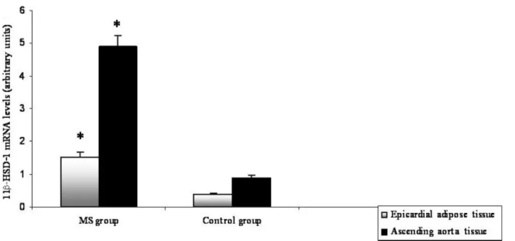
11β-HSD-1 expression in both EA and AA tissues was assessed for all subjects. Sig-nificant differences in both EA and AA tissues of the MS and control groups were found; 11β-HSD-1 expression of EA and AA of the MS group was increased 4.1- and 5.5-fold, re-spectively. These are clearly depicted in Figure 1. In addition, macrophage infiltration in both tissues was also investigated. The expression of CD68, a macrophage marker, was shown to be nearly 2-fold higher in AA tissue compared to EA tissue of the MS group. CD68 expression in both tissues was significantly increased in the MS group compared to the control group (P < 0.05, respectively).
Categorical variables are represented as numbers (%) and continuous variables as means ± SD; *P < 0.05 and **P < 0.01 are the significant values and the comparisons were made between MS and control groups; NS = not significant. MS = metabolic syndrome; BMI = body mass index; SBP = systolic blood pressure; DBP = diastolic blood pressure; LDL = low-density lipoprotein; HDL = high-density lipoprotein; HbA1c = hemoglobin A1c.
MS group (N = 20) Control group (N = 10) P value
Age 64.3 ± 5.1 63.3 ± 10.6 NS BMI (kg/m2) 29.9 ± 1.6 21.7 ± 3.5 <0.05* Waist circumference (cm) 101.6 ± 8.3 78.5 ± 17.5 <0.05* Hip circumference (cm) 109.3 ± 8.6 96.0 ± 8.6 <0.05* Waist-to-hip ratio 0.9 ± 0.1 0.8 ± 0.1 NS SBP (mmHg) 115 ± 8.5 102.5 ± 5.0 <0.05* DBP (mmHg) 74 ± 6.9 60 ± 0.0 <0.01** Ejection fraction (%) 48.5 ± 1.8 55.3 ± 2.1 NS Fasting glucose (mg/dL) 148.3 ± 56.9 92.8 ± 10.2 <0.05* Total cholesterol (mg/dL) 190.0 ± 40.9 158.4 ± 28.5 NS HDL cholesterol (mg/dL) 41.8 ± 9.9 61.0 ± 11.1 <0.01** LDL cholesterol (mg/dL) 120.4 ± 32.6 91.5 ± 3.3 NS Triglycerides (mg/dL) 162.7 ± 64.2 84.1 ± 11.5 <0.05* Uric acid (mg/dL) 5.9 ± 1.5 3.1 ± 1.2 <0.05* HbA1c (%) 5.9 ± 1.8 5.3 ± 0.7 NS Diabetes (%) 60 (12/20) 0 (0/10) <0.05* Medications Statin (%) 100 (20/20) 0 (0/10) <0.05* Beta-blocker (%) 100 (20/20) 50 (5/10) <0.05* Acetylsalicylic acid (%) 90 (18/20) 20 (2/10) <0.05* Coumadin (%) 75 (15/20) 50 (5/10) NS Angiotensin-converting enzyme inhibitor (%) 70 (14/20) 40 (4/10) NS Oral antidiabetics (%) 60 (12/20) 0 (0/10) <0.05* Thiazide (%) 50 (10/20) 40 (4/10) NS Clopidogrel (%) 30 (6/20) 0 (0/10) <0.05* Furosemide (%) 30 (6/20) 40 (4/10) NS Calcium antagonist (%) 20 (4/20) 30 (3/10) NS Fenofibrate (%) 10 (2/20) 0 (0/10) <0.05* Amiodarone (%) 10 (2/20) 0 (0/10) <0.05*
Table 1. The anthropometric, biochemical and clinical characteristics of the study group.
Figure 1. Differential expression of the 11β-HSD1 gene in epicardial adipose and ascending aorta tissues of
metabolic syndrome (MS) and control groups. Values are reported as means ± standard deviation. *Statistical significance was taken at P < 0.05. Comparisons were made between MS and control groups.
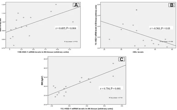
In addition to these findings, a positive correlation was found between mRNA levels of 11β-HSD-1 in EA tissue and waist-to-hip ratio (r = 0.695, P < 0.004), while a negative cor-relation was observed between mRNA levels of 11β-HSD-1 in EA tissue and HDL (r = -0.561, P < 0.03; Figure 2A and B). A correlation between mRNA levels of 11β-HSD-1 in AA tissue and BMI was also found (r = 0.756, P < 0.001; Figure 2C).
Figure 2. Correlations between 11β-HSD-1 expression levels in epicardial adipose tissue (EA) and ascending
aorta tissue (AA) with obesity parameters [waist-to-hip ratio, body mass index (BMI) and high-density lipoprotein (HDL) levels]. A. 11β-HSD-1 expression levels in EA tissue were positively correlated with waist-to-hip ratio (r = 0.695; P = 0.004). B. A negative correlation was identified between 11β-HSD-1 expression levels in EA tissue and HDL levels (r = -0.561; P = 0.03). C. Correlation between 11β-HSD-1 expression levels in AA tissue and BMI (r = 0.756; P = 0.001).
DISCUSSION
Our results demonstrate for the first time the increased expression of 11β-HSD-1 in AA and EA tissues of an MS group and the positive correlation of 11β-HSD-1 expression in EA and AA tissues with the obesity markers (waist-to-hip ratio and BMI, respectively). We further showed the increased levels of CD68 expression in both tissues. CD68 expression was significantly increased in both tissues of the MS group, although the expression was 2-fold higher in AA tissue compared to EA tissue.
Being regarded as a perivascular fat, EA tissue is found to be related to weight and age and tends to express proatherogenic and proinflammatory products in subjects with car-diovascular disease. EA tissue shows a similar pattern of expression for a number of key adipocytokines compared to OM adipose tissue. A high level of macrophage infiltration in this
depot was also confirmed, which may contribute to the pathogenic gene expression profile in this tissue (Baker et al., 2006). Omental adiposity has shown to be an independent predictor of metabolic risk, with increased mRNA expression of resistin, AGT and PAI-1, representing im-portant mediators of inflammatory, fibrinolytic and thrombotic risk (Liu et al., 2006). The low levels of adiponectin, which is increasingly becoming associated with MS and cardiovascular risk (Iacobellis et al., 2005b), strengthen the argument that in CABG patients, EA tissue rep-resents a negative influence on both cardiovascular outcome and myocardial function. Silaghi et al. (2007) reported the expression of 11β-HSD-1 in paired biopsies of SAT and EA tissues obtained from patients with and without CAD, and their data showed a positive correlation between 11β-HSD-1 and adrenomedullin expressions indicating the presence of macrophages in EA tissue. Comparative pathophysiological studies in human and animals have suggested that adipose tissue 11β-HSD-1 dysregulation found in obese patients may, at least in part, be programmed by environmental factors (Boullu-Ciocca et al., 2005). Alberti et al. (2007) found a higher level of 11β-HSD-1 expression in SAT but not in VAT of obese subjects, and the association of the increased level of 11β-HSD-1 with the worsening of metabolic condi-tions through a decrease in adiponectin release, although some contradictory results have also been reported (Desbriere et al., 2006). Despite its distinct biological properties compared to the other adipose tissues; the orbital fat tissue expresses 11β-HSD-1 as well as abundant GR-α and a large amount of CD68+ population (Bujalska et al., 2007). Reape and Groot (1999) and Weber et al. (2004) characterized atherosclerosis, in which chemokines play an important role, by the accumulation of macrophagesand T lymphocytes in the wall of large arteries, and many of the chemoattractantfactors involved in the development of atherosclerosis are expressedin the atherosclerotic plaque and produced by endothelial andsmooth muscle cells.The impor-tance of chemokines in the developmentof atherosclerosis has been demonstrated in an LDL receptor-deficient mouse model, where the invalidationof the MCP-1 gene prevented lipid de-position and macrophageinfiltration in the aortic wall.The elevated expression of 11β-HSD-1 in AA tissue could be the result of inevitable macrophage infiltration in aorta tissue as a result of the proinflammatory or proatherogenic processes. In the current study, our results showed the increased expression of 11β-HSD-1 in EA and AA tissues of the MS group with athero-sclerotic events compared to the control group. 11β-HSD-1 expression of EA and AA tissues in the MS group was 4.1- and 5.5-fold higher compared to the control group, respectively. A positive correlation between mRNA levels of 11β-HSD-1 in EA tissue and waist-to-hip ratio and between mRNA levels of 11β-HSD-1 in AA tissue and BMI, and a negative correlation be-tween mRNA levels of 11β-HSD-1 in EA tissue and HDL were also evident in the MS group. To determine the presence of macrophage infiltration in AA tissue, we further demonstrated the expression of CD68 in AA tissue of the MS group compared to controls. Therefore, the increased expression of 11β-HSD-1 in AA tissue could well be enhanced by the infiltrating macrophages in AA tissue.
In visceral obese patients, increased glucocorticoid signaling subsequent to a chronic stress exposure or to enhanced glucocorticoid receptor concentrations were determined to influence 11β-HSD-1 expression (Rebuffe-Scrive et al., 1990; Dallman et al., 2003). It is un-likely that anesthesia and surgical stress are involved in the variations of the 11β-HSD-1 ex-pression because stimulation of 11β-HSD-1 mRNA by cortisol needs at least 24 h of exposure. In this study, tissue samples were taken from both groups of patients under the same condi-tions (i.e., anesthesia, surgical procedure), and thus, environmental factors would not explain
the differences in gene expression. The statistically significant correlation found between the increase in gene expression and the phenotypic characteristics of MS patients supports our data. Factors influencing 11β-HSD-1 expression include GCs, insulin, thyroid hormones, sex steroids, GH, IGF-1, cytokines and differentiation, determination and transcription factors such as PPARg2, C/EBPα, SREBP1c/ADD1, SPARC, NFkappaB, Stat5, or FOXO binding sites. PPARg agonists have indeed been found to modify the expression and the activity of 11β-HSD-1 in adipose tissue (Nakano et al., 2007; Kolak et al., 2007).
Moreover, Paulsen et al. (2007) reported that 11β-HSD-1 mRNA in human adipose tis-sue was higher in obese subjects compared to lean subjects in both women and men and in both SAT and VAT, but interestingly they also reported that 11β-HSD-1 mRNA levels in lean women was significantly lower than in lean males. Therefore, its upregulation associated with obesity might be more devastating for women and might also help to understand the higher relative risk of cardiovascular disease in women suffering from metabolic syndrome. However, unlike in SAT and VAT, our result showed that there was no significant difference in EA tissue and AA tissue 11β-HSD-1 mRNA levels between MS males and MS females with CAD (Paulsen et al., 2007).
GCs regulate multiple processes in adipose tissue influencing fat cell size and triglyc-eride content and adipose tissue metabolism; they act through GR-α, which mediates most of their hormone-induced actions. Glucocorticoid- or growth hormone-induced modulation of 11β-HSD-1 could be involved in the accumulation of adipose tissue and the dysregulation of its metabolism seen in MS, whereas part of the therapeutic effects of PPARα ligands may take place through their action on 11β-HSD-1 (Hermanowski-Vosatka et al., 2000; Staab and Maser, 2010).
It was shown that the PPARγ-dependent increase in subcutaneous fat and reduc-tion in visceral fat are accompanied by a reduced expression of 11β-HSD-1 in VAT, whereas 11β-HSD-1 mRNA levels were not changed in SAT. Thus, a decreased cortisol level in VAT may contribute to the insulin-sensitizing effects on activation of PPARγ (Hotamisligil and Spiegelman, 1994). All our subjects were on statins, and the effect of statins on PPARs, and the expression of various genes such as FABP4 has been previously demonstrated. Unlike agonists of PPARg, there are no published data about the statin effect on 11β-HSD-1 expres-sion in EA and in AA tissues. In our study, the limited number of patients prevents us from speculating on the potential effect of medications on the expression of 11β-HSD-1.
In conclusion, to the best of our knowledge, this is the first report showing the presence of 11β-HSD-1 and CD68 in AA tissue of an MS group. Our findings showing an increased ex-pression of 11β-HSD-1 and CD68 in AA tissue of an MS group compared to controls suggest a close association between 11β-HSD-1 in AA tissue and coronary atherosclerosis. Moreover, the positive correlation between obesity markers and 11β-HSD-1 expression in AA and EA tis-sues would strengthen the role of 11β-HSD-1 in obesity and metabolic syndrome. Our findings suggest the importance of tissue-specific expression of 11β-HSD-1 in metabolic syndrome and its associated cardiovascular disorders.
STUDY LIMITATIONS
These results provide preliminary data. Ethical concerns prevented us from obtaining more tissue biopsy samples in patients undergoing heart surgery for other indications. Our future plan is to investigate in a larger study group the expressions of other candidate genes and tissue localization and the effect of therapeutic agents on gene/protein expression level.
As a continuation of this study, the activities and expressions of 11β-HSD-1 associ-ated secretomes that determine the MS phenotype in AA and EA tissues and other fat tissues will be investigated.
ACKNOWLEDGMENTS
Research supported by the Turkish Diabetes Foundation, Istanbul, Turkey. We thank Dr. Mehmet Kocak (St. Jude Children’s Research Hospital, Memphis, USA) for his help in statistical analysis. We also extend our thanks to Dr. Zeliha Yazıcı (Istanbul University, Cer-rahpasa Medical Faculty, Department of Pharmacology and Clinical Pharmacology) and Iclal Ozcelik for critical reading of the manuscript.
REFERENCES
Alberti L, Girola A, Gilardini L, Conti A, et al. (2007). Type 2 diabetes and metabolic syndrome are associated with increased expression of 11β-hydroxysteroid dehydrogenase 1 in obese subjects. Int. J. Obes. 31: 1826-1831. Baker AR, Silva NF, Quinn DW, Harte AL, et al. (2006). Human epicardial adipose tissue expresses a pathogenic profile
of adipocytokines in patients with cardiovascular disease. Cardiovasc. Diabetol. 5: 1.
Balkau B, Vernay M, Mhamdi L, Novak M, et al. (2003). The incidence and persistence of the NCEP (National Cholesterol Education Program) metabolic syndrome. The French D.E.S.I.R. study. Diabetes Metab. 29: 526-532.
Boullu-Ciocca S, Dutour A, Guillaume V, Achard V, et al. (2005). Postnatal diet-induced obesity in rats upregulates systemic and adipose tissue glucocorticoid metabolism during development and in adulthood: its relationship with the metabolic syndrome. Diabetes 54: 197-203.
Bujalska IJ, Kumar S and Stewart PM (1997). Does central obesity reflect “Cushing’s disease of the omentum?” Lancet 349: 1210-1213.
Bujalska IJ, Kumar S, Hewison M and Stewart PM (1999). Differentiation of adipose stromal cells: the roles of glucocorticoids and 11β-hydroxysteroid dehydrogenase. Endocrinology 140: 3188-3196.
Bujalska IJ, Walker EA, Hewison M and Stewart PM (2002a). A switch in dehydrogenase to reductase activity of 11β-hydroxysteroid dehydrogenase type 1 upon differentiation of human omental adipose stromal cells. J. Clin.
Endocrinol. Metab. 87: 1205-1210.
Bujalska IJ, Walker EA, Tomlinson JW, Hewison M, et al. (2002b). 11β-hydroxysteroid dehydrogenase type 1 in differentiating omental human preadipocytes: from de-activation to generation of cortisol. Endocr. Res. 28: 449-461. Bujalska IJ, Durrani OM, Abbott J, Onyimba CU, et al. (2007). Characterisation of 11β-hydroxysteroid dehydrogenase
1 in human orbital adipose tissue: a comparison with subcutaneous and omental fat. J. Endocrinol. 192: 279-288. Dallman MF, Pecoraro N, Akana SF, La Fleur SE, et al. (2003). Chronic stress and obesity: a new view of “comfort food”.
Proc. Natl. Acad. Sci. U. S. A. 100: 11696-11701.
Desbriere R, Vuaroqueaux V, Achard V, Boullu-Ciocca S, et al. (2006). 11β-hydroxysteroid dehydrogenase type 1 mRNA is increased in both visceral and subcutaneous adipose tissue of obese patients. Obesity (Silver Spring) 14: 794-798. Hermanowski-Vosatka A, Gerhold D, Mundt SS, Loving VA, et al. (2000). PPARα agonists reduce 11β-hydroxysteroid
dehydrogenase type 1 in the liver. Biochem. Biophys. Res. Commun. 279: 330-336.
Hermanowski-Vosatka A, Balkovec JM, Cheng K, Chen HY, et al. (2005). 11β-HSD1 inhibition ameliorates metabolic syndrome and prevents progression of atherosclerosis in mice. J. Exp. Med. 202: 517-527.
Hotamisligil GS and Spiegelman BM (1994). Tumor necrosis factor α: a key component of the obesity-diabetes link.
Diabetes 43: 1271-1278.
Iacobellis G, Ribaudo MC, Assael F, Vecci E, et al. (2003). Echocardiographic epicardial adipose tissue is related to anthropometric and clinical parameters of metabolic syndrome: a new indicator of cardiovascular risk. J. Clin.
Endocrinol. Metab. 88: 5163-5168.
Iacobellis G, Corradi D and Sharma AM (2005a). Epicardial adipose tissue: anatomic, biomolecular and clinical relationships with the heart. Nat. Clin. Pract. Cardiovasc. Med. 2: 536-543.
Iacobellis G, Pistilli D, Gucciardo M, Leonetti F, et al. (2005b). Adiponectin expression in human epicardial adipose tissue
in vivo is lower in patients with coronary artery disease. Cytokine 29: 251-255.
IDF (International Diabetes Federation) (2005). The IDF Concensus Worldwide Definition of the Metabolic Syndrome. Available at [http://www.idf.org/webdata/docs/metac_syndrome_def.pdf]. Accessed October 19, 2005.
Kolak M, Yki-Jarvinen H, Kannisto K, Tiikkainen M, et al. (2007). Effects of chronic rosiglitazone therapy on gene expression in human adipose tissue in vivo in patients with type 2 diabetes. J. Clin. Endocrinol. Metab. 92: 720-724. Kotelevtsev Y, Holmes MC, Burchell A, Houston PM, et al. (1997). 11β-hydroxysteroid dehydrogenase type 1 knockout mice show attenuated glucocorticoid-inducible responses and resist hyperglycemia on obesity or stress. Proc. Natl.
Acad. Sci. U. S. A. 94: 14924-14929.
Lindsay RS, Wake DJ, Nair S, Bunt J, et al. (2003). Subcutaneous adipose 11β-hydroxysteroid dehydrogenase type 1 activity and messenger ribonucleic acid levels are associated with adiposity and insulinemia in Pima Indians and Caucasians. J. Clin. Endocrinol. Metab. 88: 2738-2744.
Liu J, Wang L, Zhang A, Di W, et al. (2011). Adipose tissue-targeted 11β-hydroxysteroid dehydrogenase type 1 inhibitor protects against diet-induced obesity. Endocr. J. 58: 199-209.
Liu Y, Sun WL, Sun Y, Hu G, et al. (2006). Role of 11-β-hydroxysteroid dehydrogenase type 1 in differentiation of 3T3-L1 cells and in rats with diet-induced obesity. Acta Pharmacol. Sin. 27: 588-596.
Masuzaki H and Flier JS (2003). Tissue-specific glucocorticoid reactivating enzyme, 11β-hydroxysteroid dehydrogenase type 1 (11β-HSD1) - a promising drug target for the treatment of metabolic syndrome. Curr. Drug Targets Immune.
Endocr. Metabol. Disord. 3: 255-262.
Masuzaki H, Paterson J, Shinyama H, Morton NM, et al. (2001). A transgenic model of visceral obesity and the metabolic syndrome. Science 294: 2166-2170.
Morton NM, Ramage L and Seckl JR (2004). Down-regulation of adipose 11β-hydroxysteroid dehydrogenase type 1 by high-fat feeding in mice: a potential adaptive mechanism counteracting metabolic disease. Endocrinology 145: 2707-2712.
Nakano S, Inada Y, Masuzaki H, Tanaka T, et al. (2007). Bezafibrate regulates the expression and enzyme activity of 11β-hydroxysteroid dehydrogenase type 1 in murine adipose tissue and 3T3-L1 adipocytes. Am. J. Physiol.
Endocrinol. Metab. 292: E1213-E1222.
Onat A (2004). Lipids, lipoproteins and apolipoproteins among turks, and impact on coronary heart disease. Anadolu
Kardiyol. Derg. 4: 236-245.
Paulsen SK, Pedersen SB, Fisker S and Richelsen B (2007). 11β-HSD type 1 expression in human adipose tissue: impact of gender, obesity, and fat localization. Obesity (Silver Spring) 15: 1954-1960.
Reape TJ and Groot PH (1999). Chemokines and atherosclerosis. Atherosclerosis 147: 213-225.
Rebuffe-Scrive M, Bronnegard M, Nilsson A, Eldh J, et al. (1990). Steroid hormone receptors in human adipose tissues.
J. Clin. Endocrinol. Metab. 71: 1215-1219.
Sheppard KE and Autelitano DJ (2002). 11β-hydroxysteroid dehydrogenase 1 transforms 11-dehydrocorticosterone into transcriptionally active glucocorticoid in neonatal rat heart. Endocrinology 143: 198-204.
Silaghi A, Achard V, Paulmyer-Lacroix O, Scridon T, et al. (2007). Expression of adrenomedullin in human epicardial adipose tissue: role of coronary status. Am. J. Physiol. Endocrinol. Metab. 293: E1443-E1450.
Staab CA and Maser E (2010). 11β-Hydroxysteroid dehydrogenase type 1 is an important regulator at the interface of obesity and inflammation. J. Steroid Biochem. Mol. Biol. 119: 56-72.
Stewart PM (2003). Tissue-specific Cushing’s syndrome, 11β-hydroxysteroid dehydrogenases and the redefinition of corticosteroid hormone action. Eur. J. Endocrinol. 149: 163-168.
Tomlinson JW, Walker EA, Bujalska IJ, Draper N, et al. (2004). 11β-hydroxysteroid dehydrogenase type 1: a tissue-specific regulator of glucocorticoid response. Endocr. Rev. 25: 831-866.
Trevisan M, Liu J, Bahsas FB and Menotti A (1998). Syndrome X and mortality: a population-based study. Risk Factor and Life Expectancy Research Group. Am. J. Epidemiol. 148: 958-966.
Weber C, Schober A and Zernecke A (2004). Chemokines: key regulators of mononuclear cell recruitment in atherosclerotic vascular disease. Arterioscler. Thromb. Vasc. Biol. 24: 1997-2008.
Wilson PW, Kannel WB, Silbershatz H and D’Agostino RB (1999). Clustering of metabolic factors and coronary heart disease. Arch. Intern. Med. 159: 1104-1109.
Xu H, Barnes GT, Yang Q, Tan G, et al. (2003). Chronic inflammation in fat plays a crucial role in the development of obesity-related insulin resistance. J. Clin. Invest. 112: 1821-1830.