Ther Adv Musculoskel Dis 2016, Vol. 8(6) 220 –224 DOI: 10.1177/ 1759720X16670598 © The Author(s), 2016. Reprints and permissions: http://www.sagepub.co.uk/ journalsPermissions.nav
Therapeutic Advances in Musculoskeletal Disease
Introduction
Sarcoidosis is a systemic disease of unknown eti-ology, which can involve many tissues and organs and is characterized by a noncaseating granuloma reaction [Newman et al. 1997]. Although the pathogenesis is still not entirely clear, activation of the cellular immune system and nonspecific inflammatory response can occur with the effects
of some genetic and environmental factors [Hofmann et al. 2008]. Th1-related and mac-rophage-derived proinflammatory cytokines trig-ger the inflammatory cascade and granulomas are formed as a result of tissue permeability, cellular influx and local cell proliferation [Chen and Moller, 2008]. Determining noncaseating epithe-lioid cell granulomas is an essential pathologic
The clinical characteristics of sarcoid
arthropathy based on a prospective cohort
study
Senol Kobak, Fidan Sever, Ozan Usluer, Tuncay Goksel and Mehmet Orman
Abstract
Background: Sarcoidosis is known as a Th1-mediated disease, which can mimic many primary
rheumatologic diseases or sometimes co-exist with them. Clinical characteristics of sarcoid arthropathy are not well described and the studies reported in the literature so far are mostly based on data from referrals. The aim of this study was to evaluate the incidence and clinical characteristics of sarcoid arthropathy.
Methods: All our patients were prospectively evaluated in our rheumatology outpatient center
from 2011 to 2015. A total of 114 (32 male) patients with sarcoidosis who were admitted to our clinic were included in the study. Clinical, demographical, laboratory, radiological and histological data of these patients obtained during 4-year follow-up and treatment period were compiled and analyzed.
Results: The mean patient age was 48.1 years (range, 20–82 years), and the mean disease
duration was 40.5 months (range, 1–300 months). Sarcoid arthritis was observed in 71
(62.3%), and arthralgia in 106 (92.9%) patients. Out of the 71 patients with arthritis, 61 (85.9%) had involvement of ankle, 7 (9.8%) knee, 2 (2.8%) wrist, MCP and PIP joints, and 1 (1.4%) had shoulder periarthritis. Oligoarthritis (two to four joints) was the most common pattern followed by monoarthritis and polyarthritis. Arthritis and erytjhema nodosum and arthritis and female sex was found to be correlated (p = 0.03 and p = 0.001). Again, in patients with arthritis, even higher levels of CRP/ESR as well as ANA and RF positivity were observed (p = 0.03, p = 0.01, p = 0.01, and p = 0.02, respectively). A total of 11 patients had another rheumatic pathology concurrent with sarcoidosis.
Conclusions: Inflammatory arthritis occurs in a majority of patients with sarcoidosis. Acute
arthritis with bilateral ankle involvement is the most common pattern of sarcoid arthropathy. Sarcoidosis can mimic many primary rheumatic diseases or may coexist with them.
Sarcoidosis should be considered not only as a mimicker but also as a Th1-mediated primary rheumatologic pathology.
Keywords: arthropathy, incidence, sarcoidosis
Correspondence to:
Senol Kobak, MD, PhD Department of Rheumatology, Faculty of Medicine, Istinye University, LIV Hospital, Istanbul, Turkey
Fidan Sever, MD
Department of Chest Diseases, Faculty of Medicine, Sifa University, Izmir, Turkey
Ozan Usluer, MD
Department of Chest Surgery, Suat Seren Chest Diseases Hospital, Izmir, Turkey
Tuncay Goksel, MD
Department of Chest Diseases, Faculty of Medicine, Ege University, Izmir, Turkey
Mehmet Orman, MD
Department of Statistics, Faculty of Medicine, Ege University, Izmir, Turkey
hallmark of sarcoidosis [Smith et al. 2008; Kataria and Holter, 1997]. Different prevalence, clinical signs and symptoms, and disease course deter-mined in different races and ethnic groups is indicative of the heterogeneous nature of sar-coidosis [Rybicki et al. 1997]. Generally, it is more common among women and mostly occurs between 20–40 years of age, although a second peak has been reported in women over 50 years old. The incidence of sarcoidosis is 10.9 per 100,000 in the white population in the USA, and this ratio increases to 35.5 per 100,000 with a more severe disease course in African-Americans [Milman and Selroos, 1990]. Sarcoidosis is a chronic granulomatous disease which can present with various clinical manifestations. It can mimic many primary rheumatologic diseases and it can co-exist with them [Pettersson, 1998]. The dis-ease presents most often with bilateral hilar lym-phadenopathy, pulmonary infiltrates, and skin and eye lesions. Sarcoid arthropathy is seen at a rate of 15–25% [Gumpel et al. 1967]. Two major patterns of joint involvement have been defined: acute and chronic forms. The most common is the acute form and it can be the first symptom of sar-coidosis and may present with arthralgia, arthritis or periarthritis. Chronic sarcoid arthritis is usually associated with parenchymal lung disease or other organ involvements and is relatively rare. Although rare, muscle and bone involvements are also seen [Spilberg et al. 1969]. The prevalence of spondy-loarthritis and sacroiliitis was shown to increase in patients with sarcoidosis [Erb et al. 2005; Kobak
et al. 2014]. Many studies on the clinical
charac-teristics of sarcoid arthropathy are from referral centers [Visser et al. 2002; Gran and Bøhmer, 1996; Glennas et al. 1995]. These may not reflect the disease as it occurs in the community. This study aimed to use the data from a well-defined cohort population which shows the clinical char-acteristics of sarcoid arthropathy.
Materials and methods
All our patients were prospectively evaluated in our single rheumatology outpatient center from 2011 to 2015. A cohort of 114 patients diag-nosed with sarcoidosis were included in the study. Thorough rheumatology history was taken from all the patients, and the findings of systemic examination and musculoskeletal sys-tem examinations were recorded. All patients’ data were collected and well documented using our computer system. Biochemical, serological, laboratory tests and imaging techniques were
used for diagnosis. Biopsy specimens taken from different tissues (lymph nodes, skin, parotid gland, penis) were used for histological verifica-tion of sarcoidosis, and diagnosis was confirmed as the pathologists showed the noncaseating granulomas. All our sarcoidosis patients (except those with Löfgren syndrome in which biopsy was not performed) were diagnosed according to clinical, radiological and histopathological (non-caseating granuloma) findings. If the patients had typical Löfgren syndrome they were not required to have a pathological verification of sarcoidosis. Clinical, demographical, laboratory, radiological and histological data of these patients obtained during this 4-year follow-up and treatment period were compiled and ana-lyzed. Ethics committee approval was obtained and informed consent forms were taken from all patients
Statistical analysis
Data was analyzed by the Statistical Package for the Social Sciences (SPSS) version 20.0, software for Windows (SPSS, Chicago, Illinois, USA). Cross tables were used in analysis of data and Chi-square and Fisher’s exact test analyses were performed where appropriate. The data are given as frequency and percentages. The statistical sig-nificance threshold was taken as 0.05.
Results
In this study, 114 (32 male) patients were included. The mean patient age was 48.1 years (range, 20–82 years), and the mean disease dura-tion was 40.5 months (range, 1–300 months). Joint pain occurred in 106 (92.9%) patients, and 71 (62.3%) patients had sarcoid arthritis. Out of the 71 patients with arthritis, 61 (85.9%) had ankle involvement, 7 (9.8%) had knee involvement, 2 (2.8%) had involvement of wrists, metacarpophalangeal (MCP) and proximal inter-phalangeal (PIP) joints, and 1 (1.4%) had shoul-der periarthritis (Table 1). Of the 71 cases with objective evidence of synovitis, oligoarthritis (two to four joints) was the most common pat-tern followed by monoarthritis and polyarthritis. Radiographic investigations were obtained in all patients. None of the radiographs obtained at diagnosis demonstrated any narrowing at joint space or bony erosion. Classic sarcoidal bone lesions such as bone cyst or lytic appearance were observed in one patient. In the majority of patients (78.9%), the arthritis resolved within
6 weeks. Most patients were treated with non-steroidal anti-inflammatory drugs (NSAIDs) or oral glucocorticoids for a short period (61% with NSAID monotherapy, 26.3% with oral gluco-corticoid monotherapy, 14% with no therapy). Chronic sarcoid arthritis (more than 6 weeks) occurred in 24 (21.1%) patients. These patients received courses of disease-modifying anti- rheumatic drugs (DMARDs) including hydroxy-chloroquine, methotrexate, azathioprine, sul-fasalazine and colchicine. As for system and organ involvement; erythema nodosum was seen in 53 (46.5%) patients, uveitis in 12 (10.5%) patients, myositis in 1 (0.87%) patient, and neurosarcoido-sis in 1 (0.87%) patient. Chest X-rays and thorax computerized tomography (CT) results showed stage 0 (extrapulmonary) disease in 5 (4.3%) patients, stage 1 (bilateral hilar lymphadenopathy) in 57 (50%) patients, stage 2 (bilateral hilar lymphadenopathy + pulmonary infiltrate) in 35 (30.4%) patients, stage 3 (only pulmonary infil-trate without hilar lymphadenopathy) in 7 (6.1%) patients, and stage 4 (pulmonary fibrosis) in 10 (8.7%) patients with sarcoidosis (Table 2). Histopathological verification of sarcoidosis was done by endobronchial ultrasound (EBUS) and mediastinoscopy and also by demonstrating noncaseating granulomas through biopsies of the skin, axillary lymphadenopathy, penile mass and parotid gland. Biopsy was not taken in
20 (17.5%) patients because they presented typi-cal clinitypi-cal features of Löfgren syndrome (fever, bilateral hilar lymphadenopathy, erythema nodo-sum, ankle arthralgia/arthritis). In laboratory tests, increased serum levels of angiotensin-converting enzyme (ACE) were detected in 72 (63.1%) patients, calcium in 15 (13.1%) patients, and vitamin D3 in 4 (3.5%) patients. As for the acute phase response reactants, increased C-reactive protein (CRP) level was detected in 45 (39.5%) patients and increased erythrocyte sedimentation rate (ESR) in 59 (51.7%) patients. A total of 11 patients, however, had another rheumatic pathology that coexisted with sar-coidosis (1 Sjögren’s syndrome, 3 rheumatoid arthritis, 1 Still’s disease, 1 scleroderma, 4 ankylosing spondylitis, 1 familial Mediterranean fever). When the correlation between clinical findings was considered, arthritis and erythema nodosum and arthritis and female sex were cor-related (p = 0.03, and p = 0.001). Again, in patients with arthritis, even higher levels of CRP/ESR as well as antinuclear antibody (ANA) and rheumatoid factor (RF) positivity Table 1. Frequency of the rheumatic manifestations
in patients with sarcoidosis.
Findings Patients, N = 114 (%)
Arthralgia 106 (92.9%)
Arthritis 71 (62.3%)
Ankle joint involvement 61 (85.9%) Knee joint involvement 7 (9.8%) Wrist, MCP and PIP
joint involvement 2 (2.8%) Periarthritis 1 (1.4%) Löfgren syndrome 20 (17.5%) Erythema nodosum 53 (46.5%) Raynaud’s phenomenon 1 (0.87%) Osseous lesions 1 (0.87%) Lytic lesions 0 (0%) Sclerotic lesions 1 (0.87%) Reticular pattern 0 (0%) Myopathy 1 (0.87%) Overlap syndromes 11 (9.6%) MCP, metacarpophalangeal; PIP, proximal interphalangeal.
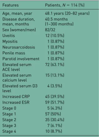
Table 2. Demographic, clinical and laboratory features in patients with sarcoidosis.
Features Patients, N = 114 (%) Age, mean, year 48.1 years (20–82 years) Disease duration,
mean, months 40.5 months (1–300 months) Sex (women/men) 82/32 Uveitis 12 (10.5%) Myositis 1 (0.87%) Neurosarcoidosis 1 (0.87%) Penile mass 1 (0.87%) Parotid involvement 1 (0.87%) Elevated serum ACE level 72 (63.1%) Elevated serum calcium level 15 (13.1%) Elevated serum D3 level 4 (3.5%) Increased CRP 45 (39.5%) Increased ESR 59 (51.7%) Stage 0 5 (4.3%) Stage 1 57 (50%) Stage 2 35 (30.4%) Stage 3 7 (6.1%) Stage 4 10 (8.7%)
ACE, angiotensin-converting enzyme; CRP, C-reactive protein; ESR erythrocyte sedimentation rate
were observed (p = 0.03, p = 0.01, p = 0.01 and
p = 0.02, respectively). Discussion
In this study of the prevalence of sarcoid arthropa-thy in a well-defined inception cohort population, joint pain was a most common clinical manifesta-tion, seen in 92.9% of patients with sarcoidosis. Acute sarcoid arthritis was seen in 71 (62.3%) patients, while chronic arthritis occurred in 24 (21.1%) patients. Oligoarthritis was the most common pattern of joint involvement, and bilat-eral ankle arthritis was the prominent feature. The prognosis of sarcoid arthropathy was generally favorable, as the arthritis resolved within 6 weeks in the majority of patients. Cutaneous manifesta-tions were observed in 46.5% of patients with sar-coid arthropathy. Erythema nodosum was the most common cutaneous finding. Other derma-tologic manifestations of sarcoidosis including lupus pernio, maculopapular rashes, psoriasis-like lesions, scars or tattoos were not observed in this cohort. A relationship between sarcoid arthritis and the female sex and acute phase reactants was detected in our cohort.
Sarcoidosis is a Th1-mediated systemic granu-lomatous disease of unknown etiology involving many different organs. It primarily starts with pulmonary symptoms, but extrapulmonary involvement is also frequent. Sarcoidosis can mimic many rheumatologic diseases or co-exist with them. It may present most frequently with clinical symptoms resembling connective tissue diseases such as primary Sjögren’s syndrome (SS), Systemic Lupus Erythematosus (SLE) and scle-roderma, as well as vasculitis and spondyloarthri-tis [Hansen et al. 2008; Fernandes et al. 2000]. Overall, two different patterns of joint involve-ment (acute /chronic) can be seen in 15–25% of patients with sarcoidosis. Acute arthritis is more common and usually affects uni/bilateral ankle, knee and wrist joints. Generalized arthralgia may also occur, in addition to some constitutional symptoms such as fatigue and fever. The inci-dence of Löfgren syndrome in our patients (17.5%) was consistent with the results reported in the literature [Sharma et al. 2012]. The high prevalence of musculoskeletal involvement detected in our study may be caused by genetic and racial reasons. Again, similar to other studies, ankle, knee and wrist were the most common joints involved. Chronic sarcoid arthritis is less common and seen in diffuse disease and in the
black race, and it involves joints such as knee, ankle, wrist, and MCP and PIP joints [Torralba and Quismorio, 2003]. It may cause Jaccoud’s-type deformative arthropathy or joint destruction. An increase is detected in synovial fluid analysis in mononuclear or polymorphonuclear cells as well as noncaseating granulomas and sometimes nonspecific signs are detected in synovial biopsy. Erosive destructive changes are rare in direct radiography, albeit soft tissue swelling, periarticu-lar osteoporosis, and joint space narrowing may be seen. In our series, 24 (21.1%) patients devel-oped chronic sarcoid arthritis. Also a positive correlation was detected between arthritis and acute phase reactants, ANA and RF positivity. Sarcoidosis can mimic many rheumatologic dis-eases or accompany them. It may present most frequently with clinical symptoms resembling connective tissue diseases such as primary SS, SLE and scleroderma, as well as vasculitis or spondyloarthritis [Kobak et al. 2013; Iannuzzi
et al. 2007]. In our series, 11 patients had another
rheumatic pathology concurrent with sarcoidosis Our study has some limitations. Restricted data from only one center and a relatively small num-ber of patients would not allow us to generalize these rheumatologic manifestations of sarcoido-sis. The clinical presentations of sarcoidosis vary with respect to ethnicity; the results of the current study might not be generalizable to other popula-tions, particularly those with a higher proportion of African-Americans. Nevertheless, our findings were similar to those reported in the literature. In conclusion, sarcoidosis is an important disease. Occasionally, diagnosis of sarcoidosis may be delayed or mistaken because it can mimic various rheumatologic diseases. Therefore, it must be con-sidered within the context of differential diagnosis for the patients admitted to the rheumatology phy-sician with complaints of the musculoskeletal sys-tem. Sarcoidosis is not just a great mimicker but it should also be considered as a Th1-mediated, pri-mary rheumatologic pathology. Further trials are needed to be performed on this subject.
Funding
This research received no specific grant from any funding agency in the public, commercial, or not-for-profit sectors.
Conflict of interest statement
The authors declare that there is no conflict of interest.
References
Chen, E. and Moller, D. (2008) Etiology of sarcoidosis. Clin Chest Med 29: 365–377.
Erb, N., Cushley, M., Kassimos, D., Shave, R. and Kitas, G. (2005) An assessment of back pain and the prevalence of sacroiliitis in sarcoidosis. Chest 127: 192–196.
Fernandes, S., Singsen, B. and Hoffman, G. (2000) Sarcoidosis and systemic vasculitis. Semin Arthritis Rheum 30: 33–46.
Glennas, A., Kvien, T., Melby, K., Refvem, O., Andrup, O., Karstensen, B. et al. (1995) Acute sarcoid arthritis: occurrence, seasonal onset, clinical features and outcome. Br J Rheumatol 34: 45–50. Gran, J. and Bøhmer, E. (1996) Acute sarcoid arthritis: a favourable outcome? A retrospective survey of 49 patients with review of the literature. Scand J Rheumatol 25: 70–73.
Gumpel, J., Johns, C. and Shulman, L. (1967) The joint disease in sarcoidosis. Ann Rheum Dis 26: 194–205.
Hansen, S., Hetta, A. and Omdal, R. (2008) Primary Sjögren’s syndrome and sarcoidosis: coexistence more than by chance? Scand J Rheumatol 37: 485–486. Hofmann, S., Franke, A., Fischer, A., Jacobs, G., Nothnagel, M., Gaede, K. et al. (2008) Genome-wide association study identifies ANXA11 as a new susceptibility locus for sarcoidosis. Nat Genet 40: 1103–1106.
Iannuzzi, M., Rybicki, B. and Teirstein, A. (2007) Sarcoidosis. N Engl J Med 357: 2153–2165. Kataria, Y. and Holter, J. (1997) Immunology of sarcoidosis. Clin Chest Med 18: 719–739.
Kobak, S., Sever, F., Ince, O. and Orman, M. (2014) The prevalence of sacroiliitis and spondyloarthritis
in patients with sarcoidosis. Int J Rheumatol 2014: 289454. DOI: 10.1155/2014/289454 [Epub ahead of print 12 May 2014].
Kobak, S., Sever, F., Sivrikoz, O. and Karaarslan, A. (2013) Coexistence of sarcoidosis and systemic sclerosis. Case Rep Rheumatol 2013: 684216. DOI: 10.1155/2013/684216 [Epub ahead of print 5 December 2013].
Milman, N. and Selroos, O. (1990) Pulmonary sarcoidosis in the Nordic countries 1950–1982: epidemiology and clinical picture. Sarcoidosis 7: 50–57. Newman, L., Rose, C. and Maier, L. (1997)
Sarcoidosis. N Engl J Med 336: 1224–1234. Pettersson, T. (1998) Rheumatic features of sarcoidosis. Curr Opin Rheumatol 10: 73–78.
Rybicki, B., Major, M., Popovich, J., Maliarik, M. and Iannuzzi, M. (1997) Racial differences in sarcoidosis incidence: a 5-year study in a health maintenance organization. Am J Epidemiol 145: 234–241.
Sharma, S., Soneja, M., Sharma, A., Sharma, M. and Hari, S. (2012) Rare manifestations of sarcoidosis in modern era of new diagnostic tools. Ind J Med Res 135: 621–629.
Smith, G., Brownell, I., Sanchez, M. and Prystowsky, S. (2008) Advances in the genetics of sarcoidosis. Clin Genet 73: 401–412.
Spilberg, I., Siltzbach, L. and McEwen, C. (1969) The arthritis of sarcoidosis. Arthritis Rheum 12: 126–137. Torralba, K. and Quismorio, F. (2003) Sarcoid arthritis: a review of clinical features, pathology, and therapy. Sarc Vasc Diff Lung Dis 20: 95–103. Visser, H., Vos, K., Zanelli, E., Verduyn, W., Schreuder, G., Speyer, I. et al. (2002) Sarcoid arthritis: clinical characteristics, diagnostic aspects, and risk factors. Ann Rheum Dis 61: 499–504. Visit SAGE journals online
http://tab.sagepub.com SAGE journals