OSTEOARTHRITIS: CAN IT BE REVERSED? A NEW BIOLOGICAL TREATMENT TECHNIQUE FOR TREATING PATIENTS WITH MODERATE TO ADVANCED GONARTHROSIS
BÜLENTKILIÇ1, MURATDEMIROĞLU2, FEYZAÜNLÜÖZKAN3, MEHMETSALIHSÖYLEMEZ4, İSMAILTÜRKMEN5, YAVUZSAĞLAM6, KORHANÖZKAN7
1Istanbul Gelişim University, Department of Health Sciences, Orthopaedic Surgeon, Istanbul, Turkey - 2Istanbul Medeniyet University, Goztepe Training and Research Hospital, Department of Orthopaedics and Traumatology, Istanbul, Turkey - 3Fatih Sultan Mehmet Training and Research Hospital, Department of Physical Therapy and Rehabilitation, Istanbul, Turkey - 4Bingöl State Hospital, Department of Orthopaedics and Traumatology, Bingöl, Turkey - 5Beykoz State Hospital, Department of Orthopaedics and Traumatology, Istanbul, Turkey - 6Biruni University, Faculty of Medicine, Department of Orthopaedics and Traumatology, Istanbul, Turkey - 7Istanbul Medeniyet University, Faculty of Medicine, Department of Orthopaedics and Traumatology, Istanbul, Turkey
Introduction
Osteoarthritis (OA) is the most prevalent form of arthritis and is a leading cause of disability, a problem that becomes more pronounced with increasing age(1). OA is found in almost 70% of the
population over 60 years of age and is characterised by a breakdown of articular cartilage and prolifera-tive changes to the surrounding bones(1-4).
Although various efforts are underway to identify different treatment modalities to prevent joint destruction and progression of the arthritis, an effective treatment protocol, particularly for advanced stages of gonartrosis, does not yet exist(2, 5). The ideal treatment of OA should focus
on prevention of articular cartilage dam¬age and many compounds are under investiga¬tion for this purpose.
Received October 30, 2016; Accepted January 02, 2017 ABSTRACT
Osteoarthritis of the knee is leading cause of disability in the elderly, all times. Although there are pharmacological and nonpharmacological treatment methods, these are generally insufficient to alleviate pain and the disability in advanced cases Although total knee arthroplasty improves quality of life and provides a generally high level of patient satisfaction for treatment of advanced gonarthrosis, it is not always without complications. Herein we present a new technique consisting of patellofemoral joint irrigation, simple osteophytectomy if needed, lateral patellar retinaculum release, subchondral drilling of the proximal tibia, percutaneous medial collateral ligament release, intra-articular injection of proximal tibial bone marrow, and the results of this treatment applied under local anesthesia in 20 knees of 13 patients. The mean VAS (Visual Analogue Scale) was 8.20 ± 0.68 prior to treatment and 3.33 ± 0.72 after treatment; the values were 18.67 ± 3.34 and 4.10 ± 3.15 for leg measurements, 7.80 ± 0.77 and 1.07 ± 0.96 for pain, 5.07 ± 2.28 and 1.80 ± 1.42 for walking, and 5.80 ± 0.92 and 1.23 ± 0.92 for daily living activities, respectively. All decreases were statistically significant (p = 0.001 for p < 0.01 in all cases). Biological treatment solutions to gonarthrosis without using foreign materials could decrease the need for prosthetic surgery and its related complications, as well as the need for further attempts at revision. In this study we presented a new biologic treatment for gonarthrosis.
Keywords: Gonarthrosis, Subchondral drilling, Bone Marrow injection, Osteophytectomy.
Here, we present a new treatment approach and accompanying results for patients with moder-ate to advanced gonarthrosis. These patients had undergone a minimally invasive treatment protocol under local anaesthesia consisting of patellofemoral joint simple osteophytectomy with joint lavage, lat-eral patellar retinaculum release, subchondral retro-grade drilling of the proximal tibia, percutaneous medial collateral ligament release, and whole bone marrow injection containing mesenchymal stem cells and supportive chondrogenic components. Materials and methods
This was an institutional Review Board-approved retrospective review of patients diagnosed with Kellgren-Lawrence grades 2-4 knee osteoarthritis who had undergone patellofemoral joint simple osteophytectomy with joint irrigation, lateral patellar retinaculum release, subchondral ret-rograde drilling of the proximal tibia, percutaneous medial collateral ligament release, and intra-articu-lar bone marrow injection at a single institution. Informed consent was obtained from all study par-ticipants and all procedures performed in this study were in accordance with the ethical standards of the institutional and/or national research committee and also with the 1964 Helsinki Declaration and its later amendments, or with comparable ethical standards.
The inclusion criteria were knee pain, gonarthrosis involving the medial compartment with or without lateral and patellar compartments of the knee, and unresponsiveness to conservative treatment.
The exclusion criteria were rheumatoid arthri-tis, blood diseases, known malignancy, immunode-ficiency, hepatitis, infection or septicemia. The Visual Analogue Score (VAS) and Lequesne Score were evaluated at initial presentation and at postop-erative months 3 and 12(6,7).
Technique
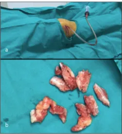
The technique was proposed by a senior sur-geon who is an expert on cartilage repair surgery and one of the authors of this paper (B.K.). An appropriate local anaesthetic (bupivacaine plus prilocaine) injection to the patellofemoral joint and around the patella was performed 10 min before the start of technique through a 0,5 cm incision created at superolateral side of patella (Fig. 1a). In cases of decreased patellofemoral joint motion due to osteo-phytes, incision was expanded to 2 cm to reach the
joint and perform simple patellofemoral joint osteo-phytectomy (Fig. 1b and 3 ). In cases of normal patellofemoral joint motion, only a lateral retinacu-lum release was performed through the same inci-sion. Then the patients underwent joint irrigation using 1500 cc sterile isotonic solution with the aid of a sterile naso-gastric feeding tube( Fig. 1a).
In the second step, local anaesthesia (bupiva-caine plus prilo(bupiva-caine) was administered to the medial collateral ligament area and medial proxi-mal tibia using injection syringes. Then a percuta-neous pie-crusting technique was used to release the medial collateral ligament with an 18-gauge needle and the application of gentle valgus force.
After completion of the second stage, sub-chondral retrograde drilling with a 5 mm drill began. The entry point of the drill was 1 cm medial to the tuberositas tibia and 3-4 cm below the joint Figure 1: a) Picture showing first incision location and
feeding tube used to deliver local injection and irrigation fluid. b) Osteophytes after patellofemoral joint osteophy-tectomy.
Figure 3: Image showing clinical appearance of the
same patients as in Figure 2 at postoperative first month. Red arrow; 2 cm lateral incision used for simple osteophytectomi. Black arrow; entrance point for retro-garde drilling.
line. Both medial and lateral plateaus of the tibia were drilled from this single hole and care was taken not to penetrate the articular cartilage (Fig 2a and 2b). Then, using an 18-gauge needle, 30 cc whole bone marrow was aspirated and the aspirate was injected into the knee joint. Range of motion exercises were started at postoperative first day and all patients were allowed to be weight-bearing as tolerated after the first day of procedure.
Statistical analysis
The 2007 Number Cruncher Statistical System (NCSS; Kaysville, Utah, USA) statistical software was used for the statistical analysis. A Wilcoxon signed-rank test was used to assess the descriptive statistical methods (mean, standard deviation, medi-an, frequency, ratio, minimum, and maximum), as well as measurements of variables without a normal distribution, before and after treatment. Significance was evaluated at a p-value < 0.01 or < 0.05
Results
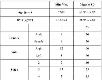
The study included 20 knees of 13 individuals, including 4 males (30%) and 9 females (70%). The ages of the individuals ranged between 52 and 85 years and the mean age was 61.90 ± 9.62 years. Twenty different measurements for each knee were recorded.
Right-sided measurements were recorded for 60% of the individuals (n = 12) and left-sided
mea-surements for 40% (n = 8). The body mass index (BMI) of the individuals ranged between 23.2 and 49.1 kg/m,2 and the mean BMI was 28.95 ± 7.68 kg/m2. Considering the osteoarthritis stages, 10 % of the individuals (n = 2) were classified as stage 2, 75 % of them (n = 15) as stage 3, and 15 % of them (n = 3) as stage 4 (Table 1 and 2).
Simple osteophytectomi was performed in 60% (n = 12) of the knees (Fig. 3 and 4).
The mean VAS was 8.20 ± 0.68 prior to treat-ment and 3.33 ± 0.72 after treattreat-ment; the values were 18.67 ± 3.34 and 4.10 ± 3.15 for leg measure-ments, 7.80 ± 0.77 and 1.07 ± 0.96 for pain, 5.07 ± 2.28 and 1.80 ± 1.42 for walking, and 5.80 ± 0.92 and 1.23 ± 0.92 for daily living activities (DLA), respectively. All decreases were statistically signifi-cant (p = 0.001 for p < 0.01 in all cases) (Graph. 1). Figure 2: İnitial anterior-posterior (a) and lateral (b)
x-rays of a 65 year-old a woman taken after procedure. Red arrows showing drilling holes.
Min-Max Mean ± SD Age (year) 52-85 61.90 ± 9.62 BMI (kg/m2) 23.2-49.1 28.95 ± 7.68 n % Gender Male 4 30 Female 9 70 Side Right 12 60 Left 8 40 Stage 2 2 10 3 15 75 4 3 15
Table 1: Distribution of descriptive characteristics.
SD: standard deviation; BMI: body mass index
n = 15 TreatmentBefore TreatmentAfter p-value
VAS Min-Max (Median) 7-9 (8) 2–4 (3) 0.001**
Mean ± SD 8.20 ± 0.68 3.33 ± 0.72
Leg Min-Max (Median) 10.5-22.5 (18) 0–8 (6) 0.001**
Mean ± SD 18.67 ± 3.34 4.10 ± 3.15
Pain Min-Max (Median) 5-8 (8) 0–2 (1) 0.001**
Mean ± SD 7.80 ± 0.77 1.07 ± 0.96
Walking Min-Max (Median) 1-8 (5) 0–4 (2) 0.001**
Mean ± SD 5.07 ± 2.28 1.80 ± 1.42
DLA
Min-Max (Median) 4-6.5 (6) 0–2 (2) 0.001**
Mean ± SD 5.80 ± 0.92 1.23 ± 0.92
Table 2: Assessments before and after treatment.
VAS: visual analogue score; SD: standard deviation; DLA: daily living activities
Statistically significant difference was not detected between osteophytectomi performed and nonper-formed individuals.
Discussion
OA affects almost 12% of adults in the United States and creates an economic burden of more than 15 billion dollars(8). Although total knee arthroplasty
improves quality of life and provides a generally high level of patient satisfaction, it is not always without complication. Unfortunately, OA can be associated with early or late infection, venous thromboemboli, neurovascular injury, septic and aseptic loosening, reflex sympathetic dystrophy, periprosthetic fractures, arthrofibrosis, wound com-plications, and even amputation(9-14).
Therefore, treatment methods that provide symptomatic relief and reversal of some aspects of gonarthrosis without the requirement for foreign materials, but rather using the body’s own regener-ative capacity, would form an innovregener-ative therapeu-tic approach to this debilitating and progressive condition.
Although subchondral bone is increasingly believed to play an important role in the disease process, there is still an ongoing debate about its role in the pathogenesis of OA. The area underlying the calcified cartilage, which is composed of sub-chondral bone plate and subsub-chondral trabecular bone, is involved in the pathogenesis of OA. Subchondral bone has two essential functions: stress absorption and maintenance of the cartilage nutrient supply. Pan et al.(16)reported the penetration
of small molecules into calcified cartilage from the subchondral bone, where the subchondral bone plate provides a direct link between articular carti-lage and subchondral bone.
In 1994, Arnoldi discussed the theory that decreased bone blood flow perfusion with decreased interstitial fluid flow in the subchondral bone results in ischemia and eventual bone death(17).
There are two potential outcomes of venous stasis in subchondral bone. First, decreased perfusion in subchondral bone results in decreased nourishment to the overlying cartilage(18). Second, the mechanical
strength of the subchondral bone could be negative-ly affected due to ischemia. In addition, venous sta-sis may lead to a loss of osteocyte viability, which stimulates a remodeling process. Subchondral bone and cartilage are complementary structures with dynamic stress-bearing features. Stiffened and less pliable subchondral bone could transmit increased loads to the overlying cartilage leading to sec-ondary cartilage damage(19).
Therefore, one of the current authors (B.K) proposed a technique using subchondral drilling to decrease the increased venous pressure to reconsti-tute the circulation, promote formation of new healthy subchondral bone instead of sclerosed bone and thus help reverse the ischemic process.
In recent years, emphasis has been given to subchondral bone marrow eudema-like lesions and subchondral bone cysts, which are presumed to play a role in the pathology of osteoarthritis(20).
Subchondral cyst-like lesions are generally seen in patients with gonarthrosis. These lesions appear as well-defined rounded areas of fluid-like intensity with no evidence of epithelial lining on histological examination.
There are two theories regarding the formation of subchondral cysts:
1) they are due to the leakage of synovial fluid through fissured or ulcerated cartilage causing the eventual development of cysts;
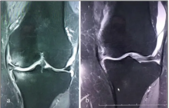
2) subchondral cysts develop from bone mar-Figure 4: Preoperative and postoperative MRI of the
same patients as in Figure 2 and 3. Coronal T2 MRI showin preoperative subchondral eudema and schlerosis (a), and coronal T2 MRI taken at postoperative 6. month showing relief of eudema and schlerosis.
row oedema-like lesions due to traumatic bone necrosis after the impacting of two opposing articu-lar surfaces. The second theory is supported by the fact that cysts are often observed in areas of the knee with concomitant bone marrow eudema-like lesions that have areas of necrosis and features of bone trauma detectable on histological examina-tion, and which in turn may be caused by decreased venous outflow with a subsequent cascade of osteoarthritis(21,22,23).
Pain in osteoarthritic joints has been associat-ed with increasassociat-ed intraosseous pressure(24,25). Thus,
drilling the subchondral bone may relieve pain by decreasing the pressure and hence increase the cir-culation.
Chondrogenic bone marrow-derived mes-enchymal stem cells are usually prepared as a buffy coat fraction of bone marrow. However, whole bone marrow retains the supportive chondrogenic com-ponents in marrow plasma and involves marrow adipocytes, which are also important in the forma-tion of chondrogenesis(26). In our patients, we used
whole bone marrow injections.
Joint lavage in OA has numerous beneficial effects. It helps to remove cartilage debris, micro-crystals, and degrading enzymes and cytokines involved in chondrolysis and disruption of intra-articular adhesions(27,28).
In releasing the medial collateral ligament in our patients, the main rationale was to unload the medial knee compartment, which in turn results in a reduced adduction moment and a reduction in the external rotation restraint in extension(29).
Biological treatment solutions to gonarthrosis without using foreign materials could decrease the need for prosthetic surgery and its related complica-tions, as well as the need for further attempts at revision (Fig. 3).
We introduced a new biological method for treating patients with moderate to advanced gonarthrosis, and the results are promising. However, further studies with control groups and a longer follow-up are necessary .
References
1) Brandt KD, Doherty M, Lohmander LS. Osteoarthritis. 2003: Oxford.
2) Farnworth L. Osteochondral defects of the knee. Orthopedics, 2000. 23(2): p. 146-57; quiz 158-159. 3) Moskowitz RW, Howell DS, Altman RD.
Osteoarthritis: Diagnosis and Medical/Surgical Management. 2001: London.
4) Prakash D, Learmonth D. Natural progression of osteo-chondral defect in the femoral condyle. Knee, 2002: 9(1); 7-10.
5) Bhosale AM, Richardson JB. Articular cartilage: struc-ture, injuries and review of management. Br Med Bull, 2008: (87); 77-95.
6) Wewers ME, Lowe NK. A critical review of visual ana-logue scales in the measurement of clinical phenomena. Research in Nursing and Health 1990: (13); 227-236. 7) Lequesne M, Mery C et al. Indexes of severity for
osteoarthritis of the hip and knee. Scand Rheumatology. 1987; (Splt65): 85-89.
8) Murphy L, Helmick CG. The impact of osteoarthritis in the United States: a population-health perspective: A population-based review of the fourth most common cause of hospitalization in U.S. adults. Orthop Nurs. 2012; 31(2): 85-91.
9) Healy WL, Della Valle CJ, Iorio R et al. Complications of total knee arthroplasty: standardized list and defini-tions of the Knee Society. Clin Orthop Relat Res. 2013; 471(1):215-220. doi: 10.1007/s11999-012-2489-y. 10) Iorio R, Della Valle CJ, Healy WL et al. Stratification
of standardized TKA complications and adverse events: a brief communication. Clin Orthop Relat Res. 2014;472(1):194-205. doi: 10.1007/s11999-013-2980-0.
11) Bozic KJ, Grosso LM, Lin Z et al. Variation in hospi-tal-level risk-standardized complication rates following elective primary total hip and knee arthroplasty. J Bone Joint Surg Am. 2014;96(8):640-647. doi: 10.2106/JBJS.L.01639.
12) Siow WM, Chin PL, Chia SL, Lo NN, Yeo SJ. Comparative demographics, ROM, and function after TKA in Chinese, Malays, and Indians. Clin Orthop Relat Res. 2013; 471(5): 1451-1457. doi: 10.1007/s11999-012-2776-7.
13) Piedade SR, Pinaroli A, Servien E, Neyret P. TKA out-comes after prior bone and soft tissue knee surgery. Knee Surg Sports Traumatol Arthrosc. 2013;21(12):2737-2743. doi: 10.1007/s00167-012-2139-7.
14) Higuera CA, Elsharkawy K, Klika AK, Brocone M, Barsoum WK. 2010 Mid-America Orthopaedic Association Physician in Training Award: predictors of early adverse outcomes after knee and hip arthroplasty in geriatric patients. Clin Orthop Relat Res. 2011;469(5):1391-1400. doi: 10.1007/s11999-011-1804-3.
15) Koh IJ, Cho WS, Choi NY, Kim TK; Kleos Korea Research Group. Causes, risk factors, and trends in fail-ures after TKA in Korea over the past 5 years: a multi-center study. Clin Orthop Relat Res. 2014;472(1):316-326. doi: 10.1007/s11999-013-3252-8.
16) Pan J, Zhou X, Li W, Novotny JE, Doty SB, Wang L. In situ measurement of transport between subchondral bone and articular cartilage. J Orthop Res. 2009; 27(10): 1347-1352
17) Arnoldi, C.C. Vascular Aspects of Degenerative Joint Disorders. A Synthesis. Acta Orthopaedica Scandinavica Suppl, 1994; (261); 1-82.
18) Imhof, H., Breitenseher, M., Kainberger, F. and Trattnig, S. Degenerative Joint Disease: Cartilageor or Vascular Disease? Skeletal Radiology, 1997: (26); 398-403.
19) Li G, Yin J, Gao J et al. Subchondral bone in osteoarthritis: insight into risk factors and microstruc-tural changes. Arthritis Res Ther. 2013; 15(6): 223. 20) Tanamas SK, Wluka AE, Pelletier JP et al. The
associa-tion between subchondral bone cysts and tibial carti-lage volume and risk of joint replacement in people with knee osteoarthritis: a longitudinal study. Arthritis Res Ther. 2010;12(2):R58. doi: 10.1186/ar2971. Epub 2010 Mar 31.
21) Davies-Tuck ML, Wluka AE, Forbes A et al. Development of bone marrow lesions is associated with adverse effects on knee cartilage while resolution is associated with improvement-a potential target for pre-vention of knee osteoarthritis: a longitudinal study. Arthritis Res Ther. 2010;12(1):R10. doi: 10.1186/ar2911.
22) Guymer E, Baranyay F, Wluka AE et al. A study of the prevalence and associations of subchondral bone mar-row lesions in the knees of healthy, middle-aged women. Osteoarthritis Cartilage. 2007; 15(12): 1437-42.
23) Tanamas SK, Wluka AE, Pelletier JP et al. The associa-tion between subchondral bone cysts and tibial carti-lage volume and risk of joint replacement in people with knee osteoarthritis: a longitudinal study. Arthritis Res Ther. 2010;12(2):R58. doi: 10.1186/ar2971. 24) Simkin PA. Bone pain and pressure in osteoarthritic
joints. Novartis Found Symp. 2004;260:179-86; discus-sion 186-190, 277-279.
25) Arnoldi CC, Lemperg K, Linderholm H. Intraosseous hypertension and pain in the knee. J Bone Joint Surg Br. 1975; 57(3): 360-363.
26) Hauser RA, Orlofsky A. Regenerative injection therapy with whole bone marrow aspirate for degenerative joint disease: a case series. Clin Med Insights Arthritis Musculoskelet Disord. 2013; 4(6): 65-72. doi: 10.4137/CMAMD.S10951.
27) Ravaud P, Moulinier L, Giraudeau B et al. Effects of joint lavage and steroid injection in patients with osteoarthritis of the knee: results of a multicenter, ran-domized, controlled trial. Arthritis Rheum. 1999; 42(3): 475-482.
28) de Rezende MU, Rosa TB, Pasqualin T, Frucchi R, Okazaki E, Villaça PR. Subjective results of joint lavage and viscosupplementation in hemophilic arthropathy. Acta Ortop Bras. 2015; 23(3): 162-166. doi: 10.1590/1413-785220152302145885.
29) Lyu SR. Arthroscopic medial release for medial com-partment osteoarthritis of the knee: the result of a single surgeon series with a minimum follow-up of four years. J Bone Joint Surg Br. 2008;90(9): 1186-92. doi: 10.1302/0301-620X.90B9.20584.
Acknowledgements
No funding or sponsorship was received for this study or publi-cation of this article.
All named authors meet the “International Committee of Medical Journal Editors” criteria for authorship for this man-uscript, take responsibility for the integrity of the work as a whole, and have given final approval to the version to be pub-lished.
Ethical Approval
All procedures performed in studies involving human partici-pants were in accordance with the ethical standards of the institutional and/or national research committee and with the 1964 Helsinki declaration and its later amendments or compa-rable ethical standards.
_________
Corresponding author
MURATDEMIROGLU, MD
Istanbul Medeniyet University, Goztepe Training and Research Hospital, Department of Orthopaedics and Traumatology, Istanbul