Original Article
Multi-assay investigation of viral etiology in pediatric central nervous
system infections
Aylin Altay-Kocak1, Gulendam Bozdayi2, Janine Michel3, Meltem Polat4, Saliha Kanik-Yuksek5, Hasan Tezer4, Aykut Ozkul6, Kamruddin Ahmed7, Andreas Nitsche3, Koray Ergunay8
1 Department of Medical Microbiology, Faculty of Medicine, Baskent University, Ankara, Turkey 2 Department of Medical Microbiology, Faculty of Medicine, Gazi University, Ankara, Turkey
3 Center for Biological Threats and Special Pathogens 1 (ZBS-1), Robert Koch Institute, Berlin, Germany 4 Paediatric Infections Unit, Department of Paediatric, Faculty of Medicine, Gazi University, Ankara, Turkey 5 Department of Paediatric, Ministry of Health Diskapi Yildirim Beyazit Training and Education Hospital, Ankara, Turkey
6 Department of Virology, Faculty of Veterinary, Ankara University, Ankara, Turkey
7 Department of Pathobiology and Medical Diagnostics, Faculty of Medicine and Health Sciences, University Malaysia Sabah, Kota Kinabalu, Malaysia
8 Department of Medical Microbiology, Faculty of Medicine, Hacettepe University, Ankara, Turkey
Abstract
Introduction: In an attempt to identify a wide spectrum of viral infections, cerebrospinal fluid (CSF) specimens were collected from pediatric cases with the preliminary diagnosis of viral encephalitis/meningoencephalitis in two reference hospitals, from October 2011 to December 2015.
Methodology: A combination of nucleic acid-based assays, including in house generic polymerase chain reaction (PCR) assays for enteroviruses, flaviviruses and phleboviruses, a commercial real-time PCR assay for herpesviruses and a commercial real time multiplex PCR, enabling detection of frequently-observed viral, bacterial and fungal agents were employed for screening.
Results: The microbial agent could be characterized in 10 (10%) of the 100 specimens. Viral etiology could be demonstrated in 7 (70%) specimens, which comprises Human Herpesvirus 6 (4/7), Herpes Simplex virus type1 (2/7) and Enteroviruses (1/7). In 3 specimens (30%), Streptococcus pneumoniae, Listeria monocytogenes and Staphylococcus aureus were detected via the multiplex PCR, which were also isolated in bacteriological media. All specimens with detectable viral nucleic acids, as well as unreactive specimens via nucleic acid testing remained negative in bacteriological cultures.
Conclusions: Herpes and enteroviruses were identified as the primary causative agents of central nervous system infections in children. Enterovirus testing must be included in the diagnostic work-up of relevant cases.
Key words: Encephalitis; meningoencephalitis; viral; enterovirus; herpesvirus. J Infect Dev Ctries 2020; 14(6):572-579. doi:10.3855/jidc.12327
(Received 16 December 2019 – Accepted 19 April 2020)
Copyright © 2020 Altay-Kocak et al. This is an open-access article distributed under the Creative Commons Attribution License, which permits unrestricted use, distribution, and reproduction in any medium, provided the original work is properly cited.
Introduction
Central nervous system infections, manifesting as meningitis or encephalitis are caused by a broad variety of infectious agents generating diagnostic and treatment challenges. The clinical presentation is referred to as meningitis, encephalitis, or meningoencephalitis, according to the presence of meningeal signs and altered brain function (mental status, behaviour changes or focal signs). A rapid etiologic diagnosis to differentiate viral and bacterial causes is crucial to guide the introduction of empirical antibiotic therapy or avoid its unnecessary use [1]. Furthermore, initiation of antiviral therapy is required in all suspected cases of
sporadic encephalitis due to Herpes simplex virus (HSV) type 1, a prevalent, debilitating and potentially fatal virally-induced central nervous system infection [2]. The conditions that are most important to recognize in the first hour are bacterial meningitis and herpetic encephalitis, as these diseases cause significant morbidity and mortality and have specific treatments that can improve patient outcome if administered rapidly [2]. Therefore, an early identification of the causative agent has a significant impact on the prognosis and individual management of the patient.
Viruses are among the leading cause of meningitis worldwide, with an estimated incidence of 12–19 cases
per 100,000 per year [3]. Moreover, around 200,000 annual viral encephalitis cases are reported globally [4]. The clinical picture caused by viral agents that elicit various neurological manifestations through different pathogenic mechanisms is not always distinct and clear-cut, but rather reflects a continuous spectrum with overlapping features of meningoencephalitis, meningomyelitis, or meningoencephalomyelitis [5]. Over 100 viruses have been implicated in the etiology of acute central nervous system infections, which further complicates differential diagnosis [4]. Although certain distinctive geographical and seasonal patterns are recognized, the most common viruses associated with encephalitis are human herpesviruses, enteroviruses and vector-borne (or arthropod-borne) viruses belonging in various families [6,7]. Although all age groups can be affected by viral central nervous system infections, some viruses are more commonly-observed among pediatric cases. Viruses of the enterovirus (EV) genus are generally considered as the primary cause of meningitis in children, but several other agents such as human herpesviruses and vector-borne viruses are also observed. Majority of the HSV type 2 encephalitis cases are documented in neonates after exposure to the virus in genital tract during delivery. Among the vector-borne viruses, several strains from the genus flavivirus, such as West Nile virus and Japanese encephalitis virus have been associated with encephalitis and meningitis [7,8]. The International Encephalitis Consortium recommends HSV, EV and human parechoviruses [in individuals younger than 3 years] as the first assessment of a probable viral etiology in children [9].
The laboratory diagnosis and identification of the etiological agent in central nervous system infections rely on the analysis of cerebrospinal fluid (CSF). As biochemical tests and direct microscopy of the CSF may provide an early insight of the ongoing pathological process, the definitive diagnosis is based on the identification of the causative microbial agent. Inoculation of the CSF specimen for bacterial and fungal isolation and subsequent antibiotic resistance analysis is widely-used in diagnostic microbiology settings, as well as rapid bacterial and fungal antigen assays. Likewise, polymerase chain reaction (PCR) based assays for frequently-observed viruses such as HSV is a common practice [10]. However, performing individual nucleic acid testing for all probable viral pathogens is often expensive and cumbersome, and beyond the scope of the majority of the hospital diagnostic services. Therefore, in some instances, precise identification of the causative agent in aseptic
meningitis/encephalitis could not be attained and the etiology may remain obscure [11]. This study was performed in order to investigate the viral etiology in pediatric cases with the preliminary diagnosis of central nervous system infections, using a combination of individual and multiplex standard and real-time assays that can detect a wide spectrum of agents reported to cause meningitis/encephalitis.
Methodology
Study design, sample collection and processing
The study was undertaken in pediatric infection outpatient or emergency clinics of two reference hospitals in Ankara, from October 2011 to December 2015. Patients with the preliminary clinical diagnosis of central nervous system infection were enrolled in the study with individual and parental informed consent. CSF specimens were obtained from patients, aliquoted and kept at -86oC. Aliquots of the specimens were
submitted for routine biochemical and bacteriological examination, including inoculation in appropriate media for bacteria causing bacterial meningitis or encephalitis. The study and associated protocols were approved by the local ethics committees of both centers (16.11.2011/341).
CSF specimens were processed identically prior to the detection pipeline. Nucleic acid purification and complementary DNA (cDNA) synthesis using random hexamers were performed by commercial assays (QIAamp® Viral RNA Mini Kit, Qiagen, Hilden, Germany; RevertAid First Strand cDNA Synthesis Kit, ThermoFisher Scientific, Waltham, Massachusetts, USA), as suggested by the manufacturers. The processed specimens were subsequently evaluated via individual and multiplexed pathogen detection assays.
Pathogen detection via individual assays
Commercial or laboratory-optimized in house polymerase chain reaction (PCR) assays were employed for screening of frequent and rare agents of viral meningoencephalitis. Herpes simplex virus (HSV) type 1 and 2 detections were undertaken via a widely-used commercial real time PCR assay (LightCycler® HSV1/2 QualKit, Roche Diagnostics, Mannheim, Germany) in a LightCycler 2.0 Instrument (software version 4.1) (Roche Diagnostics, Mannheim, Germany) according to manufacturer’s instructions.
For the screening of enteroviruses, a set of novel primers, targeting the highly-conserved 5’ untranslated region (UTR) in all enterovirus strains were designed, using the CLC Main Workbench v7.7’ (CLCBio, Aarhus, Denmark) software. The primers (Forward:
CCGGCCCCTGAATGC-3’, reverse PENV.R 5’-CACCGGATGGCCAATCCA-3’) are designed to amplify a 193-base pair (bp) stretch in the 5’-UTR and enables strain characterization after amplicon sequencing. The assay was optimized using cell culture grown Coxsackie virus B6 (Schmitt) isolate.
Previously-described generic nested PCR assays were employed for the detection of flavi- and phleboviruses. The flavivirus PCR targets the NS5 gene and amplifies all major pathogenic flaviviruses such as West Nile virus (WNV), Dengue viruses, yellow fever virus, tick-borne encephalitis virus, Murray Valley encephalitis virus, Saint Louis encephalitis virus and Usutu virus, with a detection limit of 40 TCID50 per reaction [12]. The assay produces a 960 bp amplicon in the second round, which can be sequenced for strain characterization. Vero cell-grown WNV isolate NY99-4132 was used for assay optimization and positive control during testing.
The phlebovirus assay also utilizes a nested reaction employing degenerated primers to amplify all medically-significant phleboviruses as well as Toscana virus (TOSV), the main agent of viral meningoencephalitis of the species [13]. The assay targets the RNA-dependent RNA polymerase coding region, located in the L segment of the viral genome and produces a 250 bp second round product, suitable for sequencing for strain identification. The assay was optimized using TOSV isolate Strain ISS.Phl.3, further used as positive control.
Reaction mixes for enterovirus, flavivirus and phlebovirus included 10× Buffer, 25mM Mg+2, 5U/µL
Taq polimerase (Biomatik, Wilmington, Delaware, USA), 10 mM dNTP (Fermentas, Waltham,
Massachusetts, USA) and 10 pm of the relevant primers. Amplicons of the expected size were investigated under ultraviolet light following electrophoresis in 1.5-2% agarose gels.
Pathogen detection via multiplex assay
Processed CSF specimens were evaluated in parallel using a commercial real time multiplex PCR method (MeningoFinder® 2SMART, PathoFinder BV, Maastricht, The Netherlands), enabling detection of 11 viral (HSV-1, HSV-2, Varicella-zoster virus, Epstein-Barr virus, human herpesvirus (HHV) 6, 7, HHV-8, human enteroviruses, human parechoviruses, mumps virus and measles virus), 8 bacterial (Borrelia
burgdorferi sensu lato, Echerichia coli K1, Haemophilus influenzae, Listeria monocytogenes, Neisseria meningitidis, Staphylococcus aureus,
Streptococcus agalactiae and Streptococcus
pneumoniae) and 2 fungal (Cryptococcus neoformans
s.l. and Cryptococcus gatii s.l.) agents, responsible for CNS infections. The assay employs 24 distinct primer sets combined with 14 fluorescent labelled probes to identify the targeted pathogens via melting curve analysis, performed in two reaction mixes. Purified nucleic acids from the CSF specimens were used for preparation of the reaction mixes, as the assay incorporates a reverse transcription step prior to PCR. Amplification and analysis were carried out using a LightCycler 480 instrument (Roche Diagnostics, Mannheim, Germany), with the assay controls as suggested by the manufacturer.
Results
A total of 100 patients that comprise 39 female and 61 male individuals were enrolled in the study. A predominance of 1-3 year age group (37/100) was observed in the study cohort. The distribution of individuals according to gender and age strata is provided in Figure 1.
During the clinical evaluation of the cases, 64 and 29 individuals were provided with the preliminary diagnosis of meningitis and encephalitis, respectively. Meningoencephalitic symptoms, an infection of the ventricular shunt and acute flask paralysis were noted in 7 individuals. The biochemical analysis and cell counts in the obtained CFS specimens are provided in Table 1.
The microbial pathogen, potentially associated with the clinical presentation could be identified in 10 (10%) of the CSF specimens tested. In 3 specimens, bacterial agents S. pneumoniae, L. monocytogenes and S. aureus were detected via the multiplex PCR (Table 2). Figure 1. The distribution of individuals according to gender
Table 1. CSF findings according to the preliminary diagnosis in the study cohort.
Clinical presentation
Meningitis Encephalitis Meningoencephalitis Other† Total
CSF findings
Protein Increased (> 45 mg/dL) Normal (15-45 mg/dL) 53 7 22 4 0 1 2 4 13 80
Unknown 4 3 0 0 7
Glucose
Decreased (< 45 mg/dL or lower
than ½ of blood sugar) 31 1 0 3 35
Normal (45-80 mg/dL) 27 24 1 2 54
Increased (> 80 mg/dL or higher
than 2/3 of blood sugar) 2 0 0 1 3
Unknown 4 4 0 0 8
Cell count Normal (0-5/mm
3) 2 1 0 0 3
Increased (> 5/mm3) 56 21 1 4 82
Unknown 6 7 0 2 15
Total 64 29 1 6 100
†Includes shunt infection (n:3), acute flask paralysis (n:2) and ADEM (n:1).
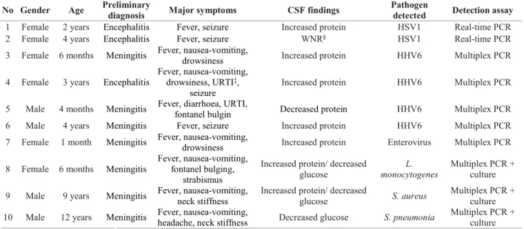
Table 2. The clinical features and main findings in individuals with detectable pathogens.
No Gender Age Preliminary diagnosis Major symptoms CSF findings Pathogen detected Detection assay
1 Female 2 years Encephalitis Fever, seizure Increased protein HSV1 Real-time PCR
2 Female 4 years Encephalitis Fever, seizure WNR§ HSV1 Real-time PCR
3 Female 6 months Meningitis Fever, nausea-vomiting, drowsiness Increased protein HHV6 Multiplex PCR 4 Female 3 years Encephalitis Fever, nausea-vomiting, drowsiness, URTI‡,
seizure Increased protein HHV6 Multiplex PCR
5 Male 4 months Meningitis Fever, diarrhoea, URTI, fontanel bulgin Decreased protein HHV6 Multiplex PCR
6 Male 4 years Meningitis Fever, seizure Increased protein HHV6 Multiplex PCR
7 Female 1 month Meningitis Fever, nausea-vomiting, drowsiness Increased protein Enterovirus Multiplex PCR 8 Female 6 months Meningitis Fever, nausea-vomiting, fontanel bulging,
strabismus
Increased protein/ decreased
glucose monocytogenes L. Multiplex PCR + culture 9 Male 9 years Meningitis Fever, nausea-vomiting, neck stiffness Increased protein/ decreased glucose S. aureus Multiplex PCR + culture 10 Male 12 years Meningitis Fever, nausea-vomiting, headache, neck stiffness Decreased glucose S. pneumonia Multiplex PCR + culture
‡ Upper respiratory tract infection; § Within normal range.
Table 3. The biochemical CSF findings in individuals with detectable pathogens.
Detectable pathogens (n)
HSV1 HHV6 EV Bacteria Total
CSF findings
Protein Increased (> 45 mg/dL) Normal (15-45 mg/dL) 1 1 3 - 1 - 1 2 2 7
Decreased (< 15 mg/dL) - 1 - - 1
Glucose
Decreased (< 45 mg/dL or lower than ½
of blood sugar) - - - - -
Normal (45-80 mg/dL) 2 4 1 - 7
Increased (> 80 mg/dL or higher than 2/3
of blood sugar) - - - 3 3
Cell count Normal (0-5/mm
3) - - - - -
Increased (> 5/mm3) 4 1 3 8
Unknown 2 - - - 2
These bacteria could also be isolated and biochemically characterized via CSF cultures, performed using separate aliquots (data not given). Viral etiology could be demonstrated in 7 of the 10 reactive specimens, that comprise HHV6 (4/7), HSV-1 (2/7) and EV (1/7). Reactive HSV-1 results were provided by the commercial real-time assay, whereas HHV6 and EV positivities were obtained via the multiplex PCR assay. All specimens with detectable viral nucleic acids, as well as unreactive specimens via nucleic acid testing remained negative in bacteriological cultures. In house assays for flavivirus, phlebovirus, and enterovirus assays were negative in all tested specimens. The clinical features and main findings in individuals with detectable pathogens are provided in Table 2 and Table 3.
The clinical outcome in individuals with detectable pathogen, as well as in 88 with negative results was favorable, with discharge from hospital without any residual neurological problem. However, an individual with preliminary diagnosis of meningitis succumbed to the disease, whereas neurological sequel remained in an individual presenting with encephalitis, after discharge. No pathogen could be identified in both cases.
Discussion
Despite the global predominance of certain agents, the etiology in virally-induced meningitis or meningoencephalitis remains unknown in a significant proportion of the cases. Rapid changes in the geographical distribution and viral emergence in new areas (Parechovirus, West Nile virus), in new hosts (West Nile in humans) or in immunocompromised patients further complicates precise laboratory diagnosis in most hospital settings [14]. The purpose of the present study was to investigate frequent as well as rare viral causes of central nervous system infections in two reference centers, located in Central Anatolia, Turkey, employing a wide spectrum of in house and commercially-available nucleic acid-based assays with individual or multiplex targets.
The causative viral agent could be identified in 7 individuals within the study cohort, comprising CSF specimens from 100 pediatric cases of central nervous system infections of presumed viral etiology (Table 2). Members of the Herpesviridae family, namely HHV6 and HSV-1, constituted the majority of the detected agents (6/7, 85.7%). This finding is in accordance with several global and local reports, where herpesviruses including HSV types 1 and 2, Varicella-Zoster virus, Cytomegalovirus, Epstein-Barr virus and HHV6 could be detected, despite variations in study groups and
detection methods [15-19]. The individuals, with detectable HSV-1 DNA, 2 and 4-year-old female children, presented with typical clinical (fever, seizure) and laboratory findings (moderately elevated or <50 mg/dl CSF protein and normal glucose) [20] (Table 2).
A relatively high incidence of HHV6 infections have been noted in the patients from both centers (4 individuals out of 7 being positive, Table 2). The well-known presentation of the primary HHV6 infection, Exanthem subitum, generally occurs in immunocompetent children aged 6 months-3 years, associated with febrile seizures with a peak incidence especially in the second year of life progress to encephalitis [21-23]. Central nervous system manifestations, mostly observed as encephalitis, are more frequent following virus reactivation usually during immunosuppressive states [22]. The individuals with detectable HHV6 DNA in CSF were immunocompetent and all presented with typical clinical and CSF findings, except for a 4-month old infant presenting mainly with meningitis accompanied by diarrhea and upper respiratory tract symptoms and decreased CSF protein (Table 2). In previously exposed individuals with latent infection, HHV6 nucleic acids can be detected in healthy brain tissue as well as in CSF and may not be indicative of an infection. Thus, it is suggested that a quantitative analysis, the detection of lytic mRNAs or viral integration and simultaneous screening of serum in addition to CSF provides better evidence for an ongoing neuroinvasive infection [7,24,25]. Although this could not be proven in the individuals we have identified, the age, medical history and clinical presentation strongly suggests a primary infection or recent exposure.
In addition to herpesviruses, we have identified EV RNA in a one month-old infant, presenting mainly with meningitic symptoms (Table 2). EV are reported to be among the most frequently-identified agent of virally-induced aseptic meningitis and standard as well as shell vial cell cultures can also be employed, as well as amplification-based nucleic acid assays for diagnosis [26]. If investigated via these assays, EVs can be readily identified in diagnostic settings [27]. For this study, we have developed and optimized an in house generic PCR for EV detection. Moreover, clinical specimens were tested via the real time multiplex PCR assay that also targets EV. The single positive EV result (weak positivity) was provided by the multiplex assay where the in house assay remained negative. Several factors may have contributed to this observation, such as processing of the different aliquots, relative difference in assay sensitivities. Moreover, low EV viral loads
have been documented in CSF an affected individuals and parallel testing of a plasma specimen is recommended to prevent false negativity [28,29]. However, we have also noted discrepant assay results for HSV-1 detection, as positive results were provided by the specific real-time PCR only. These findings can also be attributed to the issues described above. Since we have not aimed for a comprehensive evaluation of the assays and the study design have shortcomings for this purpose, the precise factors affecting assay performance could not be fully elucidated. Nevertheless, the combination of the assays we employed in this study were well-suited to overcome the major inherent problems of nucleic acid testing in diagnostic settings and could successfully detect the targeted agents.
Besides viruses, we have identified a bacterial etiology in the central nervous system infections in 3 of the evaluated individuals in CSF via the real time multiplex PCR assay (Table 2). These findings were confirmed by standard CSF culture methods, which constitute the mainstay of laboratory testing in the majority of the diagnostic service providers. The identified bacteria are frequently-detected agents of community-acquired central nervous system infections presenting as meningitis [18,30]. Compared to routine culture isolation methods, nucleic acid testing has the advantage of providing early information about nature of the causative agent, thus, facilitating the optimal use of empirical therapy regimens. However, isolation and culture-based assays are required for the antibiotic resistance testing and a precise characterization of the most bacterial and fungal pathogens. Therefore, a combination of nucleic acid assays and isolation methods, along with intrathecal antibody testing may be utilized for an optimized diagnostic strategy [31].
Despite the combination of various nucleic acid assays, the etiology in 90% of the individuals could not be identified in this study. It is documented that the etiology in encephalitis/meningoencephalitis remain obscure in up to 85% of the cases worldwide and significant regional variation can be observed in both the recognized causes and the number of undiagnosed cases [32]. Although we could cover a significant spectrum of frequent and rare microbial agents by the employed assays, bacterial infections such as tuberculosis, brucellosis and viral infections by bocavirus, metapneumovirus, previously identified as etiological agents in Turkey [33-35], were not included in the current testing algorithm. Furthermore, noninfectious causes of encephalitis, including post-infectious and immune-mediated syndromes must be
considered and ruled out via appropriate tests [31]. An appropriate specimen collection is also vital for a successful identification of the causative infectious agent. Obtaining a follow-up CSF specimen might be necessary when the initial specimen is PCR negative [31]. We have also not employed serological diagnostics in this study. Despite providing epidemiologically-relevant data based on previous or recent exposures, serology can be limiting during screening for a wide spectrum of agents, as performed in this study. Lack of standardized assays for all probable agents, requirement of complex assays such as viral neutralization and demonstration of intrathecal antibody synthesis for definitive diagnosis in certain infections are the main shortcomings of the serological approach to diagnosis in our setting. Therefore, we aimed to maintain a broad target spectrum via an arsenal of nucleic acid assays, performed on CSF specimens. Conclusion
In conclusion, this study was carried out to determine the agents of viral meningitis and encephalitis in our region, and epidemiological data were obtained even if the positivity rate was low. The causative identified agents of the patients were herpes (HSV1, HHV6) and enteroviruses. Besides HSV1, HHV6 and enteroviruses are rarely tested in clinical laboratories for CNS infections in Turkey. Since these agents may also have a role in CNS infections, they should be evaluated in diagnostic algorithms. It is very important to determine the etiology of viral meningitis and encephalitis infections and establish a rapid diagnosis, since the infection progresses rapidly and treatment should be given immediately. Therefore, more studies are needed to investigate the etiology of these infections.
Authors’ Contributions
G.B., K.E. and K.A. designed the study; M.P., S.K.Y and H.T. collected the samples and patients’ data; A.A.K. and J.M. performed experiments and analysed data; A.O. designed primers; A.A.K. and K.E. wrote the manuscript; G.B., K.E. and K.A. gave scientific support and conceptual advice.
References
1. Casaroto E, Marra AR, Coelho FMS, Pinho JRR, Sitnik R, Colombari F, Victor ES, Muto NH, Senne C, dos Santos OFP, Edmond MB (2013) Improving the diagnosis of meningitis due to enterovirus and herpes simplex virus I and II in a tertiary care hospital. BMC Infect Dis 13: 487.
2. Gaieski DF, Nathan BR, O’Brien NF (2015) Emergency neurologic life support: Meningitis and encephalitis. Neurocrit Care 23 (Suppl 2): 110–118.
3. Rudolph H, Prieto Dernbach R, Walka M, Rey-Hinterkopf P, Melichar V, Muschiol E, Schweitzer-Krantz S, Richter JW, Weiss C, Böttcher S, Diedrich S, Schroten H, Tenenbaum T. (2017) Comparison of clinical and laboratory characteristics during two major paediatric meningitis outbreaks of echovirus 30 and other non-polio enteroviruses in Germany in 2008 and 2013. Eur J Clin Microbiol Infect Dis 36: 1651–1660. 4. Xie Y, Tan Y, Chongsuvivatwong V, Wu X, Bi F, Hadler SC,
Jiraphongsa C, Sornsrivichai V, Lin M, Quan Y. (2015) A population-based acute meningitis and encephalitis syndromes surveillance in Guangxi, China, May 2007- June 2012. PLoS ONE 10: e0144366.
5. Studahl M, Lindquist L, Eriksson BM, Günther G, Bengner M, Franzen-Röhl E, Fohlman J, Bergström T, Aurelius E. (2013) Acute viral infections of the central nervous system in immunocompetent adults: Diagnosis and management. Drugs 73: 131–158.
6. Yavarian J, Gavvami N, Mamishi S (2014) Detection of human herpesvirus 6 in cerebrospinal fluid of children with possible encephalitis. Jundishapur J Microbiol 7: e11821.
7. Stahl J-P, Mailles A, Dacheux L, Morand P (2011) Epidemiology of viral encephalitis in 2011. Med Mal Infect 41: 453–464.
8. de Oliveira DB, Candiani TM, Franco-Luiz APM, Almeida GMF, Abrahão JS, Rios M, Coimbra RS, Kroon EG. (2017) Etiological agents of viral meningitis in children from a dengue-endemic area, Southeast region of Brazil. J Neurol Sci 375: 390–394.
9. Parisi SG, Basso M, Del Vecchio C, Andreis S, Franchin E, Bello FD, Pagni S, Biasolo MA, Manganelli R, Barzon L, Palu G (2016) Virological testing of cerebrospinal fluid in children aged less than 14 years with a suspected central nervous system infection: A retrospective study on 304 consecutive children from January 2012 to May 2015. Eur J Paediatr Neurol 20: 588–596.
10. Jarrin I, Sellier P, Lopes A, Morgand M, Makovec T, Delcey V, Champion K, Simoneau G, Green A, Mouly S, Bergmann J-F, Lloret-Linares C (2016) Etiologies and management of aseptic meningitis in patients admitted to an internal medicine department: Medicine 95: e2372.
11. Glaser CA, Honarmand S, Anderson LJ, Schnurr DP, Forghani B, Cossen CK, Schuster FL, Christie LJ, Tureen JH. (2006) Beyond viruses: Clinical profiles and etiologies associated with encephalitis. Clin Infect Dis 43: 1565–1577.
12. Vázquez A, Sánchez-Seco M-P, Palacios G, Molero F, Reyes N, Ruiz S, Aranda C, Marques E, Escosa R, Moreno J, Figuerola J, Tenorio A (2012) Novel flaviviruses detected in different species of mosquitoes in Spain. Vector Borne Zoonotic Dis 12: 223–229.
13. Sánchez-Seco MP, Echevarría JM, Hernández L, Estevez D, Navarro-Mari J, Tenorio A (2003) Detection and identification of Toscana and other phleboviruses by RT-nested-PCR assays with degenerated primers: Nested-RT-PCRs for Phlebovirus Detection. J Med Virol 71: 140–149.
14. Calleri G, Libanore V, Corcione S, De Rosa F, Caramello P (2017) A retrospective study of viral central nervous system infections: relationship amongst aetiology, clinical course and outcome. Infection 45: 227–231.
15. Soylar M, Altuglu I, Sertoz R, Aydin D, Akkoyun F, Zeytinoglu A. (2014) Viral agents in patients with central
nervous infections admitted to Ege University Hospital. Ege Journal of Medicine 53: 65–70 [Article in Turkish].
16. Sarınoglu RC, Saglik I, Mutlu D, Ozhak Baysan B, Ogunc D, Colak D. (2016) Viral agents identified in cerebrospinal fluid samples. Turk Mikrobiyol Cemiy Derg 46: 152-158. [Article in Turkish].
17. Erdem H, Inan A, Guven E, Hargreaves S, Larsen L, Shehata G, Pernicova E, Khan E, Bastakova L, Namani S, Harxhi A, Roganovic T, Lakatos B, Uysal S, Sipahi OR, Crisan A, Miftode E, Stebel R, Jegorovic B, Feher Z, Jekkel C, Pandak N, Moravveji A, Yılmaz H, Khalifa A, Musabak U, Yılmaz S, Jouhar A, Oztoprak N, Argemi X, Baldeyrou M, Bellaud G, Moroti RV, Hasbun R, Salazar L, Tekin R, Canestri A, Calkic L, Pratico L, Yılmaz-Karadag F, Santos L, Pinto A, Kaptan F, Bossi P, Aron J, Duissenova A, Shopayeva G, Utaganov B, Grgic S, Ersöz G, Wu AKL, Lung KC, Bruzsa A, Radic LB, Kahraman H, Momen-Heravi M, Kulzhanova S, Rigo F, Konkayeva M, Smagulova Z, Tang T, Chan P, Ahmetagic S, Porobic-Jahic H, Moradi F, Kaya S, Cag Y, Bohr A, Artuk C, Çelik I, Amsili M, Gül HC, Cascio A, Lanzafame M, Nassar M (2017) The burden and epidemiology of community-acquired central nervous system infections: a multinational study. Eur J Clin Microbiol Infect Dis 36: 1595–1611. 18. Kahraman H, Tunger A, Senol S, Gazi H, Avci M, Ormen B,
Turker N, Atalay S, Kose S, Ulusoy S, Tasbakan I, Sipahi OR, Yamazhan T, Gulay Z, Alp S, Pullukcu H (2017) Investigation of bacterial and viral etiology in community acquired central nervous system infections with molecular methods. Mikrobiyol Bul 51: 277-285. [article in Turkish].
19. Zeytinoglu A, Erensoy S, Sertoz R, Altuglu I, Cicek C, Kayin M, Sirin H, Taner S. (2017) Evaluation of viral etiology in central nervous system infections from a university hospital point of view in Izmir based on seven years data. Mikrobiyol Bul 51: 127-135. [article in Turkish].
20. Bradshaw MJ, Venkatesan A (2016) Herpes simplex virus-1 encephalitis in adults: Pathophysiology, diagnosis, and management. Neurotherapeutics 13: 493–508.
21. Epstein LG, Shinnar S, Hesdorffer DC, Nordli DR, Hamidullah A, Benn EKT, Pellock JM, Frank LM, Lewis DV, Moshe SL, Shinnar RC, Sun S, the FEBSTAT study team (2012) Human herpesvirus 6 and 7 in febrile status epilepticus: The FEBSTAT study. Epilepsia 53: 1481–1488.
22. Agut H, Bonnafous P, Gautheret-Dejean A (2017) Update on infections with human herpesviruses 6A, 6B, and 7. Med Mal Infect 47: 83–91.
23. Tyler KL (2009) Emerging viral infections of the central nervous system: Part 1. Arch Neurol 66: 939-948.
24. Fotheringham J, Akhyani N, Vortmeyer A, Donati D, Williams E, Oh U, Bishop M, Barrett J, Gea-Banacloche J, Jacobson S (2007) Detection of active human herpesvirus–6 infection in the brain: Correlation with polymerase chain reaction detection in cerebrospinal fluid. J Infect Dis 195: 450–454.
25. Ongrádi J, Ablashi DV, Yoshikawa T, Stercz B, Ogata M (2017) Roseolovirus-associated encephalitis in immunocompetent and immunocompromised individuals. J Neurovirol 23: 1–19.
26. Kilic I, Altuglu I, Cicek C, Pullukcu H, Bayram N, Sirin H, Erensoy S. (2011) Identification of enteroviruses from central nervous system infections by RT-PCR and cell culture methods. Mikrobiyol Bul 45: 468-477. [Article in Turkish]. 27. Khumalo J, Nicol M, Hardie D, Muloiwa R, Mteshana P,
Bamford C, Chaturvedi V (2017) Diagnostic accuracy of two multiplex real-time polymerase chain reaction assays for the
diagnosis of meningitis in children in a resource-limited setting. PLoS ONE 12: e0173948.
28. Cordey S, Schibler M, L’Huillier AG, Wagner N, Goncalves AR, Ambrosioni J, Asner S, Turin L, Posfay-Barbe KM, Kaiser L. (2017) Comparative analysis of viral shedding in pediatric and adult subjects with central nervous system-associated enterovirus infections from 2013 to 2015 in Switzerland. J Clin Virol 89: 22–29.
29. Harvala H, Griffiths M, Solomon T, Simmonds P (2014) Distinct systemic and central nervous system disease patterns in enterovirus and parechovirus infected children. J Infect 69: 69–74.
30. Ceyhan M, Ozsurekci Y, Gurler N, Karadag-Oncel E, Camcıoglu Y, Salman N, Celik M, Keser-Emiroglu M, Akın F, Tezer H, Ozkaya-Parlakay A, Tuygun N, Tamburacı D, Dinleyici EC, Karbuz A, Uluca U, Alhan E, Cay U, Kurugol Z, Hatipoglu N, Siraneci R, Ince T, Sensoy G, Belet N, Coskun E, Yılmaz F, Hacımustafaoglu M, Celebi S, Celik U, Ozen M, Akaslan A, Devrim I, Kuyucu N, Oz F, Bozdemir SE, Kara A (2016) Bacterial agents causing meningitis during 2013–2014 in Turkey: A multi-center hospital-based prospective surveillance study. Hum Vaccin Immunother 12: 2940–2945. 31. Ambrose HE, Granerod J, Clewley JP, Davies NWS, Keir G,
Cunningham R, Zuckerman M, Mutton KJ, Ward KN, Ijaz S, Crowcroft NS, Brown DWG, on behalf of the UK Aetiology of Encephalitis Study Group (2011) Diagnostic strategy used to establish etiologies of encephalitis in a prospective cohort of patients in England. J Clin Microbiol 49: 3576–3583.
32. Granerod J, Crowcroft NS. (2007) The epidemiology of acute encephalitis. Neuropsychol Rehabil 17: 406–428.
33. Aydın Teke T, Koyuncu H, Oz FN, Metin O, Bayhan GI, Gayretli Aydin Z, Kaman A, Tanir G. (2015) Neurobrucellosis in children: Case series from Turkey: Neurobrucellosis in children. Pediatr Int 57: 578–581.
34. Ergul AB, Altug U, Aydin K, Guven AS, Altuner Torun Y. (2017) Acute necrotizing encephalopathy causing human bocavirus. Neuroradiol J 30: 164–167.
35. Vehapoglu A, Turel O, Uygur Sahin T, Kutlu NO, Iscan A. (2015) Clinical Significance of Human Metapneumovirus in Refractory Status Epilepticus and Encephalitis: Case Report and Review of the Literature. Case Rep Neurol Med 2015 1–4. Corresponding author
Aylin Altay-Kocak, PhD Baskent University Faculty of Medicine
Department of Medical Microbiology
Baglica Campus, Fatih Sultan Mahallesi, Eskisehir Yolu 18. km TR 06790, Ankara, TURKEY
Tel: 90537 450 1217 FAX: 90 312 221 3759 Email: [email protected]