Relation of Actinomyces with Tonsillar Hypertrophy and
Antibiotic Use
Leyla Kansu
Department of Otorhinolaryngology, Başkent University School of Medicine Alanya Research and Application Hospital, Antalya, Turkey
Original Investigation
Address for Correspondence: Leyla Kansu
E-mail: [email protected] Received Date: 12.12.2016 Accepted Date: 19.01.2016
© Copyright 2017 by Official Journal of the Turkish Society of Otorhinolaryngology and Head and Neck Surgery Available online at www.turkarchotorhinolaryngol.org DOI: 10.5152/tao.2017.2176
Abstract Objective: This study aimed to investigate the
inci-dence of Actinomyces in tonsillar tissues of patients undergoing tonsillectomy and to determine the asso-ciation among tonsillar volume, preoperative antibiot-ic use, and presence of Actinomyces in tonsil tissues.
Methods: In this study, 142 patients who underwent
tonsillectomy in last four years were included. Of the total patients, 97 (66.9%) were children and 47 (33.1%) were adults. The patients’ age, sex, preoper-ative antibiotic use, tonsillar volume, and presence of actinomyces in tonsillar tissues were recorded.
Results: Actinomyces was identified in tonsillar tissues
of 16 (16.4%) pediatric and 21 (44.6%) adult patients. Of all pediatric patients positive for Actinomyces, 13
were males and three were females whereas of all adult patients positive for actinomyces, 14 were males and seven were females. Tonsillar tissue volumes in both pediatric and adult patients positive for Actinomyces were statistically higher than the Actinomyces nega-tive ones. Antibiotic use was higher and the incidence of Actinomyces was lower in pediatric patients than in adult patients positive for Actinomyces.
Conclusion: Our study results revealed that
Actino-myces was prominent in adult patients with tonsillar hypertrophy. In addition, the frequent use of antibiotic de-creased the incidence of Actinomyces in tonsillar tissues.
Keywords: Tonsillectomy, histopathological
examina-tion, actinomyces, antibiotic
Introduction
Actynomycetes are Gram-positive, spore-free, facultative anaerobic bacteria showing branch-ing filaments with varybranch-ing cellular morphology. They do not exist in the nature. Human is the only natural reservoir (1). They mostly cause acti-nomycotic infections involving cervicofacial, ab-dominopelvic, and pulmonothoracic regions (2, 3). Actinomyces israelii and Actinomyces naeslundii are the most prevalent species isolated in humans (4, 5). These bacteria are found in the gingival clefts and tonsillar crypts in normal structure of oral flora, especially in periodontal pockets, den-tal plaques, rotten teeth, and upper respiratory tracts (1, 6).
The role of Actynomycetes in the tonsillar tissue is not fully understood. Although Actynomycetes are parts of the normal flora of tonsil tissue, primary tonsillitis can cause infection if the mucosal barrier
is impaired and spread to the surrounding tissues with the proteolytic enzymes they produce (3, 7). In this study, it was aimed to investigate the in-cidence of Actinomycosis in the tonsillar tissue of patients who underwent tonsillectomy and to reveal the relationship between the presence of Actinomycosis and the age, gender, volume of tonsillar tissue, and the antibiotic use before the operation.
Methods
A total of 142 patients who underwent tonsil-lectomy with the diagnosis of recurrent acute tonsillitis or chronic tonsillitis between January 2012 and December 2015 at Başkent University Alanya Application and Research Center were included in this study. The study protocol was ap-proved by the local ethics committee (Project no: KA12/27). Patients who had an attack of acute
tonsillitis at least seven times in the last 1 year, at least five times in the last 2 years, and at least three times in the last 3 years or patients that were followed up with chronic tonsillitis infection for longer than 3 months were included in the study. Patients who underwent tonsillectomy due to malignancy were excluded from the study.
All patients underwent tonsillectomy under general anesthesia with a cold knife technique. Immediately after the operation, the tonsillar volume was calculated by measuring the three di-mensions of the tonsillar tissue without placing the specimen in the formalin solution (8). All operations were performed by the same surgeon, and all the specimens were examined by the same pathologist. The information of age, gender, tonsillar tissue size, and presence of Actinomycosis in tonsillar tissue was recorded. Whether they used antibiotics within 15 days before the operation was also noted in the patient files, and if they did, for how long and which antibiotics they used was also recorded.
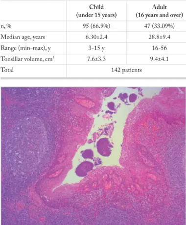
Once the tonsillar tissue was removed and its measurements were completed, it was placed in a 10% formalin solution (formaldehyde 37%-40%, Merck, Darmstadt, Germany) and sent for histopathological examination. Microscopic slices were taken after paraffin blocking. The slices were stained with hematoxylin and eosin and were examined un-der a light microscope (Axioskop 2; Carl Zeiss, Oberko-chen, Germany). Actinomycosis was investigated in all tis-sue samples (Figure 1).
The patients were divided into two groups: (i) younger than 16 years “children” and (ii) older than 16 years “adults.” In both the groups, the presence of Actinomycosis was compared with age, gender, and tonsillar tissue size.
The data obtained in the study were analyzed using the Statistical Package for Social Sciences (SPSS for Windows 22.00; IBM, NY, USA) program. The descriptive statistical data were calculated as number, percentage, mean, and stan-dard deviation when the data were assessed. Before com-parisons in age subgroups, the appropriateness of the data to the normal distribution was investigated in terms of age distribution and the tonsillar volume. Therefore, nonpara-metric Mann-Whitney U test was used for binary compar-isons of age distribution and the tonsillar volumes among the groups. The data on gender distribution (male-female), presence of Actinomyces, and antibiotic use in groups were processed using the chi-square test which we use in the sta-tistics of the data indicated by numbers. The findings were interpreted within a significance level of 0.05 and in a 95% confidence interval.
Informed consents were obtained from the adult patients themselves, and for children, they were obtained from their parents.
Results
In this study, 284 tonsillar tissues of 142 patients were exam-ined histopathologically. There were 95 patients in the pediatric group. The mean age of the children was 6.3±2.4 years (3-15 years). Of the children, 55 were boys and 40 were girls. There were 47 patients in the adult group. The mean age was 28.8±9.4 years (16-56 years). Of these patients, 27 were male and 20 were female (Table 1).
The mean volume of the tonsils in the pediatric group was 7.6±3.3 cm3 and 9.4±4.1 cm3 in the adult group. In the
pediat-ric group, while the mean volume of tonsils with Actinomyces in their tonsillar tissue was 9.6±3.9 cm3, the mean volume of
tonsils without Actinomyces was 7.2±3.0 cm3 (p=0.030). In
the adult group, the mean volume of tonsils with Actinomyces in tonsillar tissue was 9.7±4.1 cm3, whereas the volume
with-out Actinomyces was 8.0 ± 4.1 cm3 (p=0.043). In both the
pe-diatric and the adult groups, the volume of tonsils was higher in which Actinomyces was detected and this was statistically significant.
In the pediatric group, Actinomyces was detected in the ton-sils of 16 patients (16.4%). Three of them were girls and 13 were boys. Actinomyces was detected in tonsillar tissues of 21 adult patients (44.6%). Of these, 14 were male and 7 were
Figure 1. Actinomyces colonies in the tonsillar crypt lumen and leukocyte infiltration with polymorphic nucleus in the crypt epithelium. Hyperplasic lymphoid tissue with obvious germinal center is seen in subepithelial stromal area (hematoxylin and eosin, ×50).
Table 1. General descriptive demographic information Child Adult (under 15 years) (16 years and over)
n, % 95 (66.9%) 47 (33.09%)
Median age, years 6.30±2.4 28.8±9.4
Range (min-max), y 3-15 y 16-56
Tonsillar volume, cm3 7.6±3.3 9.4±4.1
female. In both the pediatric and adult groups, the number of Actinomyces-positive male patients was higher, and it was found that this difference was statistically significant in terms of gender. The mean age of children with Actinomyces was 7.6±3.0 years, whereas the mean age of the adults with Acti-nomyces was 31.2±10.3 years (Table 2). While the relation-ship between the incidence of Actinomyces and the mean age was significant in children (p=0.043), it was not significant in adults (p=0.073). While the incidence of Actinomyces in-creases as the age inin-creases in children, no such finding was found in adults.
Before the tonsillectomy operation, 42 children had a history of 1-week to 10-day antibiotic use (penicillin or clarithromy-cin) in the last 15 days. In adults, 10 patients had a history of antibiotic use (penicillin or clarithromycin) and two patients had a history of antibiotic use (ciprofloxacin). While six of the children with Actinomyces used antibiotics, three of the adults with Actinomyces used antibiotics. A statistically significant inverse relationship was found in terms of the use of antibiot-ics and the likelihood of Actinomyces in both the pediatric and adult groups (p=0.026 for children, p=0.020 for adults).
Discussion
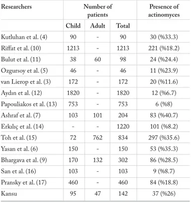
The presence of Actinomyces in the tonsillar tissue was first detected in 1896 (9). Since then, numerous studies were con-ducted on this subject, and various data were reported on the frequency of Actinomyces in tonsillar tissue in different publica-tions (6.7%-40.7%) (Table 3). While Aydin et al. (12) examined 1820 tonsillectomy materials with the highest patient data and found Actinomyces in only 6.7% of them, Riffat and Walker (10) studied tonsillar tissues in 1213 patients and found Acti-nomyces in 18.2% of them. In our study, tonsillectomy materials of 142 patients were examined and Actinomyces was detected in 37 (26%) of them.
Considering the gender factor, there are many studies that show that Actinomyces is more common in men. However, there is no definite consensus on this issue. Although San et al. (16) published in their article that Actinomyces was more common in men, in some other studies, it was reported that there was no difference in terms of gender (3, 4, 9). Erkiliç et al. (14) found Actinomyces in 101 of the tonsillectomy materials of 1220 pa-tients and found that there was a clear female predominance. Yasan et al. (6) also found more Actinomyces in women in the same way. However, in our study, significant male predominance was found in both the pediatric and adult groups.
Compared with children, it is stated that adults had more Ac-tinomyces in the tonsillar tissue (7, 12, 15). Moreover, it was reported that tonsils in children younger than 3 years had no Actinomyces (16). In our study, the incidence of Actinomyces was significantly higher in the adult group than the pediatric group.
The role of Actinomyces found in the tonsillar tissue was not fully understood. Özgürsoy et al. (5) made histological examina-tion of the tonsillar specimens and found that there was a signif-icant increase in lymphoepithelial squamous cell metaplasia and secondary follicles in the Actinomycosis-positive tonsillar tissue. Takasaki et al. (18) published a 75-year-old case with unilater-al excessive tonsillar hypertrophy that could be confused with malignancy and emphasized that Actinomyces causes lymphoid hypertrophy in the tonsils.
There are a number of studies investigating Actinomyces in ton-sillectomy materials obtained for both recurrent tonsillitis and obstructive sleep apnea. In the majority of these publications, patients with obstructive sleep apnea had a higher incidence of Actinomyces in the tonsillectomy specimens than in those with recurrent attacks of tonsillitis (9, 17). As a result, it is thought that Actinomyces settled in the tonsillar tissue can play a role in tonsillar hypertrophy. It is thought that the microorganisms settled in the tonsil produce some toxins, resulting in tonsil hy-pertrophy (3, 10). On the other hand, Toh et al. (15) found no significant relationship between the size of the tonsil and the presence of Actinomyces. Likewise, Yasan et al. (6) reported that Actinomyces did not cause tonsillar hypertrophy. However,
Table 2. Presence of actinomyces in child and adult groups Child Adult Actinomyces Actinomyces Actinomyces Actinomyces
(+) (-) p (+) (-) p
n (%) 16 (16.84%) 79 (83.15%) 0.000 21 (44.6%) 26 (55.3%) 0.000 Median age, y 7.62±3.00 6.11±2.25 0.043 31.28±10.32 26.88±8.90 0.073
Table 3. Rates of actinomyces detection in tonsillar tissue according
to publications
Researchers Number of Presence of patients actinomyces Child Adult Total
Kutluhan et al. (4) 90 - 90 30 (%33.3)
Riffat et al. (10) 1213 - 1213 221 (%18.2)
Bulut et al. (11) 38 60 98 24 (%24.4)
Ozgursoy et al. (5) 46 - 46 11 (%23.9)
van Lierop et al. (3) 172 - 172 20 (%11.6)
Aydın et al. (12) 1820 - 1820 12 (%6.7) Papouliakos et al. (13) 753 - 753 6 (%8) Ashraf et al. (7) 103 101 204 83 (%40.7) Erkılıç et al. (14) - - 1220 101 (%8.2) Toh et al. (15) 72 762 834 297 (%35.6) Yasan et al. (6) 150 - 150 53 (%35.3) Bhargava et al. (9) 170 132 302 86 (%28.5) San et al. (16) 103 - 103 9 (%8.7) Pransky et al. (17) 460 - 460 84 (%18.8) Kansu 95 47 142 37 (%26)
no tonsil size was measured in any study except one (4). In our study, the size of the tonsils was compared and it was found that tonsils with Actinomyces in both the pediatric and the adult groups were significantly larger than those without Actinomy-ces.
One of the most important complications of Actinomyces settled in the tonsillar tissue, except the tonsil hypertrophy is bleeding in the late postoperative period. Schrock et al. (19) ex-amined 1522 patients who underwent tonsillectomy and found a significant relationship between postoperative bleeding and Actinomycosis. In this study, 15 of the 113 patients with Acti-nomycosis were found to have bleeding in the late postoperative period. Although we experienced bleeding in three patients in the late period after tonsillectomy, they were not related to Ac-tinomyces.
Considering that the patients with recurrent attacks of ton-sillitis and that less Actinomyces was found in the tonsils of patients using antibiotics, Pransky et al. (17) suggested that administering penicillin therapy to the patients with obstruc-tive sleep apnea for 12 weeks would reduce the size of tonsils and eventually eliminate the obstructive symptoms. But there is no study conducted on this subject. On the other hand, Bhargava et al. (9) found that patients with sickle cell ane-mia had a high rate of Actinomyces in their tonsils despite receiving long-term antibiotic treatment. Riffat and Walk-er (10) examined 1213 children who had tonsillectomy and found that children who underwent surgery for obstructive sleep apnea had a higher rate of Actinomyces colonization in the tonsillar tissues than children who underwent surgery for recurrent tonsillitis. They also interpreted the result that the frequent use of antibiotics reduced the prevalence of Ac-tinomyces in tonsillar crypts. It was found that our patients who used antibiotics in the last 2 weeks before surgery had less Actinomyces and that there was a significant negative correlation between antibiotic use and the presence of Acti-nomyces in tonsillar specimens.
Some systemic diseases increase the incidence of Actinomyces. Bhargava et al. (9) found that Actinomyces in the tonsillar tis-sues of people with sickle cell anemia, thalassemia, and bronchi-al asthma was at higher rate and the probability of disease for-mation was also higher. It was thought that poor dental hygiene caused this situation.
Conclusion
Actinomyces are microorganisms that can be found in the ton-sillar tissues opportunistically. Their presence in tonsillectomy material does not always indicate active infection. There is a higher incidence in adult tonsillar tissue than in children. Simi-larly, there is a higher rate in men than in women. It is thought that they cause lymphoid hypertrophy with the enzymes they produce. Frequent use of antibiotics reduces the frequency of Actinomyces in tonsillar tissue.
Ethics Committee Approval: Ethics committee approval was
re-ceived for this study from the local ethics committee (Project no: KA12/27).
Informed Consent: Written informed consent was obtained from
pa-tients and papa-tients’ parents who participated in this study.
Peer-review: Externally peer-reviewed.
Conflict of Interest: No conflict of interest was declared by the
aut-hor.
Financial Disclosure: The author declared that this study has received
no financial support.
References
1. Can Ş, Bayındır T, Kuzucu Ç, Bayındır Y, Kızılay A. Serviko-fa-siyal aktinomikoz: Akut süpüratif seyir gösteren bir olgu sunumu. Bozok Tıp Derg 2014; 1: 12-6.
2. Hasan M, Kumar A. Actinomycosis and tonsillar disease. BMC Case Rep 2011; 12: 20111. [CrossRef]
3. van Lierop AC, Prescott CA, Sinclair-Smith CC. An investigation of the significance of actinomycosis in tonsil disease. Int J Pediatr Otorhinolaryngol 2007; 71: 1883-8. [CrossRef]
4. Kutluhan A, Şalvız M, Yalçıner G, Kandemir O, Yeşil C. The role of the actinomyces in obstructive tonsillar hypertrophy and recur-rent tonsillitis in pediatric population. Int J Pediatr Otorhinolary-ngol 2011; 75: 391-4. [CrossRef]
5. Ozgursoy OB, Kemal O, Saatci MR, Tulunay O. Actinomycosis in the etiology of recurrent tonsillitis and obstructive tonsillar hy-pertrophy: answer from a histopathologic point of view. J Otolar-yngol Head Neck Surg 2008; 37: 865-9.
6. Yasan H, Çiriş M, Özel BF, Doğru H, Çandır Ö. The significance of histpathologic tonsillar actinomycosis in pediatric patients with recurrent acute tonsillitis. KBB-Forum 2006; 5: 1-4.
7. Ashraf MJ, Azarpira N, Khademi B, Hashemi B, Shishegar M. Relation between actinomycosis and histopathological and clinical features of the palatine tonsils: An Iranian experience. Iran Red Crescent Med J 2011; 13: 499-502.
8. Prim MP, De Diego JI, Garcia-Bermudez C, Perez-Fernan-dez E, Hardisson D. A method to calculate the volume of palatine tonsils. Anat Rec (Hoboken) 2010; 293: 2144-6.
[CrossRef ]
9. Bhargava D, Bhusnurmath B, Sundaram KR, Raman R, Al Okbi HM, Al Abri R, et al. Tonsillar actinomycosis: a clinicopathologi-cal study. Acta Trop 2001; 80: 163-8. [CrossRef]
10. Riffat F, Walker P. Prevalence of tonsillar actinomyces in children undergoing tonsillectomy for sleep disordered breathing compared with recurrent tonsillitis. Int J Pediatr Otorhinolaryngol 2009; 73: 1111-3. [CrossRef]
11. Bulut AŞ. The presence of actinomyces spp. in tonsillectomy speci-mens. Flora 2012; 17: 148-50.
12. Aydin A, Erkiliç S, Beyazit YA, Koçer NE, Ozer E, Kanlikama M. Relation between actinomycosis and histopathological and clinical features of the palatine tonsils: a comparative study between adults and pediatric patients. Rev Laryngol Otol Rhinol (Bord) 2005; 126: 95-8.
13. Papouliakos S, Karkos PD, Korres G, Karatzias G, Sastry A, Riga M. Comparison of clinical and histopathological evaluation of tonsils in pediatric and adult patients. Eur Arch Otorhinolaryngol 2009; 266: 1309-13. [CrossRef]
14. Erkılıç S, Aydın A, Koçer NE. Tonsilla palatinanın benign histo-patolojik lezyonları: 1220 olgunun retrospektif incelenmesi. Turk J Pathol 2002; 18: 20-1.
15. Toh ST, Yuen HW, Goh YH. Actinomycetes colonization of ton-sils: a comparative study between patients with and without recur-rent tonsillitis. J laryngol Otol 2007; 121: 775-8. [CrossRef]
16. San T, Gürkan E, Erdoğan B, Özkanlı Ş. Does actinomyces have any role in tonsiller diseases in children? İstanbul Med J 2014; 15: 209-12.
17. Pransky SM, Feldman JI, Kearns DB, Seid AB, Billman GF. Acti-nomycosis in obstructive tonsillar hypertrophy and recurrent tonsilli-tis. Arch Otolaryngol Head Neck Surg 1991; 117: 883-5. [CrossRef]
18. Takasaki K, Kitaoka K, Kaieda S, Hayashi T, Abe K, Takahashi H. A case of actinomycosis causing unilateral tonsillar hypertrophy. Acta Otolaryngol 2006; 126: 1001-4. [CrossRef]
19. Schrock A, Send T, Heukamp L, Gerstner AO, Bootz F, Jacob M. The role of histology and other risk factors for post-tonsillectomy haemor-rhage. Eur Arch Otorhinolaryngol 2009; 266: 1983-7. [CrossRef]