Epigenetic Inactivation of 14-3-3
in Oral Carcinoma: Association with p16
INK4aSilencing and Human Papillomavirus Negativity
1Milena Gasco, Alexandra K. Bell, Victoria Heath, Alex Sullivan, Paul Smith, Louise Hiller, Isik Yulug,
Gianmauro Numico, Marco Merlano, Paul J. Farrell, Mahvash Tavassoli, Barry Gusterson, and Tim Crook
2UO Oncologia Medica, Azienda Ospedaliera S Croce e Carle, 12100 Cuneo, Italy [M. G., G. N., M. M.]; Ludwig Institute for Cancer Research, Imperial College of Science and Medicine, St. Mary’s Campus, Norfolk Place, London W2 1PG, United Kingdom [A. S., P. J. F., T. C.]; Institute of Cancer Genetics, Brunel University, Uxbridge UB8 4SP, United Kingdom [P. S.]; CRC Trials Unit, Institute for Cancer Studies, Edgbaston, Birmingham B15 2TT, United Kingdom [L. H.]; Department of Molecular Biology and Genetics, Bilkent University, 06533 Ankara, Turkey [I. Y.]; Cancer Gene Therapy Group, King’s College London, The Rayne Institute, London SE5 9NU [M. T.]; and University Department of Pathology, Western Infirmary, Glasgow G11 6NT, Scotland [A. K. B., V. H., B. G.]
ABSTRACT
In vitro studies have identified 14-3-3 as a regulator of senescence in human keratinocytes. To assess its contribution to squamous neoplasia, we have analyzed genetic and epigenetic changes in this gene in squamous cell carcinomas (SCCs) and dysplastic lesions of the oral cavity. No mutations were detected in the coding sequence of 14-3-3 in 20 oral carcinomas, and there was loss of heterozygosity in only 7 of 40 informative cases. In contrast to the absence of genetic change, aberrant methylation within 14-3-3 was detected in 32 of 92 squamous cell carcinomas and in 3 of 6 oral dysplasias and was associated with reduced or absent expression at both mRNA and protein levels. Methylation was not detected in matched, normal epithelial tissue controls. Carcinomas in which 14-3-3 was meth-ylated were significantly more likely to lack DNA sequences from human papillomavirus and to have coincident methylation of p16INK4athan cases that expressed 14-3-3. Methylation was detected in SCC, both wild-type and mutant for p53, but was more commonly detected in cancers with wild-type p53. These results implicate coincident epigenetic abrogation of function in both and p16INK4ain a subset of SCCs of the oral cavity.
INTRODUCTION SCC3
of the head and neck, including carcinoma of the oral cavity, is the sixth most common cancer worldwide. Epidemiological data strongly link smoking and alcohol consumption to the development of this malignancy (1). There is also evidence that a subset of oral SCCs is associated with infection by oncogenic HPV; a recent study iden-tified HPV 16 in⬃25% of cases (2).
(also known as stratifin) was first identified as a gene expressed specifically in stratified squamous epithelium (3). In that study, it was shown that expression of was absent from a few breast carcinoma cell lines but was not down-regulated in other cancer cell lines. Down-regulation of was also reported in a head and neck SCC cell line (4). is a regulator of senescence in epithelial cells. Down-regulation of allows keratinocytes to escape from replicative senes-cence (5). Steady-state levels of p16INK4a
increase as keratinocytes approach replicative senescence, and conversely, p16INK4a
is almost always inactivated in immortalized keratinocytes (6, 7). Consistent with these observations, there is loss of p16INK4a
expression in human keratinocytes immortalized after transduction by retroviruses express-ing antisense (5). Thus, in vitro evidence suggests that may have an important function in malignant development in epithelial tissues. is induced by p53 in response to DNA damage (8) and mediates
a G2checkpoint. Such a mechanistic model might imply that down-regulation of would not occur in cancers with p53 mutation or in HPV-associated carcinomas, because p53 is targeted by HPV-encoded E6 (9).
The involvement of in human cancer has been established in studies of breast cancer in which methylation-dependent silencing of the gene was observed in a majority of cases of ductal carcinoma (10). Furthermore, loss of occurs early in neoplastic development in breast epithelium (11). Silencing of expression has also been re-ported in 43% of primary gastric adenocarcinomas (12) and 89% of hepatocellular carcinomas (13).
To determine the contribution of changes in to carcinogenesis in squamous epithelium, we have analyzed genetic and epigenetic changes in the gene in a series of malignant and premalignant neo-plastic oral lesions.
MATERIALS AND METHODS
Tissues and Nucleic Acid Isolation. Cancers, with patient-matched
nor-mal epithelium where available, were obtained at operation. Tissues were snap frozen and stored in liquid nitrogen before analysis. The presence of a majority of neoplastic tissue was verified in each carcinoma by histopathological analysis of tissue sections. Genomic DNA was isolated by proteinase K digestion, and total RNA was isolated by phenol/guanidinium. cDNA was synthesized from 5g of total RNA using the Prostar system (Stratagene). Fifty-six SCCs were available for analysis from this series. A second series of cancers, comprising 36 paraffin-embedded tissue sections, was also analyzed. For isolation of genomic DNA, sections were treated with xylene to remove paraffin, dehydrated in ethanol, and then subjected to extended digestion in proteinase K.
Analysis of Structure and Expression of. Mutation analysis and
anal-ysis of LOH were performed using primers and conditions essentially as described by Ferguson et al. (10). Analysis of SCC for mutations was performed by amplification of the open reading frame with Pfx DNA poly-merase and sequencing of clones in the vector pCRblunt (Invitrogen). For analysis of methylation, genomic DNA (1 g) was modified with sodium bisulfite using the CpG modification system (Intergen) as directed by the manufacturers. Bisulfite sequencing was done with primers described previ-ously (10). After PCR, reactions were cleaned with the Qiagen PCR purifica-tion kit and then directly sequenced on an ABI sequencer. MSP was done according to Ferguson et al. (10) with the exception that for DNA isolated from paraffin sections, PCR was performed for 40 rather than 35 cycles. PCR products were resolved on 10% polyacrylamide gels and visualized by staining with ethidium bromide. For analysis of expression, cDNA was synthesized as above from RNA isolated from matched pairs of normal and tumor. This was used as substrate for PCR with primers as described (10). PCR was performed for 28 cycles, under which conditions the reaction is in the expo-nential phase of amplification. PCR products were resolved on 2% agarose gels, transferred to nylon, and then hybridized with oligonucleotides comple-mentary to the amplified sequences, end-labeled with [␥-32P]ATP by
polynu-cleotide kinase. For immunocytochemical analysis, 3m unstained sections were taken from 38 samples also analyzed for HPV, p53 mutations, and sequence and methylation in. Tissue sections were dewaxed in xylene, rinsed well in ethanol, and then washed in tap water. Antigen retrieval was performed
Received 10/23/01; accepted 1/30/02.
The costs of publication of this article were defrayed in part by the payment of page charges. This article must therefore be hereby marked advertisement in accordance with 18 U.S.C. Section 1734 solely to indicate this fact.
1Research in the laboratory of B. A. G. is supported by Breakthrough Breast Cancer. 2To whom requests for reprints should be addressed, at Ludwig Institute for Cancer
Research, Imperial College Faculty of Medicine, St. Mary’s Campus, Norfolk Place, W2 1PG London, United Kingdom. Phone: 0207-563-7721; Fax: 0207-724-8586; E-mail: [email protected].
3The abbreviations used are: SCC, squamous cell carcinoma; HPV, human
papillo-mavirus;, 14-3-3; MSP, methylation-specific PCR; LOH, loss of heterozygosity; RT-PCR, reverse transcription-PCR.
polyacrylamide gels and visualized under UV light after staining with ethidium bromide. RT-PCR analysis of expression was done as described by Gonzalez-Zuluetta et al. (16).
p53 Analysis. Mutations in p53 were sought using single-strand
confor-mation analysis. Suspected mutations were identified by reamplification with
Pfx polymerase, ligation into pCRblunt (Invitrogen), and sequencing of
mul-tiple plasmid clones.
HPV Typing. HPV sequences were sought in DNA from frozen tissue
using the MY09/MY11 (HPV) and PC04/GP20 (globin) primers (17). For detection of HPV in DNA from paraffin sections, each genomic DNA was initially checked by amplification with the PCO3/PCO4 primers (18) and then analyzed with the CPI/CPIIG consensus primer pair (19). HPV type was determined by direct sequencing.
Statistical Analysis. All Ps were obtained from2tests with continuity
corrections.
RESULTS
Inactivation of in Oral Cancer Occurs Predominantly by Epigenetic Silencing. LOH in was sought using a microsatellite described previously (10). LOH was detected in 7 of 40 informa-tive cases. To determine the presence of mutations, the open reading frame was amplified as a single fragment from genomic DNA of 20 SCCs, and the sequence of individual plasmid clones was determined. No mutations were detected in these cases. Next, evidence for epigenetic change in was sought in DNA from all 92 primary oral SCCs, 56 from fresh-frozen SCCs, and 36 from
formed immunocytochemical analysis of expression on the 36 paraffin-embedded cases. Expression of was reduced or absent in each case with methylation (Fig. 2).
Methylation of Is Detected in Premalignant Oral Lesions. We were interested to determine whether methylation was present in oral premalignant lesions. Using MSP, methylated DNA was de-tected in 3 of 6 oral dysplasias (Fig. 1).
Silencing of Predominantly Targets Oral Cancers Lacking HPV DNA. The etiological association between oral cancer and HPV is well established (2). We therefore analyzed each case for HPV DNA. HPV DNA sequences were detected in 17 of 92 (18%) cases (Fig. 3). HPV 16 was detected in 13 cases, HPV 6 in 3 cases, and 1 case contained types 6 and 16. Of the 17 HPV-positive cancers, there was methylation of in only 1 case (containing HPV 16; 6%), whereas methylation was detected in 31 of 75 (41%) HPV-negative cases (P⫽ 0.013; Table 1). The 3 oral dysplasias with methylation were all negative for HPV DNA. These results indicate that methyl-ation of is significantly more common in HPV-negative than in HPV-positive oral cancers.
Methylation of Occurs More Commonly in Cancers with Wild-Type p53. expression is induced by p53 (8), and we inves-tigated the hypothesis that inactivation of would be less likely to occur in cancers with mutant p53. Mutations in p53 were identified in a total of 37 of 92 (40%) cancers. Methylation of was detected in 24 of 55 (44%) cases with wild-type p53 and 8 of 37 (22%) cases with
Fig. 1. Methylation of in oral carcinoma. A, bisulfite se-quencing of in oral carcinoma. Representative sequencing anal-ysis from different regions of the sequenced fragment of is shown. Arrows, presence of methylated CpG dinucleotides. Note the presence of unmethylated (T) residues in some CpGs. These indicate either hemimethylation or the presence of normal tissue.
B, MSP analysis of methylation in (upper panel) and p16INK4a
(lower panel) in oral neoplasia. Lanes 1–3, oral SCC; Lanes 4 and
5, oral dysplasias. M, methylated reaction; U, unmethylated
re-action.
p53 mutations (Table 1; P⫽ 0.074). Although this P just fails to reach statistical significance, these results nevertheless indicate that silenc-ing of occurs more commonly in cases lacking p53 mutations.
Concomitant Methylation of and p16INK4a
in Oral Cancer. Down-regulation of is accompanied by loss of p16INK4a
expression during keratinocyte immortalization (5). We determined whether there was methylation in the p16INK4a
gene in the series of oral SCCs characterized for. Methylated p16INK4a
DNA was detected in 38 of 92 (41%) SCCs (Fig. 1); these comprised 5 of 17 HPV-positive and 33 of 75 HPV-negative cases. Methylation of p16INK4a
was associated with reduced expression (Fig. 2). Of the 32 SCCs with methylated DNA, there was concomitant methylation of p16INK4a
in 25 cases (78%; Table 1), whereas of the 60 SCCs with unmethylated, there was methylation of p16INK4a
in 13 (22%; P ⫽ 0.001). Of the 3 dysplasias with methylation, there was concomitant methylation of
p16INK4a
in 1 case, whereas a further dysplasia had methylated
p16INK4a
but not . The previously described polymorphism (Ala3Thr) at codon 148 occurred in 6 cases. Mutations in p16INK4a
were detected in 3 of 92 SCCs, all of which were negative for HPV DNA and unmethylated in (Table 2).
Taken together, these results indicate a significant association between epigenetic silencing of and p16INK4a
in HPV-negative oral SCCs.
DISCUSSION
The importance of as a regulator of senescence in human kera-tinocytes has been demonstrated clearly (5). In the present study, we demonstrate that is subject to methylation-dependent transcriptional silencing in primary oral SCC and in premalignant oral dysplastic lesions.
The first conclusion to be drawn from our studies is that genetic changes in are uncommon in oral cancer, with a complete absence of mutations and a relatively low (18%) frequency of LOH in the gene. Inactivation of in oral neoplasia, therefore, appears to occur almost exclusively by epigenetic, transcriptional silencing. Absence
of mutations and low frequency of LOH are consistent with studies of breast cancer (10, 11) and other adenocarcinomas (12, 13). The common silencing of seen strongly implies that loss of expression is an important event in malignant transformation in a proportion of oral SCC and suggests that functions as a tumor suppressor gene in squamous as well as glandular epithelium. Furthermore, detection of methylated DNA in dysplastic oral lesions suggests that epigenetic silencing of the gene occurs as an early event in a subset of oral SCCs.
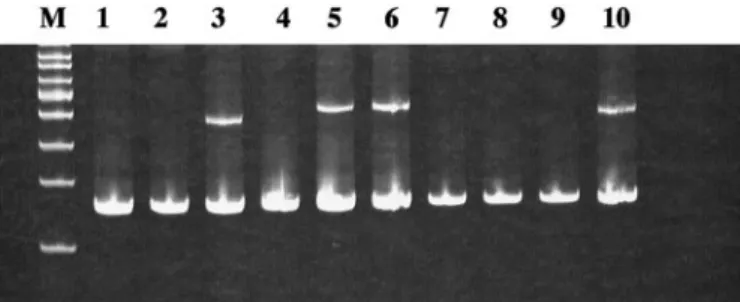
Fig. 3. Detection of HPV in oral neoplasia. Duplex, degenerate PCR was performed with primers for HPV (upper band present in Lanes 3, 5, 6, and 10) and globin (lower
band) as described in “Materials and Methods.” Amplified products were resolved on 10%
acrylamide gels. M, molecular weight markers.
Table 1 Characteristics of oral SCC with and without methylation of p53 WTa(n⫽ 55) p53 MTa(n⫽ 37) P methylated (n ⫽ 32) 24/55 (0.44) 8/37 (0.56) 0.074 unmethylated (n ⫽ 60) 31/55 (0.56) 24/55 (0.44) HPV⫹ve (n ⫽ 17) HPV⫺ve (n ⫽ 75) P methylated (n ⫽ 32) 1/17 (0.06) 31/75 (0.41) 0.013 unmethylated (n ⫽ 60) 16/17 (0.94) 44/75 (0.59) p16 meth. (n⫽ 38) p16 unmeth. (n⫽ 54) methylated (n ⫽ 32) 25/32 (0.78) 7/32 (0.22) unmethylated (n ⫽ 60) 13/60 (0.22) 47/60 (0.78) P 0.001
ap53WT, wild-type p53; p53MT, mutant p53;⫹ve, HPV positive; ⫺ve, HPV
nega-tive; meth., methylated; unmeth., unmethylated. Fig. 2. Down-regulation of expression in oral
neopla-sia. Upper panel, RT-PCR analysis of and p16INK4a
mRNA in matched pairs of normal (N) and tumor (T).
Lanes 1– 4, oral SCCs; Lanes 5 and 6, oral dysplasias. Lower panel, immunocytochemical analysis of
expres-sion. A, is expressed in the cytoplasm of normal oral epithelium. B, loss of expression in oral carcinoma.
a significant proportion of ductal carcinomas in situ (11). There is compelling evidence that inactivation of p16INK4a
is an important event in immortalization of keratinocytes (6, 7). Consistent with this, immortalization of primary keratinocytes by antisense is accompa-nied by down-regulation of p16INK4a
expression (5). In our series, the majority of oral SCCs with methylation also had methylation of
p16INK4a
. HPV sequences were detected in⬃20% of the SCCs in our series, comparable with a previous large study (2), and cancers with concomitant methylation of and p16INK4a
were almost invariably HPV negative. In contrast, cases in which only p16INK4a
was meth-ylated were both HPV positive and HPV negative. Taken together, these observations are consistent with the hypothesis that down-regulation of has effects equivalent to expression of E6 and E7 proteins of HPV (5), because these proteins cooperate to immortalize primary keratinocytes (20).
The observation that expression is induced by p53 (8) suggested that silencing of might represent a response to the presence of the wild-type protein in cancers lacking a mechanism for inactivating p53. One recognized mechanism of p53 inactivation is expression of HPV 16 E6 protein, which mediates ubiquitin-dependent proteolysis via E6-AP. It is of interest that cancers with methylated sequences were predominantly those lacking HPV DNA. This observation supports the hypothesis that abrogation of p53-dependent induction is, at least in part, the mechanistic basis for silencing of to predominantly target HPV-negative oral cancers. Analysis of the p53 sequence of the cases also supported this; SCC with methylation was more com-monly wild-type for p53.
This raises the question of why some cancers containing p53 mutations also have methylation. One likely explanation is that mutation of p53 is required to abrogate transcriptional induction of other genes in the p53 pathway that cannot be or are not epige-netically silenced. p53 regulates expression of a large number of effector proteins including mediators of cell cycle arrest, differen-tiation, and apoptosis (21). Inactivation of p53, by mutation or other means, will abrogate induction of the entire p53-dependent program of gene expression and thereby have more profound effects than mutation or inactivation of individual downstream genes (21). An alternative but not mutually exclusive explanation is that silencing of is an early event in a subset of oral SCCs. Selection for p53 mutation would then be proposed to operate later during neoplastic progression to favor outgrowth of clones unable to undergo p53-dependent apoptosis or perhaps those expressing “gain of function” p53 mutants. Supporting this possibility, loss of is detectable in a high proportion of early breast lesions (11) and was detected in oral premalignant lesions in our series. What then is the role of down-regulation in squamous carcinomas? One obvious possibility is that loss is sufficient to immortalize squamous epithelium. As such, the role of silencing in squamous neoplasia would be analogous to that observed in vitro in primary keratinocytes (5). Detection of methylation in preneoplastic oral epithelium and preneoplastic vulval intraepithelial neoplasia is consistent with such a hypothesis (22). A further possibility is that silencing of is not required for immortalization but occurs at a stage after immortalization. Loss at this stage may result in
im- in oral cancer, it will be of interest to determine whether cases with silencing show differences in response to treatment regimens based on radiotherapy or chemotherapy. Moreover, the increased radio- and chemosensitivity of ⫺/⫺ cells may facilitate identification of patients likely to derive greater benefit from specific treatment strategies.
REFERENCES
1. Franceschi, S., Talamini, R., Barra, S., Baron, A. E., Negri, E., Bidoli, E., Serraino, D., and La Vecchia, C. Smoking and drinking in relation to cancers of the oral cavity, pharynx, larynx and esophagus in northern Italy. Cancer Res., 50: 6502– 6507, 1990.
2. Gillison, M. L., Koch, W. M., Capone, R. B., Spafford, M., Westra, W. H., Wu, L., Zahurak, M. L., Daniel, R. W., Viglione, M., Symer, D. E., Shah, K. V., and Sidransky, D. Evidence for a causal association between human papillomavirus and a subset of head and neck cancers. J. Natl. Cancer Inst., 92: 709 –720, 2000. 3. Prasad, G. L., Valverius, E. M., McDuffie, E., and Cooper, H. L. Complementary
DNA cloning of a novel epithelial cell marker protein, HME1, that may be down-regulated in neoplastic mammary cells. Cell Growth Differ., 3: 507–513, 1992. 4. Vellucci, V. F., Germino, F. J., and Reiss, M. Cloning of putative regulatory genes
from primary human keratinocytes by subtractive hybridisation. Gene (Amst.), 166: 213–220, 1995.
5. Dellambra, E., Golisano, O., Bondanza, S., Siviero, E., Lacal, P., Molinari, M., D’Atri, S., and De Luca, M. Down-regulation of 14-3-3 prevents clonal evolution and leads to immortalization of primary human keratinocytes. J. Cell Biol., 149: 1117–1129, 2000.
6. Loughran, O., Malliri, A., Owens, D., Gallimore, P. H., Stanley, M. A., Ozanne, B., Frame, M. C., and Parkinson, E. K. Association of CDKN2A/p16INK4Awith human
head and neck keratinocyte replicative senescence: relationship of the dysfunction to immortality and neoplasia. Oncogene, 13: 561–568, 1996.
7. Munro, J., Stott, F. J., Vousden, K. H., Peters, G., and Parkinson, E. K. Role of the alternative INK4A proteins in human keratinocyte senescence: evidence for the specific inactivation of p16INK4aupon immortalization. Cancer Res., 59: 2516 –2521,
1999.
8. Hermeking, H., Lengauer, C., Polyak, K., He, T. C., Zhang, L., Thiagalingam, S., Kinzler, K. W., and Vogelstein, B. 14-3-3 is a p53-regulated inhibitor of G2/M. Mol. Cell, 1: 3–11, 1997.
9. Werness, B. A., Levine, A. J., and Howley, P. M. Association of human papil-lomavirus types 16 and 18 E6 proteins with p53. Science (Wash. DC), 248: 76 –79, 1990.
10. Ferguson, A. T., Evron, E., Umbricht, C. B., Pandita, T. K., Chan, T. A., Hermeking, H., Marks, J. R., Lambers, A. R., Futreal, P. A., Stampfer, M. R., and Sukumar, S. High frequency of hypermethylation at the 14-3-3 locus leads to gene silencing in breast cancer. Proc. Natl. Acad. Sci. USA, 97: 6049 – 6054, 2000.
11. Umbricht, C. B., Evron, E., Gabrielson, E., Ferguson, A., Marks, J., and Sukumar, S. Hypermethylation of 14-3-3 sigma (stratifin) is an early event in breast cancer. Oncogene, 20: 3348 –3353, 2001.
12. Suzuki, H., Itoh, F., Toyota, M., Kikuchi, T., Kakiuchi, H., and Imai, K. Inactivation of the 14-3-3 gene is associated with 5⬘ CpG island hypermethylation in human cancers. Cancer Res., 60: 4353– 4357, 2000.
13. Iwata, N., Yamamoto, H., Sasaki, S., Itoh, F., Suzuki, H., Kikuchi, T., Kaneto, H., Iku, S., Ozeki, I., Karino, Y., Satoh, T., Toyota, J., Satoh, M., Endo, T., and Imai, K. Frequent hypermethylation of CpG islands and loss of expression of the 14-3-3 gene in human hepatocellular carcinoma. Oncogene, 19: 5298 –5302, 2000.
14. Zhang, S. Y., Klein-Szanto, A. J., Sauter, E. R., Shafarenko, M., Mitsunaga, S., Nobori, T., Carson, D. A., Ridge, J. A., and Goodrow, T. L. Higher frequency of alterations in the p16/CDKN2 gene in squamous cell carcinoma cell lines than in primary tumors of the head and neck. Cancer Res., 54: 5050 –5053, 1994. 15. Herman, J. G., Graff, J. R., Myohanen, S., Nelkin, B. D., and Baylin, S. B.
Methy-lation-specific PCR: a novel assay for methylation status of CpG islands. Proc. Natl. Acad. Sci. USA, 93: 9821–9826, 1996.
16. Gonzalez-Zulueta, M., Bender, C. M., Yang, A. S., Nguyen, T., Beart, R. W., Van Tornout, J. M., and Jones, P. A. Methylation of the 5⬘ CpG island of the p16/CDKN2 tumor suppressor gene in normal and transformed human tissues correlates with gene silencing. Cancer Res., 55: 4531– 4535, 1995.
17. Resnick, R. M., Cornelissen, M. T., Wright, D. K., Eichinger, G. H., Fox, H. S., ter Schegget, J., and Manos, M. M. Detection and typing of human papillomavirus in archival cervical cancer specimens by DNA amplification with consensus primers. J. Natl. Cancer Inst., 82: 1477–1484, 1990.
18. Greer, C. E., Peterson, S. L., Kiviat, N. B., and Manos, N. M. PCR amplification from paraffin-embedded tissues: effects of fixative and fixation time. Am. J. Clin. Pathol.,
95: 117–124, 1991.
19. Smits, H. L., Tieben, L. M., Tjong-A-Hung, S. P., Jebbink, M. F., Minnaar, R. P., Jansen, C. L., and ter Schegget, J. Detection and typing of human papillomaviruses present in fixed and stained archival cervical smears by a consensus polymerase chain reaction and direct sequence analysis allow the identification of a broad spectrum of human papillomavirus types. J. Gen. Virol., 73: 3263–3268, 1992.
20. Hawley-Nelson, P., Vousden, K. H., Hubbert, N. L., Lowy, D. R., and Schiller, J. T. HPV 16E6 and E7 proteins co-operate to immortalize human foreskin keratinocytes. EMBO J., 8: 3905–3910, 1987.
21. Vogelstein, B., Lane, D., and Levine, A. J. Surfing the p53 network. Nature (Lond.),
408: 307–310, 2000.
22. Gasco, M., Sullivan, A., Repellin, C., Brooks, L., Farrell, P. J., Tidy, J. A., Dunne, B., Gusterson, B., Evans, D. J., and Crook, T. Co-incident inactivation of 14-3-3 and p16INK4ais an early event in vulval squamous neoplasia. Oncogene, in press,
2002.
23. Subramanian, R. R., Masters, S. C., Zhang, H., and Fu, H. Functional conservation of 14-3-3 isoforms in inhibiting bad-induced apoptosis. Exp. Cell Res., 271: 142–151, 2001.
24. Laronga, C., Yang, H. Y., Neal, C., and Lee, M. H. Association of the cyclin-dependent kinases and 14-3-3 negatively regulates cell cycle progression. J. Biol. Chem., 275: 23106 –23112, 2000.
25. Chan, T. A., Hwang, P. M., Hermeking, H., Kinzler, K. W., and Vogelstein, B. Co-operative effects of genes controlling the G2/M checkpoint. Genes Dev., 14:
1584 –1588, 2000.